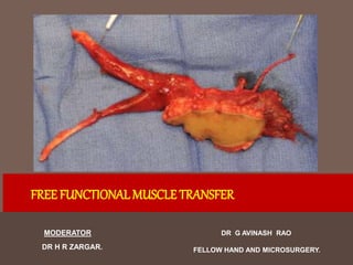
SlideShare a Scribd company logo
1 of 110
DR G AVINASH RAO
FELLOW HAND AND MICROSURGERY.
FREE FUNCTIONALMUSCLE TRANSFER
MODERATOR
DR H R ZARGAR.
Introduction
 Loss of upper extremity function secondary to brachial plexus injuries /
severe trauma / established VIC / Congenital absence of muscle / Muscle
loss after tumor surgery / Peripheral nerve injuries are a challenging
problems in reconstructive hand Surgery.
 These cases have profound functional loss with limited reconstructive
options.
 Advances in microsurgery offered a new approach in the management of
these injuries.
 Vascularized free muscle flaps are indicated for complex reconstruction of:
 Defects requiring filling of dead space
 Coverage of exposed vital structures
 Treatment of osteomyelitis
 Functional reconstruction of muscle loss or absence in congenital conditions
 Coverage of exposed orthopedic hardware
……………………………………………………………………………………..
 fFMT provide useful function apart from the above advantages
mentioned for VfMT
 Tamai et al. Free muscle transplants in dogs, with microsurgical neurovascular
anastomoses. Plast Reconstr Surg. 1970.
 Stevanovic, Seaber, Urbaniak Canine experimental free muscle transplantation.
Microsurgery. 1986.
………………………………………………………………………
 Terzis, Suggested that muscle bulk decreases with muscle transplantation to
25-50%. J Hand Surg, 1978.
 Transplanted muscles regained full strength, sometimes stronger
than pre-transplanted power. Doi K , Clin Plast Surg, 2002 •
Functional Free Muscle
 Indications
 Deficiency of critical motor function with no suitable
tendon transfer options.
 No suitable rotational muscle transfer - available
 Soft tissue defect requiring coverage in combination with
functional loss.
Functional Free Muscle -
Indications
 Functional reconstruction after:
▪▪ Volkmann ischemic contracture
▪▪ Severe brachial plexus preganglionic injury
▪▪ Long-standing neurologic injury
▪▪ Loss of muscle from trauma
▪▪ Loss of muscle from tumor resection
▪▪ Electrical injuries to the upper extremity
▪▪ Congenital muscle absence
FFMT - Upper Extremity
Indications
▪▪ Deltoid reconstruction
▪▪ Triceps reconstruction
▪▪ Elbow flexion
▪▪ Finger flexion
▪▪ Finger extension
▪▪ Thumb opposition
Contraindications - Absolute
▪▪ Medical comorbidities that would not allow a patient to undergo a long
surgical procedure safely
▪▪ No acceptable donor nerve to innervate the transferred Muscle / Inadequate
recipient vessels for microvascular anastamosis
▪▪ Patient unable to participate in and complete the time-consuming
rehabilitation program for the FFMT
Contraindications - Relative
▪▪ Poor passive range of motion of the involved joints across which the
muscle is to act: The patient may require staged procedures to prepare the
upper extremity for the FFMT, such as tenolysis, capsulotomies, and/or
contracture release. This requires attentive postoperative physiotherapy before
the staged FFMT.
▪▪ Lack of antagonistic muscle function: This can be reconstructed with a double
transfer or a second FFMT, or function can be augmented with additional
procedures, such as total wrist arthrodesis.
▪▪ Poor soft-tissue coverage: The FFMT can include a skin paddle to address
both a soft-tissue loss as well as a functional loss.
▪▪ Poor sensation in the hand of the extremity requiring reconstruction: This
can be addressed with sensory nerve reconstruction either before or at the same
time as the FFMT.
Functional Free Muscle Goals
(Manktelow)
 • Supply a useful range of motion.
 • Provide adequate strength for functional activities.
 • FMT must be under volitional (ones will) control.
 Manktelow, Zuker, McKee. Functioning free muscle transplantation. J Hand
Surg [Am]. 1984
Ideal Candidates for FFMT
▪▪ Excellent results in children
▪▪ Adult patients ideally under the age of 45
▪▪ Compliant and motivated patient
▪▪ Healthy, with no comorbidities that would put the patient at risk during the
FFMT or that would compromise the ability to complete the extensive
rehabilitation Post-op.
▪▪ Access to skilled therapists, knowledgeable in upperextremity and FFMT
rehabilitation
Functional Free Muscle - Pre-
requisites
▪▪ Stable soft-tissue coverage at the reconstruction site.
▪▪ Full passive range of motion of the joints across which the transfer will act
▪▪ Tendons with adequate gliding
▪▪ Antagonistic muscle function required.
▪▪Reconstructive site with reliable recipient vessels for microvascular
anastomosis
▪▪ Reconstructive site with a pure, undamaged motor nerve to innervate the
FFMT
▪▪ Less complicated options for reconstruction either not available or
unsuccessful
 There is no fixed time limits for the procedure from
the time of injury or loss of function – Provided all
the prerequisites are fulfilled.
Mathes Nahai – Myocutaneous
Flap
Free muscle transfer
• Type of blood supply
I. One vascular pedicle - Rectus femoris,Tensor fascia lata, AbdDM.
II. Dominant pedicles and minor pedicles - Gracilis, Biceps femoris, Soleus,
Trapezius.
III.Two Dominant Vessels – Rectus Abdominis, G.Maximus, Serratus,
Temporalis.
IV. Segmental Supply – Sartorius, T.Anterior, FHL.
V. One dominant pedicle and secondary segmental pedicles - Latissimus
dorsi, Pectoralis major.
Donor Muscle Options
 Gracilis
 Latissimus
 Rectus femoris
 Pectoralis Major
 Medial gastrocnemius
 Tensor fascia lata
 Serratus
Donor Muscle - General
Considerations
• Expendible donor muscle
– sacrificed with acceptable donor site Morbidity
• Adequate length and excursion for new function
• Vascular pedicle permits transfer
 Muscle Type – pennate (stronger) Rectus femoris – strap (better
excursion) Gracilis, Latissimus dorsi
• Cross sectional area – pennate - greater cross sectional area results in
greater strength.
Muscle Excursion – Ideally 6-7 cm of muscle excursion to produce
functional range of flexion of fingers and elbow.
Examination/Imaging
 Physical Exam
▪▪ A detailed preoperative physical exam is important to evaluate which nerves are
functioning in the upper extremity, and to identify the options for innervating the
FFMT.
▪▪ A focused vascular exam of the upper extremity is also important. This involves
identifying palpable arteries and those that can be identified by Doppler. It is also
important to perform a preoperative Allen test for planning reconstructive
procedures in the forearm.
▪▪ The soft-tissue coverage for the site of the transfer should be assessed to ensure
it is adequate.
▪▪ Passive range of motion of the joints across which the muscle is to act should be
evaluated, and the excursion of the recipient tendons should be tested.
 Investigation
▪▪ Nerve conduction studies are usually not helpful for preoperative planning.
▪▪ Electromyography can be useful to evaluate pronator quadratus, which can
give useful information about the functional status of the anterior
interosseous nerve.
Surgical Technique
▪▪ Free functional muscle transfers are complex reconstructive procedures that
often require prolonged operative time, and skilled anesthesiologists
experienced in the care of patients undergoing these procedures.
▪▪ It is important that the patient maintain excellent peripheral perfusion and a
normal body temperature during the procedures so as not to compromise the
FFMT. It is also important the patients do not receive long-acting paralytic
medication, to enable intraoperative stimulation of donor nerves.
.
▪▪Well-maintained microsurgical instruments are important for the microsurgical
component of the procedure.
▪▪ The operating microscope is used.
▪▪ 9.0 or 10.0 nylon suture is used with 70–100 micron needle for the vascular
anastomosis and nerve coaptation.
▪▪ A nerve stimulator is needed.
▪▪ Fibrin glue is used to augment the nerve coaptation.
Patient Positioning
▪▪ For free functional gracilis muscle transfers, patients are most commonly
placed supine. This allows access to the upper extremity for reconstruction
as well as to the leg for gracilis muscle harvest.
▪▪ The gracilis muscle is harvested with the patient in a frog-leg position.
▪▪ The preference is to use the contralateral gracilis for elbow flexion and
finger extension reconstruction and the ipsilateral gracilis for finger
flexion reconstruction. This orientation is chosen based on the position of
the recipient vessels.
▪▪Free functional latissimus dorsi muscle transfer requires planning for patient
positioning.
▪▪ The latissimus dorsi muscle can be harvested with the patient either prone or
in the lateral decubitus position.
▪▪ Muscle transfer and origin and insertion creation require repositioning the
patient into the supine position.
▪▪ Occasionally, for finger flexion or extension reconstruction, the entire
procedure can be performed in the lateral decubitus position.
▪▪ A tourniquet is used on the upper extremity during the preparation of the
forearm for functional finger flexion or extension reconstruction.
▪▪ It is important to ensure the tourniquet is deflated, before performing the
microvascular anastomosis
Surgical Sequence
▪▪ Before harvesting of a free functional muscle, the recipient site should be
dissected and prepared. This is done to ensure there is adequate arterial
inflow and venous outflow for the transferred muscle, as well as a suitable
donor nerve.
▪▪ The dissection can often be difficult, depending on the amount of scar tissue
in the site for reconstruction. This can be made easier by working in a
proximal to distal direction along the damaged structures and going from
normal to abnormal tissue.
▪▪ The new origin and insertion for the free functional muscle should be
prepared before transfer. This may include placement of strong,
nonabsorbable (PDS) suture to secure the muscle once it is harvested.
▪▪ Plan incisions for exposure and for tendon coverage.
▪▪ Prepare tendon for muscle insertion – to maintaining normal cascade.
▪▪ Select healthy vessels close to the muscle pedicle.
▪▪ Select healthy motor nerve.
▪▪ Perform a nerve repair as close to the muscle as possible to minimize time of denervation.
▪▪ Secure fixation at origin and insertion to minimize stretching.
▪▪ Ensure correct resting length of the muscle.
▪▪ In obese patients, deltoid reconstruction and elbow flexion can have less than optimal
results due to the weight that the transferred muscle has to control.
▪▪ If there is any question on the health of the donor nerve to power the FFMT, an
intraoperative biopsy should be performed before starting the functional muscle
dissection.
Pitfalls
 Acute surgical complications
▪▪ Infection.
▪▪Wound breakdown.
▪▪ Arterial inflow or venous outflow failure (even if the patient can be brought back to
the operating room and the problem is corrected, the functional outcome is
significantly compromised, especially if the ischemic period is greater than 1
hour).
 Delayed
▪▪ Tendon adhesions.
▪▪Wrist flexion deformities caused by weak antagonistic extensor muscles or from
ongoing growth in the pediatric population (these can be prevented with diligent
night splinting until bony maturity, or corrected with wrist arthrodesis).
Gracilis
 Relevant Anatomy
▪▪ The gracilis is a strap muscle, with an average muscle fiber length of 24 cm.
The muscle fibers insert sequentially into its tendon.
▪▪ An adult gracilis muscle shortens 12 to 16 cm when stimulated to maximal
contraction; hence, the useful range of powerful muscle excursion is ~ 8 to
10 cm.
▪▪ The gracilis is anatomically located in the medial thigh, posterior to the
adductor longus muscle and superior to the adductor magnus muscle.
▪▪ The sartorius muscle is lateral.
▪▪ The semimembranosus and semitendinosus muscles are posterior.
▪▪ The origin is the pubic tubercle.
▪▪ The insertion is over the medial aspect of the tibial tubercle (pes anserinus).
▪▪The gracilis muscle is classified as a type II muscle, with a dominant pedicle
and minor pedicles.
▪▪ Its dominant blood supply is the ascending branch of the medial femoral
circumflex artery, originating from the profunda femoral artery, and entering
the superior third of the muscle. The pedicle enters 8–12 cm distal to the
origin of the muscle at the pubic tubercle. It is 1–2 mm in diameter and can
be dissected to 4–6 cm in length.
▪▪ Minor blood supply: 1–2 perforators from the superficial femoral artery
entering the distal half of the muscle.
▪▪ The dominant pedicle has two venae comitantes, each measuring 1 to 4 mm
in diameter. They commonly converge to one main vena comitans at the
level of the profunda femoral vein.
▪▪ Innervation is from the obturator nerve (anterior division).
▪▪ There is a single motor nerve, the anterior branch of the obturator nerve,
composed of two or three fascicles. The nerve enters the muscle
immediately proximal to the vascular pedicle and lies under the adductor
longus. With nerve stimulation, the adult gracilis muscle shortens more than
50% of its extended length, for a functional contraction of 12 to 15 cm.
▪▪ By using a nerve stimulator with frequency and voltage control, it is usually
possible to separate the muscle into longitudinal, separately functioning
neuromuscular territories.
**Ninety percent of the time, a single fascicle controls the anterior 20% to
50% of the muscle, with the remaining portion controlled by the remaining
fascicles.
▪▪ This functional separation is useful when the muscle is used to provide
independent thumb and finger flexion.
Gracilis
▪▪ The axis for a myocutaneous flap is marked 2–3 cm posterior to a line between
the pubic tubercle and the medial femoral condyle.
▪▪ The gracilis is usually more posterior in the medial thigh than initially thought, and
palpation of the adductor longus can help to ensure the designed incision is in
the correct position.
▪▪ A skin paddle is can be designed over the center of this axis. This can cover the
entire length of the muscle and remain viable by including an extended amount
of the fascia from the surrounding muscles.
▪▪ The skin is first incised distally to isolate the gracilis tendon. Once it is identified,
it is freed circumferentially and a Penrose drain is placed around it. With traction
on the muscle, the central axis of the flap can be confirmed.
▪▪ The skin is incised around the skin paddle, and the subcutaneous tissue is
beveled outward down to the fascia.
▪▪ Once the fascia is encountered, the subcutaneous tissue is elevated widely
both anteriorly and posteriorly to maximize the blood supply to the skin
paddle. This is particularly important with large skin paddles.
▪▪ A superficial branch of the saphenous vein is included with the cutaneous
paddle proximally to augment venous outflow from the flap once it is
transferred if needed. The saphenous vein can also be used as a landmark
during dissection through the subcutaneous tissue, and if it is encountered,
this suggests dissection is too anterior and should proceed posteriorly and
inferiorly down to the gracilis muscle fascia.
▪▪ Once the surrounding fascia is divided widely, it is sutured to the overlying skin
paddle to prevent shearing injury during the remainder of the dissection.
▪▪ The muscle is isolated on its neurovascular pedicle in a distal to proximal
direction.
▪▪ The minor pedicles are encountered during this dissection and need to be
divided.
▪▪ Once the dominant pedicle is identified, it can be dissected back to branches
traveling upward to the adductor longus muscle. and the vessels are traced to
their origin from the profunda femoral artery. At this level, the two venae
comitantes usually converge to form a single vein originating from the profunda
femoral vein. A pedicle length of 4–6 cm should be expected.
▪▪ The obturator nerve is found just proximal to the vascular pedicle and should be dissected
to an adequate length. This is done to ensure no nerve grafts are needed for coaptation in
the upper extremity.
▪▪ Before division of the origin and insertion of the gracilis, the resting length of the muscle
should be marked. The hip is maximally abducted and the knee extended. Silk sutures are
placed at 5 cm intervals.
▪▪ The insertion of the muscle is divided first, followed by the origin. The gracilis muscle
should be completely surrounded by fascia after completion of the dissection.
▪▪ The neurovascular pedicle is divided only once the microscope has been brought into the
operative field for the upper extremity and the donor vessels and nerve have been
properly prepared. In addition, the new origin and insertion for the muscle in the upper
extremity should be ready to receive the gracilis muscle before the pedicle is divided.
Achieve optimal muscle resting length
Revascularization
▪▪ Once the gracilis neurovascular pedicle is divided, it is brought to the upper
extremity and prepared under the microscope.
▪▪ The muscle should be placed in the position of full stretch before
anastomosis to ensure the pedicle does not kink or become stretched with
motion.
▪▪ Coaptation of the nerve should be performed as close to the muscle as
possible to minimize the time for reinnervation.
▪▪ The nerve repair is performed using the surgical microscope with minimal
suturing using 9.0 or 10.0 nylon and augmented with fibrin glue.
▪▪ The vascular anastomosis is completed using 9.0 or 10.0 nylon suture.
▪▪ Approximately 5 minutes following the arterial and venous anastomosis is
allowed to observe revascularization of the muscle. Following this, the venous
flow though the comitant vein should be assessed and can indicate the
adequacy of perfusion. The vein should not appear engorged or have a dark
color.
▪▪ The edge of the skin paddle, or muscle, can also be used to assess the success
of the anastomosis, where bright blood should be seen from its divided edge.
▪▪ The goal is to limit the ischemia time to 30 minutes.
▪▪ Outcomes are less successful if there are problems with reperfusion or if
establishment of good arterial inflow and venous outflow to the muscle takes
longer than 2 hours.
Latissimus Dorsi
▪▪ The latissiumus dorsi is anatomically located in the posterior-inferior trunk.
▪▪ The majority of the muscle is superior to the posterior trunk musculature
(erector spinae, serratus posterior inferior, and serratus anterior).
▪▪ The lateral border can have adhesions with the serratus anterior muscle, and
it is important to recognize and divide these during flap harvest.
▪▪ The origin is the T6–T12 vertebrae, lower four ribs, posterior iliac crest, and
minor attachments to the scapula.
▪▪ The insertion is the medial border of the intertubercular groove of the
humerus.
▪▪ The latissimus dorsi muscle is classified as a type V muscle, as it has one
dominant artery and multiple segmental perforators.
▪▪ Its dominant blood supply is the thoracodorsal artery, originating from the
subscapular artery. It is 1.5–3.0 mm in diameter and enters the muscle ~ 10–
15 cm inferior to the muscle insertion on the humerus.
▪▪ Its minor blood supply consists of posterior lumbar perforating vessels,
medially, and posterior intercostal perforating vessels, laterally.
▪▪ The dominant artery is accompanied by two venae comitantes, which usually
join to form a single vein as they approach the subscapular vein. The single
vena comitans usually varies in size from 3 to 5 mm.
▪▪ Innervation is from the thoracodorsal nerve.
▪▪ The nerve travels with the vascular pedicle.
▪▪ Like the obturator nerve supplying the gracilis, the thoracodorsal nerve divides
into two motor territories that can be isolated for independent functional
reconstruction. One division supplies the lateral portion of the muscle, and the
other supplies the medial portion, allowing two functionally separate
neuromuscular territories in 80% of muscles.
▪▪ An oblique incision is designed from the posterior axillary line superiorly, to the
posterior inferior iliac crest inferiorly. This parallels the lateral border of the
latissimus dorsi muscle.
▪▪ The skin paddle is centered on the muscle and should start 6–8 cm inferior to the
axilla.
▪▪ The skin paddle can extend as far distally as needed to cover the transferred
muscle completely.
▪▪ The ideal skin paddle width should be 8–10 cm or less to ensure the donor site
can be closed primarily. This is influenced by the laxity of the patient’s skin and
the patient’s body habitus.
▪▪ The incision is made around the skin paddle and the subcutaneous tissue is
beveled outward to preserve as much blood supply as possible.
▪▪ The fascia overlying the muscle should be preserved, as this facilitates muscle
contracture and glide once transferred to the upper extremity.
▪▪ Once the subcutaneous tissue has been completely elevated from the muscle
and its fascia, the resting length of the latissimus dorsi is marked with the
arm in 180 degrees of abduction and forward flexion. Silk sutures are placed
at 5 cm intervals starting from the musculotendinous junction
▪▪ The origin of the muscle is divided, and the muscle is then elevated toward the
insertion.
▪▪ Minor pedicle perforators from the lumbar and intercostal vessels should be
ligated during the dissection.
▪▪ During elevation, the vascular pedicle to the underlying serratus anterior is
identified lying on the superficial surface of the muscle, and this is used as a key
landmark to identify common thoracodorsal pedicle.
▪▪ The serratus pedicle is ligated close to its division with the thoracodorsal artery to
allow the dissection to continue superiorly.
▪▪ The thoracodorsal pedicle can then be traced to its origin from the subscapular
artery. This requires division of several other branches, such as those to the
teres major, as well as the circumflex scapular artery and venae comitantes.
▪▪ The thoracodorsal nerve travels with the vascular Pedicle, and careful attention
should be paid to ensure no injury occurs to the nerve during the dissection.
▪▪ Once the neurovascular pedicle is identified and protected, the dissection
superior to this can proceed rapidly. This allows the isolation of the latissimus
dorsi tendinous insertion on the humerus. The tendinous portion of the muscle
should be divided as close to the insertion as possible.
▪▪ The same principles outlined for the gracilis muscle harvest apply for the
latissimus dorsi, where the neurovascular pedicle should be divided only when
the recipient site has been prepared completely to receive the muscle.
▪▪ The revascularization principles outlined for the gracilis muscle are identical for
the latissimus dorsi in upper extremity reconstruction.
Site-Specific Reconstruction
Principles
 Deltoid –
▪▪ The acromion and distal half of the clavicle are used for the origin of the
FFMT
▪▪ The insertion is usually into the humerus directly or into remnants of the
deltoid anatomic insertion on the anterolateral portion of the humerus.
▪▪ If possible, the thoracodorsal artery is used as the recipient artery. The venae
comitantes are used for venous outflow.
▪▪ The muscle is sutured into position, with the origin firmly fixed to the
acromion and clavicle by sutures or anchors. After anastomosis of the artery
and the vein, the motor nerve (branch of Accesory nerve) is coapted.
Gracilis muscle inset as FFMT for deltoid reconstruction. Origin is created from the
lateral half of the clavicle, with the insertion into the humerus at the deltoid
▪▪ The resting length of the muscle is recreated with the shoulder in
hyperextension posteriorly, and the muscle is then stretched until the marks
are 5 cm apart.
** This length is noted, and then insertion repair is performed with the arm
forward flexed and abducted to remove tension.
▪▪ The arm is immobilized in this position for 8 weeks.
 Elbow Flexion
▪▪ The musculocutaneous nerve is usually used in cases of biceps and brachialis
muscle loss. Approximately 50% of the cross-sectional area of the
musculocutaneous nerve is sensory, and the cross-sectional diameter is much
larger than the gracilis motor nerve. It is therefore necessary to identify the
motor component of the musculocutaneous nerve to obtain good innervation.
This can be done by identifying branches that lead to remnants of the biceps
and brachialis.
▪▪ The most suitable donors in cases of multiple root avulsions are the
suprascapular nerve, or the second to fourth intercostal nerves, which can be
directly coapted to the gracilis recipient nerve in the upper arm without a nerve
graft. Phrenic nerve palsy is a relative contraindication.
▪▪ The intercostal nerves travel in the intercostal space inferior to the intercostal
artery and vein. The intercostal nerves contain between 1,200 and 1,300
myelinated fibers. Each nerve divides into a motor branch, a lateral sensory
branch to the chest wall, and a collateral branch. Each intercostal nerve ends in
anterior cutaneous branch.
▪▪ The motor branch is usually deep to the sensory branch and can be followed
beyond the mid-clavicular line.
▪▪ An anterior thoracic exposure facilitates direct suture of the intercostal nerves to
the gracilis recipient nerve. A semicircular incision is extended from the usual
upper arm incision at the anterior border of the axilla onto the chest wall. The
nerve is located between the intercostales intimi and the internal intercostal
muscles, taking care not to perforate the pleura.
▪▪ Insetting of the origin for the FFMT is performed into the lateral third of the
clavicle to the acromion using either sutures directly or suture anchors.
▪▪ The brachial artery, profunda brachii, humeral circumflex arteries, and ulnar
recurrent artery are suitable donor vessels for the FFMT. Venae comitantes are
usually available for venous anastomosis.
▪▪ The distal insertion is ideally into the biceps tendon. Alternatively, suture anchors
can be used in the radial tuberosity, or a drill hole at the same level in the ulna, if
the biceps tendon is unavailable.
▪▪ The resting length of the muscle is recreated with the arm in full extension at the
elbow. Once this is marked, the insetting is performed with the elbow flexed to
90 degrees to remove tension from the repair site.
▪▪ The arm is immobilized with the elbow flexed at 90 degrees for 7–8 weeks.
Elbow flexion reconstruction with free functional gracilis muscle. Origin is created at the
lateral third of the clavicle and the acromion. The distal insertion is ideally into the biceps
tendon.
Triceps Reconstruction
▪▪ Ideally, branches of the radial nerve are used for for reinnervation. Any suitable
local vessels can be used for revascularization, including the brachial artery and
axillary vein.
▪▪ If this is not possible, intercostal nerves or the spinal accessory nerve can be
used.
▪▪ The origin for the transferred free function muscle is inset into the superior-lateral
aspect of the scapula or the posterior aspect of the acromion.
▪▪ The insertion is created either into the triceps tendon distally at its insertion to the
olecranon, or into the olecranon directly using drill holes and suture anchors.
▪▪ The resting length for the muscle is recreated with the elbow in full flexion.
▪▪ The distal insertion is sutured with the elbow reduced to ~ 20 degrees of flexion
to remove tension from the repair site.
Triceps reconstruction with gracilis FFMT.
Finger Flexion
▪▪ The prerequisites for this transfer have been mentioned and include supple
finger joints, undamaged tendons in the hand, intrinsic muscle function,
motors for finger extension and wrist stabilization, good skin coverage in the
distal forearm, and an undamaged motor nerve.
▪▪ The origin for a free functional muscle used to restore finger flexion should be
the medial epicondyle.
▪▪ Insertion is placed into the flexor digitorum profundus (FDP) tendons for the
index, long, ring and small fingers. These need to be prepared before repair
with side-to-side suturing to allow them to function as a single unit. The
flexor digitorum superficialis (FDS) tendons are excised at the wrist to help
minimize adhesion formation.
▪▪ The FFMT is secured to the FDP tendons using a Pulvertaft weave for
maximal strength. Tensioning of this weave should be performed so there is
a slight progressive increased flexion of the digits from radial to ulnar.
▪▪ The resting length for the muscle is recreated with the wrist and fingers in full
extension. Once the location for the tendon suturing is marked, this is then
performed with the wrist and fingers in flexion to minimize tension on the
repair.
▪▪ Thumb flexion restoration requires special mention.
▪▪ If there are no tendon transfer options, the flexor pollicis longus (FPL) tendon
can be woven with the FDP tendons to the gracilis tendon to allow for
combined finger and thumb flexion.
▪▪ It is important that the FPL not be tensioned as tight as the finger flexors in
order for the fingers to flex before the thumb, and to allow for the thumb to
pinch against the index for key pinch.
▪▪ If a gracilis muscle is used, this can be divided into separate neuromuscular
territories to allow for independent finger and thumb flexion. The obturator
nerve fascicles are individually stimulated before the muscle is
harvested to mark the territories. Two donor nerves are required for 2
separate nerve coaptations.
▪▪ Postoperatively, the hand is splinted with the wrist at 20–30° of flexion, the
MCP joints at 70–90° flexion, and the IP joints straight.
▪▪ The thumb is splinted in abduction with the MCP and IP joints in slight flexion.
▪▪ For the first 4 weeks postoperatively, the elbow is also splinted in flexion of ~
90 degrees. Passive range of motion flexor tendon protocols are employed
during this time period.
Finger Extension
▪▪ The posterior interosseous nerve is the nerve of choice for finger extension
after it exits from the supinator.
▪▪ The arteries on the extensor aspect of the forearm are usually inadequate for
anastomoses; hence, the radial artery can be used as an end-to-side repair
or the radial recurrent branch of the radial artery. Routing of the gracilis
artery to the radial artery can be either under or over the extensor carpi
radialis brevis and longus and the brachioradialis. The deep location is
preferred because it is better protected than when lying on the surface. A
superficial or deep vein in the forearm is usually used for venous outflow.
▪▪ The origin of the muscle is reattached to the lateral epicondyle and
surrounding fascia.
▪▪ The extensor digitorum communis tendons are woven together to allow for
coordinated finger extension. The extensor pollicis longus (EPL) tendon can
also be rerouted and incorporated into the tendon coaptation with the FFMT.
This is performed if there are no good tendon transfer option for thumb
extension/abduction.
▪▪ The correct resting length of the muscle is restored with the fingers and wrist
placed in full flexion. The position of tendon overlap is noted, and the wrist is
then brought into extension to remove tension from the repair site.
▪▪ Tendon-to-tendon repairs are performed with a Pulvertaft weave.
▪▪ Postoperative care involves maintaining the wrist in 30–45 degrees of
extension, with the MCP joints flexed at 70 degrees, and the proximal
interphalangeal (PIP) and distal interphalangeal (DIP) joints in full extension.
▪▪ In addition, the elbow is maintained flexed at 90 degrees for 4 weeks.
Dressings and Postoperative Monitoring and Care
▪▪ The initial postoperative care is similar to that for other free tissue transfers
and involves close monitoring for 3–5 days.
▪▪ The first 24 hours is the most critical time, and flap checks should be
performed every 30 minutes to 1 hour.
▪▪ Therapists involved in the care of these patients post operativelyare
important to the success of the procedure. The first few weeks to months
involve passive stretching of the upper extremity to ensure that contractures
do not develop, and for the tendons to maintain their gliding.
▪▪ Electrical stimulation of the muscle is performed with a transcutaneous
muscle simulation device twice a day, either by the therapist or by the patient
and family at home.
▪▪ When spontaneous muscle contraction is observed, the patient is
encouraged to contract the muscle actively throughout the day.
▪▪ Resistance exercises are started 3–4 months after surgery to ensure that
rupture of the muscle origin or insertion does not occur. A graduated strength
program is then initiated.
▪▪ Swim therapy programs are excellent for range of motion and strength.
Outcomes
▪▪ Muscle contraction in the transferred muscle can be expected to be observed 3–
6 months after surgery.
▪▪ Functional outcomes can continue to improve up to 2 years following transfer.
▪▪ Patients requiring free FFMT have a great variability in their initial presentation
and functional deficits, and this can make objective comparisons of outcomes
difficult.
▪▪ For shoulder reconstruction, an excellent outcome would include shoulder
abduction of 80 degrees or greater.
▪▪ Patients undergoing FFMT to restore deltoid anterior flexion and active shoulder
flexion to ~ 60 degrees, and it may correct chronic glenohumeral subluxation.
▪▪ FFMT for elbow flexion should result in the ability to flex at least 90–120
degrees.
▪▪ Goals for the transfer are British Medical Research Council (BMRC)
strength 4 or greater, and the ability to lift a 5-pound (2.25 kg) weight.
▪▪ Kay et al found 70% of their patients to achieve a > 1 BMRC gain in strength
to BMRC 3 or greater.5
▪▪ Chuang et al achieved BMRC strength of 4 or more in 100% of their patients
using the musculocutaneous nerve as a donor to power the FFMT. With
intercostal nerves used for donor nerves, BMRC > 4 was achieved in 78% of
their patients (18/23).
▪▪ For finger flexion reconstruction, results are considered excellent when the
fingers can flex to touch the proximal palmar crease and coordinated pinch
can be achieved.
Serratus Anterior
Rectus Femoris
Free gracilis transfer to reconstruct finger flexion
after rhabdomyosarcoma resection
Illustrative case:
Free gracilis for finger flexion
tumor
Free gracilis for finger flexion
Free serratus anterior to reconstruct opposition 3 yrs
after crush left hand and thenar muscle debridement.
 .
Illustrative case:
Imaging
Loss of opposition function and significant
loss of thenar bulk preoperatively.
Operativ
e
Preparation of recurrent motor branch of median nerve to innervate FFMT
Operative
Preparation of origin and insertion in
hypothenar and thenar pockets in the
hand.
Operative
Serratus anterior
Operative
Operative
Operative
Operative
Opposition retraining postoperatively, 3
months posttransfer.
Reconstruction of thumb
opposition with free functional
serratus anterior muscle transfer
Immediate Reconstruction
of
finger flexion after severe
Compartment Syndrome
with liquifactive muscle
necrosis
Immediate Functional
Reconstruction
Flexor Tendons
Median Nerve
Functioning Free Gracilis Muscle Transfer for
Restoration of Elbow Flexion in Adult Brachial Plexus
Palsy – (Ganga Hospital)
Right sided global palsy,with failed spinal
accessory to musculocutaneous nerve transfer.
Vascular anastomosis between the thoracoacromial
trunk and the gracilis pedicle
Pilot holes being drilled in the clavicle with a
1.5
mm k wire.
Nerve co-aptation between the obturator and Spinal
accessory nerves.
PDS sutures being passed through the clavicle
PRE Op 18 month post
operative showing MRC
Grade 4 outcome
Summary
 Free functional muscle transfers are innovative procedures that are at the
cutting edge of upper extremity reconstruction.
 When they are performed successfully, the functional outcome for severely
debilitating injuries can be greatly improved, resulting in a rewarding result
for the patients and the surgical teams involved in their care.
References
 Zuker RM, Manktelow RT. Functioning free muscle transfers. Hand Clin 2007;23(1):57–72 PubMed
 Seal AN, Stevanovic M. Free functional muscle transfer for the upper extremity. Clin Plast Surg 2011;38(4):561–575
 Doi K, Muramatsu K, Hattori Y, et al. Restoration of prehension with the double free muscle technique following
complete avulsion of the brachial plexus. Indications and long-term results.J Bone Joint Surg Am 2000;82(5):652–666
PubMed
 Manktelow RT, Zuker RM, McKee NH. Functioning free muscle transplantation. J Hand Surg Am 1984;9A(1):32–39
PubMed
 Kay S, Pinder R, Wiper J, Hart A, Jones F, Yates A. Microvascular free functioning gracilis transfer with nerve transfer to
establish elbow flexion. J Plast Reconstr Aesthet Surg 2010;63(7): 1142–1149 PubMed
 Chuang DC, Epstein MD, Yeh MC, Wei FC. Functional restoration of elbow flexion in brachial plexus injuries: results in
167 patients (excluding obstetric brachial plexus injury). J Hand Surg Am 1993;18(2):285–291 PubMed
 Functioning Free Gracilis Muscle Transfer for Restoration of Elbow Flexion in Adult Brachial Plexus Palsy - The Ganga
Hospital Approach - Hari Venkatramani, Praveen Bhardwaj, S Raja Sabapathy.
Free Functional Muscle Transfer

More Related Content

What's hot

Rectus abdominis flap
Rectus abdominis flapRectus abdominis flap
Rectus abdominis flapVivek Gs
 
Reverse peroneal artery flap
Reverse peroneal artery flapReverse peroneal artery flap
Reverse peroneal artery flapSumer Yadav
 
Antero Lateral thigh flap
Antero Lateral thigh flapAntero Lateral thigh flap
Antero Lateral thigh flapSatish Kumar
 
Thumb reconstruction by microvascular methods
Thumb reconstruction by microvascular methodsThumb reconstruction by microvascular methods
Thumb reconstruction by microvascular methodsDr. Suiyibangbe
 
Flaps Basics and Important Leg Flaps and Trauma to Leg management
Flaps Basics and Important Leg Flaps and Trauma to Leg managementFlaps Basics and Important Leg Flaps and Trauma to Leg management
Flaps Basics and Important Leg Flaps and Trauma to Leg managementDr. Hardik Dodia
 
Thumb reconstruction
Thumb reconstructionThumb reconstruction
Thumb reconstructionAkashah Ambar
 
anterolateral thigh flap
anterolateral thigh flapanterolateral thigh flap
anterolateral thigh flapSumer Yadav
 
Free Fibula Osteocutaneous Flap
Free Fibula Osteocutaneous FlapFree Fibula Osteocutaneous Flap
Free Fibula Osteocutaneous FlapHimanshu Soni
 
DIEP Flap - For Breast Reconstruction
DIEP Flap - For Breast ReconstructionDIEP Flap - For Breast Reconstruction
DIEP Flap - For Breast ReconstructionStamatis Sapountzis
 
Basic Principles Of Local Flap In Plastic Surgery
Basic Principles Of Local Flap In Plastic SurgeryBasic Principles Of Local Flap In Plastic Surgery
Basic Principles Of Local Flap In Plastic SurgeryShamendra Sahu
 
Free fibula flap OMFS 2021 journal club presentation
Free fibula flap OMFS 2021 journal club  presentationFree fibula flap OMFS 2021 journal club  presentation
Free fibula flap OMFS 2021 journal club presentationRobertMbaluka
 

What's hot (20)

Scapular flap
Scapular flapScapular flap
Scapular flap
 
Rectus abdominis flap
Rectus abdominis flapRectus abdominis flap
Rectus abdominis flap
 
Reverse peroneal artery flap
Reverse peroneal artery flapReverse peroneal artery flap
Reverse peroneal artery flap
 
Scalp reconstruction
Scalp reconstructionScalp reconstruction
Scalp reconstruction
 
Antero Lateral thigh flap
Antero Lateral thigh flapAntero Lateral thigh flap
Antero Lateral thigh flap
 
Biogeometry of flaps.ppt
Biogeometry of flaps.pptBiogeometry of flaps.ppt
Biogeometry of flaps.ppt
 
Classification of flaps
Classification of flapsClassification of flaps
Classification of flaps
 
Nose reconstruction
Nose reconstructionNose reconstruction
Nose reconstruction
 
Thumb reconstruction by microvascular methods
Thumb reconstruction by microvascular methodsThumb reconstruction by microvascular methods
Thumb reconstruction by microvascular methods
 
Perforator flaps
Perforator flapsPerforator flaps
Perforator flaps
 
Local flaps seminar
Local flaps seminarLocal flaps seminar
Local flaps seminar
 
Flaps Basics and Important Leg Flaps and Trauma to Leg management
Flaps Basics and Important Leg Flaps and Trauma to Leg managementFlaps Basics and Important Leg Flaps and Trauma to Leg management
Flaps Basics and Important Leg Flaps and Trauma to Leg management
 
Anterolateral thigh flap
Anterolateral thigh flap Anterolateral thigh flap
Anterolateral thigh flap
 
Thumb reconstruction
Thumb reconstructionThumb reconstruction
Thumb reconstruction
 
anterolateral thigh flap
anterolateral thigh flapanterolateral thigh flap
anterolateral thigh flap
 
Oberlin Transfer
Oberlin TransferOberlin Transfer
Oberlin Transfer
 
Free Fibula Osteocutaneous Flap
Free Fibula Osteocutaneous FlapFree Fibula Osteocutaneous Flap
Free Fibula Osteocutaneous Flap
 
DIEP Flap - For Breast Reconstruction
DIEP Flap - For Breast ReconstructionDIEP Flap - For Breast Reconstruction
DIEP Flap - For Breast Reconstruction
 
Basic Principles Of Local Flap In Plastic Surgery
Basic Principles Of Local Flap In Plastic SurgeryBasic Principles Of Local Flap In Plastic Surgery
Basic Principles Of Local Flap In Plastic Surgery
 
Free fibula flap OMFS 2021 journal club presentation
Free fibula flap OMFS 2021 journal club  presentationFree fibula flap OMFS 2021 journal club  presentation
Free fibula flap OMFS 2021 journal club presentation
 

Similar to Free Functional Muscle Transfer

Treatment For Sarcoma Cancer
Treatment For Sarcoma Cancer Treatment For Sarcoma Cancer
Treatment For Sarcoma Cancer tim joseph
 
Knee stiffness dr anil k jain
Knee stiffness dr anil k jainKnee stiffness dr anil k jain
Knee stiffness dr anil k jainvaruntandra
 
Percutaneous treatment of communited patellar fracture.pptx
Percutaneous treatment of communited patellar fracture.pptxPercutaneous treatment of communited patellar fracture.pptx
Percutaneous treatment of communited patellar fracture.pptxAyalewKomande1
 
ARTHROPLASTY
ARTHROPLASTYARTHROPLASTY
ARTHROPLASTYRIA
 
JC Acromioclavicular joint injuries.pptx
JC Acromioclavicular joint injuries.pptxJC Acromioclavicular joint injuries.pptx
JC Acromioclavicular joint injuries.pptxNiraj Raj
 
(Knee plc surg tech) la prade rf
(Knee plc surg tech) la prade rf(Knee plc surg tech) la prade rf
(Knee plc surg tech) la prade rfRicardo Vieta
 
Open Anterior Capsular Reconstruction of the Shoulder for Chronic Instability...
Open Anterior Capsular Reconstruction of the Shoulder for Chronic Instability...Open Anterior Capsular Reconstruction of the Shoulder for Chronic Instability...
Open Anterior Capsular Reconstruction of the Shoulder for Chronic Instability...Peter Millett MD
 
Rotator cuff-repair-study
Rotator cuff-repair-studyRotator cuff-repair-study
Rotator cuff-repair-studySoulderPain
 
Rotator cuff-repair-study
Rotator cuff-repair-studyRotator cuff-repair-study
Rotator cuff-repair-studySoulderPain
 
Journal club: Etiopathology and Management of Stiff Knees: A Current Concept ...
Journal club: Etiopathology and Management of Stiff Knees: A Current Concept ...Journal club: Etiopathology and Management of Stiff Knees: A Current Concept ...
Journal club: Etiopathology and Management of Stiff Knees: A Current Concept ...Dr.Anandu Mathews Anto
 
Mitral valve repair and related aspects
Mitral valve repair and related aspectsMitral valve repair and related aspects
Mitral valve repair and related aspectsDheeraj Sharma
 
Thumb reconstruction
Thumb reconstructionThumb reconstruction
Thumb reconstructionAsapulu Chou
 
TOE to THUMB transfer ppt.pptx
TOE to THUMB transfer ppt.pptxTOE to THUMB transfer ppt.pptx
TOE to THUMB transfer ppt.pptxShubhanshu Gaurav
 
Principles of tendon transfer surgeries in rehabilitation
Principles of tendon transfer surgeries in rehabilitationPrinciples of tendon transfer surgeries in rehabilitation
Principles of tendon transfer surgeries in rehabilitationJoe Antony
 
Open Operative Treatment for Anterior Shoulder Instability | Orthopedic Surge...
Open Operative Treatment for Anterior Shoulder Instability | Orthopedic Surge...Open Operative Treatment for Anterior Shoulder Instability | Orthopedic Surge...
Open Operative Treatment for Anterior Shoulder Instability | Orthopedic Surge...Peter Millett MD
 
Techniques for the management of failed surgery for
Techniques for the management of failed surgery forTechniques for the management of failed surgery for
Techniques for the management of failed surgery forPonnilavan Ponz
 
Acs0620 Lower Extremity Amputation For Ischemia
Acs0620 Lower Extremity Amputation For IschemiaAcs0620 Lower Extremity Amputation For Ischemia
Acs0620 Lower Extremity Amputation For Ischemiamedbookonline
 

Similar to Free Functional Muscle Transfer (20)

Treatment For Sarcoma Cancer
Treatment For Sarcoma Cancer Treatment For Sarcoma Cancer
Treatment For Sarcoma Cancer
 
Knee stiffness dr anil k jain
Knee stiffness dr anil k jainKnee stiffness dr anil k jain
Knee stiffness dr anil k jain
 
Tendon tranfer
Tendon tranferTendon tranfer
Tendon tranfer
 
Percutaneous treatment of communited patellar fracture.pptx
Percutaneous treatment of communited patellar fracture.pptxPercutaneous treatment of communited patellar fracture.pptx
Percutaneous treatment of communited patellar fracture.pptx
 
ARTHROPLASTY
ARTHROPLASTYARTHROPLASTY
ARTHROPLASTY
 
JC Acromioclavicular joint injuries.pptx
JC Acromioclavicular joint injuries.pptxJC Acromioclavicular joint injuries.pptx
JC Acromioclavicular joint injuries.pptx
 
(Knee plc surg tech) la prade rf
(Knee plc surg tech) la prade rf(Knee plc surg tech) la prade rf
(Knee plc surg tech) la prade rf
 
Open Anterior Capsular Reconstruction of the Shoulder for Chronic Instability...
Open Anterior Capsular Reconstruction of the Shoulder for Chronic Instability...Open Anterior Capsular Reconstruction of the Shoulder for Chronic Instability...
Open Anterior Capsular Reconstruction of the Shoulder for Chronic Instability...
 
Rotator cuff-repair-study
Rotator cuff-repair-studyRotator cuff-repair-study
Rotator cuff-repair-study
 
Rotator cuff-repair-study
Rotator cuff-repair-studyRotator cuff-repair-study
Rotator cuff-repair-study
 
Journal club: Etiopathology and Management of Stiff Knees: A Current Concept ...
Journal club: Etiopathology and Management of Stiff Knees: A Current Concept ...Journal club: Etiopathology and Management of Stiff Knees: A Current Concept ...
Journal club: Etiopathology and Management of Stiff Knees: A Current Concept ...
 
Tkr
TkrTkr
Tkr
 
Mitral valve repair and related aspects
Mitral valve repair and related aspectsMitral valve repair and related aspects
Mitral valve repair and related aspects
 
Distal humeral fracture
Distal humeral fractureDistal humeral fracture
Distal humeral fracture
 
Thumb reconstruction
Thumb reconstructionThumb reconstruction
Thumb reconstruction
 
TOE to THUMB transfer ppt.pptx
TOE to THUMB transfer ppt.pptxTOE to THUMB transfer ppt.pptx
TOE to THUMB transfer ppt.pptx
 
Principles of tendon transfer surgeries in rehabilitation
Principles of tendon transfer surgeries in rehabilitationPrinciples of tendon transfer surgeries in rehabilitation
Principles of tendon transfer surgeries in rehabilitation
 
Open Operative Treatment for Anterior Shoulder Instability | Orthopedic Surge...
Open Operative Treatment for Anterior Shoulder Instability | Orthopedic Surge...Open Operative Treatment for Anterior Shoulder Instability | Orthopedic Surge...
Open Operative Treatment for Anterior Shoulder Instability | Orthopedic Surge...
 
Techniques for the management of failed surgery for
Techniques for the management of failed surgery forTechniques for the management of failed surgery for
Techniques for the management of failed surgery for
 
Acs0620 Lower Extremity Amputation For Ischemia
Acs0620 Lower Extremity Amputation For IschemiaAcs0620 Lower Extremity Amputation For Ischemia
Acs0620 Lower Extremity Amputation For Ischemia
 

More from Dr.Avinash Rao Gundavarapu

A prospective observational study on comparing the outcome of patellar resurf...
A prospective observational study on comparing the outcome of patellar resurf...A prospective observational study on comparing the outcome of patellar resurf...
A prospective observational study on comparing the outcome of patellar resurf...Dr.Avinash Rao Gundavarapu
 
A Prospective Comparative Study Correlating Arthroscopic Findings And Magneti...
A Prospective Comparative Study Correlating Arthroscopic Findings And Magneti...A Prospective Comparative Study Correlating Arthroscopic Findings And Magneti...
A Prospective Comparative Study Correlating Arthroscopic Findings And Magneti...Dr.Avinash Rao Gundavarapu
 
Study of functional outcome following arthroscopic anatomical ACL reconstruct...
Study of functional outcome following arthroscopic anatomical ACL reconstruct...Study of functional outcome following arthroscopic anatomical ACL reconstruct...
Study of functional outcome following arthroscopic anatomical ACL reconstruct...Dr.Avinash Rao Gundavarapu
 
Birth Brachial Plexus Palsy. & Split Weld Technique for Nerve Repair
Birth Brachial Plexus Palsy. & Split Weld Technique for Nerve RepairBirth Brachial Plexus Palsy. & Split Weld Technique for Nerve Repair
Birth Brachial Plexus Palsy. & Split Weld Technique for Nerve RepairDr.Avinash Rao Gundavarapu
 

More from Dr.Avinash Rao Gundavarapu (20)

Examination of Hand
Examination of HandExamination of Hand
Examination of Hand
 
Tendon reconstruction
Tendon reconstructionTendon reconstruction
Tendon reconstruction
 
Avinash clavicle
Avinash clavicleAvinash clavicle
Avinash clavicle
 
Polydactyly
PolydactylyPolydactyly
Polydactyly
 
A prospective observational study on comparing the outcome of patellar resurf...
A prospective observational study on comparing the outcome of patellar resurf...A prospective observational study on comparing the outcome of patellar resurf...
A prospective observational study on comparing the outcome of patellar resurf...
 
Tendon transfers for radial nerve palsy
Tendon transfers for radial nerve palsyTendon transfers for radial nerve palsy
Tendon transfers for radial nerve palsy
 
Avinash bioscrew
Avinash bioscrewAvinash bioscrew
Avinash bioscrew
 
Ijoro femur paper
Ijoro femur paper Ijoro femur paper
Ijoro femur paper
 
Avinash ilizarov paper
Avinash ilizarov paperAvinash ilizarov paper
Avinash ilizarov paper
 
Lymphoedema of upper limb
Lymphoedema of upper limbLymphoedema of upper limb
Lymphoedema of upper limb
 
Management of displaced_patella_fracture
Management of displaced_patella_fractureManagement of displaced_patella_fracture
Management of displaced_patella_fracture
 
pronator syndrome
pronator syndromepronator syndrome
pronator syndrome
 
A Prospective Comparative Study Correlating Arthroscopic Findings And Magneti...
A Prospective Comparative Study Correlating Arthroscopic Findings And Magneti...A Prospective Comparative Study Correlating Arthroscopic Findings And Magneti...
A Prospective Comparative Study Correlating Arthroscopic Findings And Magneti...
 
Study of functional outcome following arthroscopic anatomical ACL reconstruct...
Study of functional outcome following arthroscopic anatomical ACL reconstruct...Study of functional outcome following arthroscopic anatomical ACL reconstruct...
Study of functional outcome following arthroscopic anatomical ACL reconstruct...
 
Correction of club foot by Ponseti method
Correction of club foot by Ponseti methodCorrection of club foot by Ponseti method
Correction of club foot by Ponseti method
 
Macrodactyly presentation
Macrodactyly presentationMacrodactyly presentation
Macrodactyly presentation
 
Arthrogryposis
ArthrogryposisArthrogryposis
Arthrogryposis
 
Birth Brachial Plexus Palsy. & Split Weld Technique for Nerve Repair
Birth Brachial Plexus Palsy. & Split Weld Technique for Nerve RepairBirth Brachial Plexus Palsy. & Split Weld Technique for Nerve Repair
Birth Brachial Plexus Palsy. & Split Weld Technique for Nerve Repair
 
carpal tunnel syndrome - hand surgery
carpal tunnel syndrome - hand surgerycarpal tunnel syndrome - hand surgery
carpal tunnel syndrome - hand surgery
 
Principles of microsurgery
Principles of microsurgeryPrinciples of microsurgery
Principles of microsurgery
 

Recently uploaded

Call Now ☎ 9999965857 !! Call Girls in Hauz Khas Escort Service Delhi N.C.R.
Call Now ☎ 9999965857 !! Call Girls in Hauz Khas Escort Service Delhi N.C.R.Call Now ☎ 9999965857 !! Call Girls in Hauz Khas Escort Service Delhi N.C.R.
Call Now ☎ 9999965857 !! Call Girls in Hauz Khas Escort Service Delhi N.C.R.ktanvi103
 
Vip sexy Call Girls Service In Sector 137,9999965857 Young Female Escorts Ser...
Vip sexy Call Girls Service In Sector 137,9999965857 Young Female Escorts Ser...Vip sexy Call Girls Service In Sector 137,9999965857 Young Female Escorts Ser...
Vip sexy Call Girls Service In Sector 137,9999965857 Young Female Escorts Ser...Call Girls Noida
 
pOOJA sexy Call Girls In Sector 49,9999965857 Young Female Escorts Service In...
pOOJA sexy Call Girls In Sector 49,9999965857 Young Female Escorts Service In...pOOJA sexy Call Girls In Sector 49,9999965857 Young Female Escorts Service In...
pOOJA sexy Call Girls In Sector 49,9999965857 Young Female Escorts Service In...Call Girls Noida
 
No Advance 9053900678 Chandigarh Call Girls , Indian Call Girls For Full Ni...
No Advance 9053900678 Chandigarh  Call Girls , Indian Call Girls  For Full Ni...No Advance 9053900678 Chandigarh  Call Girls , Indian Call Girls  For Full Ni...
No Advance 9053900678 Chandigarh Call Girls , Indian Call Girls For Full Ni...Vip call girls In Chandigarh
 
Jalandhar Female Call Girls Contact Number 9053900678 💚Jalandhar Female Call...
Jalandhar  Female Call Girls Contact Number 9053900678 💚Jalandhar Female Call...Jalandhar  Female Call Girls Contact Number 9053900678 💚Jalandhar Female Call...
Jalandhar Female Call Girls Contact Number 9053900678 💚Jalandhar Female Call...Call Girls Service Chandigarh Ayushi
 
Udaipur Call Girls 📲 9999965857 Call Girl in Udaipur
Udaipur Call Girls 📲 9999965857 Call Girl in UdaipurUdaipur Call Girls 📲 9999965857 Call Girl in Udaipur
Udaipur Call Girls 📲 9999965857 Call Girl in Udaipurseemahedar019
 
Bangalore call girl 👯‍♀️@ Simran Independent Call Girls in Bangalore GIUXUZ...
Bangalore call girl  👯‍♀️@ Simran Independent Call Girls in Bangalore  GIUXUZ...Bangalore call girl  👯‍♀️@ Simran Independent Call Girls in Bangalore  GIUXUZ...
Bangalore call girl 👯‍♀️@ Simran Independent Call Girls in Bangalore GIUXUZ...Gfnyt
 
❤️♀️@ Jaipur Call Girl Agency ❤️♀️@ Manjeet Russian Call Girls Service in Jai...
❤️♀️@ Jaipur Call Girl Agency ❤️♀️@ Manjeet Russian Call Girls Service in Jai...❤️♀️@ Jaipur Call Girl Agency ❤️♀️@ Manjeet Russian Call Girls Service in Jai...
❤️♀️@ Jaipur Call Girl Agency ❤️♀️@ Manjeet Russian Call Girls Service in Jai...Gfnyt.com
 
Hot Call Girl In Chandigarh 👅🥵 9053'900678 Call Girls Service In Chandigarh
Hot  Call Girl In Chandigarh 👅🥵 9053'900678 Call Girls Service In ChandigarhHot  Call Girl In Chandigarh 👅🥵 9053'900678 Call Girls Service In Chandigarh
Hot Call Girl In Chandigarh 👅🥵 9053'900678 Call Girls Service In ChandigarhVip call girls In Chandigarh
 
(Sonam Bajaj) Call Girl in Jaipur- 09257276172 Escorts Service 50% Off with C...
(Sonam Bajaj) Call Girl in Jaipur- 09257276172 Escorts Service 50% Off with C...(Sonam Bajaj) Call Girl in Jaipur- 09257276172 Escorts Service 50% Off with C...
(Sonam Bajaj) Call Girl in Jaipur- 09257276172 Escorts Service 50% Off with C...indiancallgirl4rent
 
Call Girls Service Chandigarh Grishma ❤️🍑 9907093804 👄🫦 Independent Escort Se...
Call Girls Service Chandigarh Grishma ❤️🍑 9907093804 👄🫦 Independent Escort Se...Call Girls Service Chandigarh Grishma ❤️🍑 9907093804 👄🫦 Independent Escort Se...
Call Girls Service Chandigarh Grishma ❤️🍑 9907093804 👄🫦 Independent Escort Se...High Profile Call Girls Chandigarh Aarushi
 
Call Girls in Mohali Surbhi ❤️🍑 9907093804 👄🫦 Independent Escort Service Mohali
Call Girls in Mohali Surbhi ❤️🍑 9907093804 👄🫦 Independent Escort Service MohaliCall Girls in Mohali Surbhi ❤️🍑 9907093804 👄🫦 Independent Escort Service Mohali
Call Girls in Mohali Surbhi ❤️🍑 9907093804 👄🫦 Independent Escort Service MohaliHigh Profile Call Girls Chandigarh Aarushi
 
Call Girls Chandigarh 👙 7001035870 👙 Genuine WhatsApp Number for Real Meet
Call Girls Chandigarh 👙 7001035870 👙 Genuine WhatsApp Number for Real MeetCall Girls Chandigarh 👙 7001035870 👙 Genuine WhatsApp Number for Real Meet
Call Girls Chandigarh 👙 7001035870 👙 Genuine WhatsApp Number for Real Meetpriyashah722354
 
Call Girls Service Chandigarh Gori WhatsApp ❤7710465962 VIP Call Girls Chandi...
Call Girls Service Chandigarh Gori WhatsApp ❤7710465962 VIP Call Girls Chandi...Call Girls Service Chandigarh Gori WhatsApp ❤7710465962 VIP Call Girls Chandi...
Call Girls Service Chandigarh Gori WhatsApp ❤7710465962 VIP Call Girls Chandi...Niamh verma
 
indian Call Girl Panchkula ❤️🍑 9907093804 Low Rate Call Girls Ludhiana Tulsi
indian Call Girl Panchkula ❤️🍑 9907093804 Low Rate Call Girls Ludhiana Tulsiindian Call Girl Panchkula ❤️🍑 9907093804 Low Rate Call Girls Ludhiana Tulsi
indian Call Girl Panchkula ❤️🍑 9907093804 Low Rate Call Girls Ludhiana TulsiHigh Profile Call Girls Chandigarh Aarushi
 

Recently uploaded (20)

#9711199012# African Student Escorts in Delhi 😘 Call Girls Delhi
#9711199012# African Student Escorts in Delhi 😘 Call Girls Delhi#9711199012# African Student Escorts in Delhi 😘 Call Girls Delhi
#9711199012# African Student Escorts in Delhi 😘 Call Girls Delhi
 
Call Now ☎ 9999965857 !! Call Girls in Hauz Khas Escort Service Delhi N.C.R.
Call Now ☎ 9999965857 !! Call Girls in Hauz Khas Escort Service Delhi N.C.R.Call Now ☎ 9999965857 !! Call Girls in Hauz Khas Escort Service Delhi N.C.R.
Call Now ☎ 9999965857 !! Call Girls in Hauz Khas Escort Service Delhi N.C.R.
 
Call Girl Dehradun Aashi 🔝 7001305949 🔝 💃 Independent Escort Service Dehradun
Call Girl Dehradun Aashi 🔝 7001305949 🔝 💃 Independent Escort Service DehradunCall Girl Dehradun Aashi 🔝 7001305949 🔝 💃 Independent Escort Service Dehradun
Call Girl Dehradun Aashi 🔝 7001305949 🔝 💃 Independent Escort Service Dehradun
 
Vip sexy Call Girls Service In Sector 137,9999965857 Young Female Escorts Ser...
Vip sexy Call Girls Service In Sector 137,9999965857 Young Female Escorts Ser...Vip sexy Call Girls Service In Sector 137,9999965857 Young Female Escorts Ser...
Vip sexy Call Girls Service In Sector 137,9999965857 Young Female Escorts Ser...
 
pOOJA sexy Call Girls In Sector 49,9999965857 Young Female Escorts Service In...
pOOJA sexy Call Girls In Sector 49,9999965857 Young Female Escorts Service In...pOOJA sexy Call Girls In Sector 49,9999965857 Young Female Escorts Service In...
pOOJA sexy Call Girls In Sector 49,9999965857 Young Female Escorts Service In...
 
No Advance 9053900678 Chandigarh Call Girls , Indian Call Girls For Full Ni...
No Advance 9053900678 Chandigarh  Call Girls , Indian Call Girls  For Full Ni...No Advance 9053900678 Chandigarh  Call Girls , Indian Call Girls  For Full Ni...
No Advance 9053900678 Chandigarh Call Girls , Indian Call Girls For Full Ni...
 
Russian Call Girls in Dehradun Komal 🔝 7001305949 🔝 📍 Independent Escort Serv...
Russian Call Girls in Dehradun Komal 🔝 7001305949 🔝 📍 Independent Escort Serv...Russian Call Girls in Dehradun Komal 🔝 7001305949 🔝 📍 Independent Escort Serv...
Russian Call Girls in Dehradun Komal 🔝 7001305949 🔝 📍 Independent Escort Serv...
 
Jalandhar Female Call Girls Contact Number 9053900678 💚Jalandhar Female Call...
Jalandhar  Female Call Girls Contact Number 9053900678 💚Jalandhar Female Call...Jalandhar  Female Call Girls Contact Number 9053900678 💚Jalandhar Female Call...
Jalandhar Female Call Girls Contact Number 9053900678 💚Jalandhar Female Call...
 
Udaipur Call Girls 📲 9999965857 Call Girl in Udaipur
Udaipur Call Girls 📲 9999965857 Call Girl in UdaipurUdaipur Call Girls 📲 9999965857 Call Girl in Udaipur
Udaipur Call Girls 📲 9999965857 Call Girl in Udaipur
 
Bangalore call girl 👯‍♀️@ Simran Independent Call Girls in Bangalore GIUXUZ...
Bangalore call girl  👯‍♀️@ Simran Independent Call Girls in Bangalore  GIUXUZ...Bangalore call girl  👯‍♀️@ Simran Independent Call Girls in Bangalore  GIUXUZ...
Bangalore call girl 👯‍♀️@ Simran Independent Call Girls in Bangalore GIUXUZ...
 
❤️♀️@ Jaipur Call Girl Agency ❤️♀️@ Manjeet Russian Call Girls Service in Jai...
❤️♀️@ Jaipur Call Girl Agency ❤️♀️@ Manjeet Russian Call Girls Service in Jai...❤️♀️@ Jaipur Call Girl Agency ❤️♀️@ Manjeet Russian Call Girls Service in Jai...
❤️♀️@ Jaipur Call Girl Agency ❤️♀️@ Manjeet Russian Call Girls Service in Jai...
 
Hot Call Girl In Chandigarh 👅🥵 9053'900678 Call Girls Service In Chandigarh
Hot  Call Girl In Chandigarh 👅🥵 9053'900678 Call Girls Service In ChandigarhHot  Call Girl In Chandigarh 👅🥵 9053'900678 Call Girls Service In Chandigarh
Hot Call Girl In Chandigarh 👅🥵 9053'900678 Call Girls Service In Chandigarh
 
(Sonam Bajaj) Call Girl in Jaipur- 09257276172 Escorts Service 50% Off with C...
(Sonam Bajaj) Call Girl in Jaipur- 09257276172 Escorts Service 50% Off with C...(Sonam Bajaj) Call Girl in Jaipur- 09257276172 Escorts Service 50% Off with C...
(Sonam Bajaj) Call Girl in Jaipur- 09257276172 Escorts Service 50% Off with C...
 
Call Girls Service Chandigarh Grishma ❤️🍑 9907093804 👄🫦 Independent Escort Se...
Call Girls Service Chandigarh Grishma ❤️🍑 9907093804 👄🫦 Independent Escort Se...Call Girls Service Chandigarh Grishma ❤️🍑 9907093804 👄🫦 Independent Escort Se...
Call Girls Service Chandigarh Grishma ❤️🍑 9907093804 👄🫦 Independent Escort Se...
 
Call Girl Guwahati Aashi 👉 7001305949 👈 🔝 Independent Escort Service Guwahati
Call Girl Guwahati Aashi 👉 7001305949 👈 🔝 Independent Escort Service GuwahatiCall Girl Guwahati Aashi 👉 7001305949 👈 🔝 Independent Escort Service Guwahati
Call Girl Guwahati Aashi 👉 7001305949 👈 🔝 Independent Escort Service Guwahati
 
Call Girls in Mohali Surbhi ❤️🍑 9907093804 👄🫦 Independent Escort Service Mohali
Call Girls in Mohali Surbhi ❤️🍑 9907093804 👄🫦 Independent Escort Service MohaliCall Girls in Mohali Surbhi ❤️🍑 9907093804 👄🫦 Independent Escort Service Mohali
Call Girls in Mohali Surbhi ❤️🍑 9907093804 👄🫦 Independent Escort Service Mohali
 
Call Girls Chandigarh 👙 7001035870 👙 Genuine WhatsApp Number for Real Meet
Call Girls Chandigarh 👙 7001035870 👙 Genuine WhatsApp Number for Real MeetCall Girls Chandigarh 👙 7001035870 👙 Genuine WhatsApp Number for Real Meet
Call Girls Chandigarh 👙 7001035870 👙 Genuine WhatsApp Number for Real Meet
 
Call Girls Service Chandigarh Gori WhatsApp ❤7710465962 VIP Call Girls Chandi...
Call Girls Service Chandigarh Gori WhatsApp ❤7710465962 VIP Call Girls Chandi...Call Girls Service Chandigarh Gori WhatsApp ❤7710465962 VIP Call Girls Chandi...
Call Girls Service Chandigarh Gori WhatsApp ❤7710465962 VIP Call Girls Chandi...
 
Call Girls in Lucknow Esha 🔝 8923113531 🔝 🎶 Independent Escort Service Lucknow
Call Girls in Lucknow Esha 🔝 8923113531  🔝 🎶 Independent Escort Service LucknowCall Girls in Lucknow Esha 🔝 8923113531  🔝 🎶 Independent Escort Service Lucknow
Call Girls in Lucknow Esha 🔝 8923113531 🔝 🎶 Independent Escort Service Lucknow
 
indian Call Girl Panchkula ❤️🍑 9907093804 Low Rate Call Girls Ludhiana Tulsi
indian Call Girl Panchkula ❤️🍑 9907093804 Low Rate Call Girls Ludhiana Tulsiindian Call Girl Panchkula ❤️🍑 9907093804 Low Rate Call Girls Ludhiana Tulsi
indian Call Girl Panchkula ❤️🍑 9907093804 Low Rate Call Girls Ludhiana Tulsi
 

Free Functional Muscle Transfer

  • 1. DR G AVINASH RAO FELLOW HAND AND MICROSURGERY. FREE FUNCTIONALMUSCLE TRANSFER MODERATOR DR H R ZARGAR.
  • 2. Introduction  Loss of upper extremity function secondary to brachial plexus injuries / severe trauma / established VIC / Congenital absence of muscle / Muscle loss after tumor surgery / Peripheral nerve injuries are a challenging problems in reconstructive hand Surgery.  These cases have profound functional loss with limited reconstructive options.  Advances in microsurgery offered a new approach in the management of these injuries.
  • 3.
  • 4.
  • 5.  Vascularized free muscle flaps are indicated for complex reconstruction of:  Defects requiring filling of dead space  Coverage of exposed vital structures  Treatment of osteomyelitis  Functional reconstruction of muscle loss or absence in congenital conditions  Coverage of exposed orthopedic hardware ……………………………………………………………………………………..  fFMT provide useful function apart from the above advantages mentioned for VfMT
  • 6.  Tamai et al. Free muscle transplants in dogs, with microsurgical neurovascular anastomoses. Plast Reconstr Surg. 1970.  Stevanovic, Seaber, Urbaniak Canine experimental free muscle transplantation. Microsurgery. 1986. ………………………………………………………………………  Terzis, Suggested that muscle bulk decreases with muscle transplantation to 25-50%. J Hand Surg, 1978.  Transplanted muscles regained full strength, sometimes stronger than pre-transplanted power. Doi K , Clin Plast Surg, 2002 •
  • 7. Functional Free Muscle  Indications  Deficiency of critical motor function with no suitable tendon transfer options.  No suitable rotational muscle transfer - available  Soft tissue defect requiring coverage in combination with functional loss.
  • 8. Functional Free Muscle - Indications  Functional reconstruction after: ▪▪ Volkmann ischemic contracture ▪▪ Severe brachial plexus preganglionic injury ▪▪ Long-standing neurologic injury ▪▪ Loss of muscle from trauma ▪▪ Loss of muscle from tumor resection ▪▪ Electrical injuries to the upper extremity ▪▪ Congenital muscle absence
  • 9. FFMT - Upper Extremity Indications ▪▪ Deltoid reconstruction ▪▪ Triceps reconstruction ▪▪ Elbow flexion ▪▪ Finger flexion ▪▪ Finger extension ▪▪ Thumb opposition
  • 10. Contraindications - Absolute ▪▪ Medical comorbidities that would not allow a patient to undergo a long surgical procedure safely ▪▪ No acceptable donor nerve to innervate the transferred Muscle / Inadequate recipient vessels for microvascular anastamosis ▪▪ Patient unable to participate in and complete the time-consuming rehabilitation program for the FFMT
  • 11. Contraindications - Relative ▪▪ Poor passive range of motion of the involved joints across which the muscle is to act: The patient may require staged procedures to prepare the upper extremity for the FFMT, such as tenolysis, capsulotomies, and/or contracture release. This requires attentive postoperative physiotherapy before the staged FFMT. ▪▪ Lack of antagonistic muscle function: This can be reconstructed with a double transfer or a second FFMT, or function can be augmented with additional procedures, such as total wrist arthrodesis. ▪▪ Poor soft-tissue coverage: The FFMT can include a skin paddle to address both a soft-tissue loss as well as a functional loss. ▪▪ Poor sensation in the hand of the extremity requiring reconstruction: This can be addressed with sensory nerve reconstruction either before or at the same time as the FFMT.
  • 12. Functional Free Muscle Goals (Manktelow)  • Supply a useful range of motion.  • Provide adequate strength for functional activities.  • FMT must be under volitional (ones will) control.  Manktelow, Zuker, McKee. Functioning free muscle transplantation. J Hand Surg [Am]. 1984
  • 13. Ideal Candidates for FFMT ▪▪ Excellent results in children ▪▪ Adult patients ideally under the age of 45 ▪▪ Compliant and motivated patient ▪▪ Healthy, with no comorbidities that would put the patient at risk during the FFMT or that would compromise the ability to complete the extensive rehabilitation Post-op. ▪▪ Access to skilled therapists, knowledgeable in upperextremity and FFMT rehabilitation
  • 14. Functional Free Muscle - Pre- requisites ▪▪ Stable soft-tissue coverage at the reconstruction site. ▪▪ Full passive range of motion of the joints across which the transfer will act ▪▪ Tendons with adequate gliding ▪▪ Antagonistic muscle function required. ▪▪Reconstructive site with reliable recipient vessels for microvascular anastomosis ▪▪ Reconstructive site with a pure, undamaged motor nerve to innervate the FFMT ▪▪ Less complicated options for reconstruction either not available or unsuccessful
  • 15.  There is no fixed time limits for the procedure from the time of injury or loss of function – Provided all the prerequisites are fulfilled.
  • 16. Mathes Nahai – Myocutaneous Flap
  • 17. Free muscle transfer • Type of blood supply I. One vascular pedicle - Rectus femoris,Tensor fascia lata, AbdDM. II. Dominant pedicles and minor pedicles - Gracilis, Biceps femoris, Soleus, Trapezius. III.Two Dominant Vessels – Rectus Abdominis, G.Maximus, Serratus, Temporalis. IV. Segmental Supply – Sartorius, T.Anterior, FHL. V. One dominant pedicle and secondary segmental pedicles - Latissimus dorsi, Pectoralis major.
  • 18. Donor Muscle Options  Gracilis  Latissimus  Rectus femoris  Pectoralis Major  Medial gastrocnemius  Tensor fascia lata  Serratus
  • 19. Donor Muscle - General Considerations • Expendible donor muscle – sacrificed with acceptable donor site Morbidity • Adequate length and excursion for new function • Vascular pedicle permits transfer  Muscle Type – pennate (stronger) Rectus femoris – strap (better excursion) Gracilis, Latissimus dorsi • Cross sectional area – pennate - greater cross sectional area results in greater strength. Muscle Excursion – Ideally 6-7 cm of muscle excursion to produce functional range of flexion of fingers and elbow.
  • 20. Examination/Imaging  Physical Exam ▪▪ A detailed preoperative physical exam is important to evaluate which nerves are functioning in the upper extremity, and to identify the options for innervating the FFMT. ▪▪ A focused vascular exam of the upper extremity is also important. This involves identifying palpable arteries and those that can be identified by Doppler. It is also important to perform a preoperative Allen test for planning reconstructive procedures in the forearm. ▪▪ The soft-tissue coverage for the site of the transfer should be assessed to ensure it is adequate. ▪▪ Passive range of motion of the joints across which the muscle is to act should be evaluated, and the excursion of the recipient tendons should be tested.
  • 21.  Investigation ▪▪ Nerve conduction studies are usually not helpful for preoperative planning. ▪▪ Electromyography can be useful to evaluate pronator quadratus, which can give useful information about the functional status of the anterior interosseous nerve.
  • 22. Surgical Technique ▪▪ Free functional muscle transfers are complex reconstructive procedures that often require prolonged operative time, and skilled anesthesiologists experienced in the care of patients undergoing these procedures. ▪▪ It is important that the patient maintain excellent peripheral perfusion and a normal body temperature during the procedures so as not to compromise the FFMT. It is also important the patients do not receive long-acting paralytic medication, to enable intraoperative stimulation of donor nerves. .
  • 23. ▪▪Well-maintained microsurgical instruments are important for the microsurgical component of the procedure. ▪▪ The operating microscope is used. ▪▪ 9.0 or 10.0 nylon suture is used with 70–100 micron needle for the vascular anastomosis and nerve coaptation. ▪▪ A nerve stimulator is needed. ▪▪ Fibrin glue is used to augment the nerve coaptation.
  • 24. Patient Positioning ▪▪ For free functional gracilis muscle transfers, patients are most commonly placed supine. This allows access to the upper extremity for reconstruction as well as to the leg for gracilis muscle harvest. ▪▪ The gracilis muscle is harvested with the patient in a frog-leg position. ▪▪ The preference is to use the contralateral gracilis for elbow flexion and finger extension reconstruction and the ipsilateral gracilis for finger flexion reconstruction. This orientation is chosen based on the position of the recipient vessels. ▪▪Free functional latissimus dorsi muscle transfer requires planning for patient positioning.
  • 25. ▪▪ The latissimus dorsi muscle can be harvested with the patient either prone or in the lateral decubitus position. ▪▪ Muscle transfer and origin and insertion creation require repositioning the patient into the supine position. ▪▪ Occasionally, for finger flexion or extension reconstruction, the entire procedure can be performed in the lateral decubitus position. ▪▪ A tourniquet is used on the upper extremity during the preparation of the forearm for functional finger flexion or extension reconstruction. ▪▪ It is important to ensure the tourniquet is deflated, before performing the microvascular anastomosis
  • 26. Surgical Sequence ▪▪ Before harvesting of a free functional muscle, the recipient site should be dissected and prepared. This is done to ensure there is adequate arterial inflow and venous outflow for the transferred muscle, as well as a suitable donor nerve. ▪▪ The dissection can often be difficult, depending on the amount of scar tissue in the site for reconstruction. This can be made easier by working in a proximal to distal direction along the damaged structures and going from normal to abnormal tissue. ▪▪ The new origin and insertion for the free functional muscle should be prepared before transfer. This may include placement of strong, nonabsorbable (PDS) suture to secure the muscle once it is harvested.
  • 27. ▪▪ Plan incisions for exposure and for tendon coverage. ▪▪ Prepare tendon for muscle insertion – to maintaining normal cascade. ▪▪ Select healthy vessels close to the muscle pedicle. ▪▪ Select healthy motor nerve. ▪▪ Perform a nerve repair as close to the muscle as possible to minimize time of denervation. ▪▪ Secure fixation at origin and insertion to minimize stretching. ▪▪ Ensure correct resting length of the muscle. ▪▪ In obese patients, deltoid reconstruction and elbow flexion can have less than optimal results due to the weight that the transferred muscle has to control. ▪▪ If there is any question on the health of the donor nerve to power the FFMT, an intraoperative biopsy should be performed before starting the functional muscle dissection.
  • 28. Pitfalls  Acute surgical complications ▪▪ Infection. ▪▪Wound breakdown. ▪▪ Arterial inflow or venous outflow failure (even if the patient can be brought back to the operating room and the problem is corrected, the functional outcome is significantly compromised, especially if the ischemic period is greater than 1 hour).  Delayed ▪▪ Tendon adhesions. ▪▪Wrist flexion deformities caused by weak antagonistic extensor muscles or from ongoing growth in the pediatric population (these can be prevented with diligent night splinting until bony maturity, or corrected with wrist arthrodesis).
  • 29. Gracilis  Relevant Anatomy ▪▪ The gracilis is a strap muscle, with an average muscle fiber length of 24 cm. The muscle fibers insert sequentially into its tendon. ▪▪ An adult gracilis muscle shortens 12 to 16 cm when stimulated to maximal contraction; hence, the useful range of powerful muscle excursion is ~ 8 to 10 cm. ▪▪ The gracilis is anatomically located in the medial thigh, posterior to the adductor longus muscle and superior to the adductor magnus muscle. ▪▪ The sartorius muscle is lateral. ▪▪ The semimembranosus and semitendinosus muscles are posterior. ▪▪ The origin is the pubic tubercle.
  • 30.
  • 31. ▪▪ The insertion is over the medial aspect of the tibial tubercle (pes anserinus). ▪▪The gracilis muscle is classified as a type II muscle, with a dominant pedicle and minor pedicles. ▪▪ Its dominant blood supply is the ascending branch of the medial femoral circumflex artery, originating from the profunda femoral artery, and entering the superior third of the muscle. The pedicle enters 8–12 cm distal to the origin of the muscle at the pubic tubercle. It is 1–2 mm in diameter and can be dissected to 4–6 cm in length. ▪▪ Minor blood supply: 1–2 perforators from the superficial femoral artery entering the distal half of the muscle.
  • 32. ▪▪ The dominant pedicle has two venae comitantes, each measuring 1 to 4 mm in diameter. They commonly converge to one main vena comitans at the level of the profunda femoral vein. ▪▪ Innervation is from the obturator nerve (anterior division). ▪▪ There is a single motor nerve, the anterior branch of the obturator nerve, composed of two or three fascicles. The nerve enters the muscle immediately proximal to the vascular pedicle and lies under the adductor longus. With nerve stimulation, the adult gracilis muscle shortens more than 50% of its extended length, for a functional contraction of 12 to 15 cm.
  • 33. ▪▪ By using a nerve stimulator with frequency and voltage control, it is usually possible to separate the muscle into longitudinal, separately functioning neuromuscular territories. **Ninety percent of the time, a single fascicle controls the anterior 20% to 50% of the muscle, with the remaining portion controlled by the remaining fascicles. ▪▪ This functional separation is useful when the muscle is used to provide independent thumb and finger flexion.
  • 34. Gracilis ▪▪ The axis for a myocutaneous flap is marked 2–3 cm posterior to a line between the pubic tubercle and the medial femoral condyle. ▪▪ The gracilis is usually more posterior in the medial thigh than initially thought, and palpation of the adductor longus can help to ensure the designed incision is in the correct position. ▪▪ A skin paddle is can be designed over the center of this axis. This can cover the entire length of the muscle and remain viable by including an extended amount of the fascia from the surrounding muscles. ▪▪ The skin is first incised distally to isolate the gracilis tendon. Once it is identified, it is freed circumferentially and a Penrose drain is placed around it. With traction on the muscle, the central axis of the flap can be confirmed.
  • 35.
  • 36. ▪▪ The skin is incised around the skin paddle, and the subcutaneous tissue is beveled outward down to the fascia. ▪▪ Once the fascia is encountered, the subcutaneous tissue is elevated widely both anteriorly and posteriorly to maximize the blood supply to the skin paddle. This is particularly important with large skin paddles. ▪▪ A superficial branch of the saphenous vein is included with the cutaneous paddle proximally to augment venous outflow from the flap once it is transferred if needed. The saphenous vein can also be used as a landmark during dissection through the subcutaneous tissue, and if it is encountered, this suggests dissection is too anterior and should proceed posteriorly and inferiorly down to the gracilis muscle fascia.
  • 37. ▪▪ Once the surrounding fascia is divided widely, it is sutured to the overlying skin paddle to prevent shearing injury during the remainder of the dissection. ▪▪ The muscle is isolated on its neurovascular pedicle in a distal to proximal direction. ▪▪ The minor pedicles are encountered during this dissection and need to be divided. ▪▪ Once the dominant pedicle is identified, it can be dissected back to branches traveling upward to the adductor longus muscle. and the vessels are traced to their origin from the profunda femoral artery. At this level, the two venae comitantes usually converge to form a single vein originating from the profunda femoral vein. A pedicle length of 4–6 cm should be expected.
  • 38. ▪▪ The obturator nerve is found just proximal to the vascular pedicle and should be dissected to an adequate length. This is done to ensure no nerve grafts are needed for coaptation in the upper extremity. ▪▪ Before division of the origin and insertion of the gracilis, the resting length of the muscle should be marked. The hip is maximally abducted and the knee extended. Silk sutures are placed at 5 cm intervals. ▪▪ The insertion of the muscle is divided first, followed by the origin. The gracilis muscle should be completely surrounded by fascia after completion of the dissection. ▪▪ The neurovascular pedicle is divided only once the microscope has been brought into the operative field for the upper extremity and the donor vessels and nerve have been properly prepared. In addition, the new origin and insertion for the muscle in the upper extremity should be ready to receive the gracilis muscle before the pedicle is divided.
  • 39. Achieve optimal muscle resting length
  • 40. Revascularization ▪▪ Once the gracilis neurovascular pedicle is divided, it is brought to the upper extremity and prepared under the microscope. ▪▪ The muscle should be placed in the position of full stretch before anastomosis to ensure the pedicle does not kink or become stretched with motion. ▪▪ Coaptation of the nerve should be performed as close to the muscle as possible to minimize the time for reinnervation. ▪▪ The nerve repair is performed using the surgical microscope with minimal suturing using 9.0 or 10.0 nylon and augmented with fibrin glue. ▪▪ The vascular anastomosis is completed using 9.0 or 10.0 nylon suture.
  • 41. ▪▪ Approximately 5 minutes following the arterial and venous anastomosis is allowed to observe revascularization of the muscle. Following this, the venous flow though the comitant vein should be assessed and can indicate the adequacy of perfusion. The vein should not appear engorged or have a dark color. ▪▪ The edge of the skin paddle, or muscle, can also be used to assess the success of the anastomosis, where bright blood should be seen from its divided edge. ▪▪ The goal is to limit the ischemia time to 30 minutes. ▪▪ Outcomes are less successful if there are problems with reperfusion or if establishment of good arterial inflow and venous outflow to the muscle takes longer than 2 hours.
  • 42. Latissimus Dorsi ▪▪ The latissiumus dorsi is anatomically located in the posterior-inferior trunk. ▪▪ The majority of the muscle is superior to the posterior trunk musculature (erector spinae, serratus posterior inferior, and serratus anterior). ▪▪ The lateral border can have adhesions with the serratus anterior muscle, and it is important to recognize and divide these during flap harvest. ▪▪ The origin is the T6–T12 vertebrae, lower four ribs, posterior iliac crest, and minor attachments to the scapula. ▪▪ The insertion is the medial border of the intertubercular groove of the humerus. ▪▪ The latissimus dorsi muscle is classified as a type V muscle, as it has one dominant artery and multiple segmental perforators.
  • 43. ▪▪ Its dominant blood supply is the thoracodorsal artery, originating from the subscapular artery. It is 1.5–3.0 mm in diameter and enters the muscle ~ 10– 15 cm inferior to the muscle insertion on the humerus. ▪▪ Its minor blood supply consists of posterior lumbar perforating vessels, medially, and posterior intercostal perforating vessels, laterally. ▪▪ The dominant artery is accompanied by two venae comitantes, which usually join to form a single vein as they approach the subscapular vein. The single vena comitans usually varies in size from 3 to 5 mm. ▪▪ Innervation is from the thoracodorsal nerve. ▪▪ The nerve travels with the vascular pedicle.
  • 44.
  • 45. ▪▪ Like the obturator nerve supplying the gracilis, the thoracodorsal nerve divides into two motor territories that can be isolated for independent functional reconstruction. One division supplies the lateral portion of the muscle, and the other supplies the medial portion, allowing two functionally separate neuromuscular territories in 80% of muscles. ▪▪ An oblique incision is designed from the posterior axillary line superiorly, to the posterior inferior iliac crest inferiorly. This parallels the lateral border of the latissimus dorsi muscle. ▪▪ The skin paddle is centered on the muscle and should start 6–8 cm inferior to the axilla. ▪▪ The skin paddle can extend as far distally as needed to cover the transferred muscle completely.
  • 46.
  • 47. ▪▪ The ideal skin paddle width should be 8–10 cm or less to ensure the donor site can be closed primarily. This is influenced by the laxity of the patient’s skin and the patient’s body habitus. ▪▪ The incision is made around the skin paddle and the subcutaneous tissue is beveled outward to preserve as much blood supply as possible. ▪▪ The fascia overlying the muscle should be preserved, as this facilitates muscle contracture and glide once transferred to the upper extremity. ▪▪ Once the subcutaneous tissue has been completely elevated from the muscle and its fascia, the resting length of the latissimus dorsi is marked with the arm in 180 degrees of abduction and forward flexion. Silk sutures are placed at 5 cm intervals starting from the musculotendinous junction
  • 48. ▪▪ The origin of the muscle is divided, and the muscle is then elevated toward the insertion. ▪▪ Minor pedicle perforators from the lumbar and intercostal vessels should be ligated during the dissection. ▪▪ During elevation, the vascular pedicle to the underlying serratus anterior is identified lying on the superficial surface of the muscle, and this is used as a key landmark to identify common thoracodorsal pedicle. ▪▪ The serratus pedicle is ligated close to its division with the thoracodorsal artery to allow the dissection to continue superiorly. ▪▪ The thoracodorsal pedicle can then be traced to its origin from the subscapular artery. This requires division of several other branches, such as those to the teres major, as well as the circumflex scapular artery and venae comitantes.
  • 49.
  • 50. ▪▪ The thoracodorsal nerve travels with the vascular Pedicle, and careful attention should be paid to ensure no injury occurs to the nerve during the dissection. ▪▪ Once the neurovascular pedicle is identified and protected, the dissection superior to this can proceed rapidly. This allows the isolation of the latissimus dorsi tendinous insertion on the humerus. The tendinous portion of the muscle should be divided as close to the insertion as possible. ▪▪ The same principles outlined for the gracilis muscle harvest apply for the latissimus dorsi, where the neurovascular pedicle should be divided only when the recipient site has been prepared completely to receive the muscle. ▪▪ The revascularization principles outlined for the gracilis muscle are identical for the latissimus dorsi in upper extremity reconstruction.
  • 51. Site-Specific Reconstruction Principles  Deltoid – ▪▪ The acromion and distal half of the clavicle are used for the origin of the FFMT ▪▪ The insertion is usually into the humerus directly or into remnants of the deltoid anatomic insertion on the anterolateral portion of the humerus. ▪▪ If possible, the thoracodorsal artery is used as the recipient artery. The venae comitantes are used for venous outflow. ▪▪ The muscle is sutured into position, with the origin firmly fixed to the acromion and clavicle by sutures or anchors. After anastomosis of the artery and the vein, the motor nerve (branch of Accesory nerve) is coapted.
  • 52. Gracilis muscle inset as FFMT for deltoid reconstruction. Origin is created from the lateral half of the clavicle, with the insertion into the humerus at the deltoid
  • 53. ▪▪ The resting length of the muscle is recreated with the shoulder in hyperextension posteriorly, and the muscle is then stretched until the marks are 5 cm apart. ** This length is noted, and then insertion repair is performed with the arm forward flexed and abducted to remove tension. ▪▪ The arm is immobilized in this position for 8 weeks.
  • 54.  Elbow Flexion ▪▪ The musculocutaneous nerve is usually used in cases of biceps and brachialis muscle loss. Approximately 50% of the cross-sectional area of the musculocutaneous nerve is sensory, and the cross-sectional diameter is much larger than the gracilis motor nerve. It is therefore necessary to identify the motor component of the musculocutaneous nerve to obtain good innervation. This can be done by identifying branches that lead to remnants of the biceps and brachialis. ▪▪ The most suitable donors in cases of multiple root avulsions are the suprascapular nerve, or the second to fourth intercostal nerves, which can be directly coapted to the gracilis recipient nerve in the upper arm without a nerve graft. Phrenic nerve palsy is a relative contraindication.
  • 55. ▪▪ The intercostal nerves travel in the intercostal space inferior to the intercostal artery and vein. The intercostal nerves contain between 1,200 and 1,300 myelinated fibers. Each nerve divides into a motor branch, a lateral sensory branch to the chest wall, and a collateral branch. Each intercostal nerve ends in anterior cutaneous branch. ▪▪ The motor branch is usually deep to the sensory branch and can be followed beyond the mid-clavicular line. ▪▪ An anterior thoracic exposure facilitates direct suture of the intercostal nerves to the gracilis recipient nerve. A semicircular incision is extended from the usual upper arm incision at the anterior border of the axilla onto the chest wall. The nerve is located between the intercostales intimi and the internal intercostal muscles, taking care not to perforate the pleura.
  • 56. ▪▪ Insetting of the origin for the FFMT is performed into the lateral third of the clavicle to the acromion using either sutures directly or suture anchors. ▪▪ The brachial artery, profunda brachii, humeral circumflex arteries, and ulnar recurrent artery are suitable donor vessels for the FFMT. Venae comitantes are usually available for venous anastomosis. ▪▪ The distal insertion is ideally into the biceps tendon. Alternatively, suture anchors can be used in the radial tuberosity, or a drill hole at the same level in the ulna, if the biceps tendon is unavailable. ▪▪ The resting length of the muscle is recreated with the arm in full extension at the elbow. Once this is marked, the insetting is performed with the elbow flexed to 90 degrees to remove tension from the repair site. ▪▪ The arm is immobilized with the elbow flexed at 90 degrees for 7–8 weeks.
  • 57. Elbow flexion reconstruction with free functional gracilis muscle. Origin is created at the lateral third of the clavicle and the acromion. The distal insertion is ideally into the biceps tendon.
  • 58. Triceps Reconstruction ▪▪ Ideally, branches of the radial nerve are used for for reinnervation. Any suitable local vessels can be used for revascularization, including the brachial artery and axillary vein. ▪▪ If this is not possible, intercostal nerves or the spinal accessory nerve can be used. ▪▪ The origin for the transferred free function muscle is inset into the superior-lateral aspect of the scapula or the posterior aspect of the acromion. ▪▪ The insertion is created either into the triceps tendon distally at its insertion to the olecranon, or into the olecranon directly using drill holes and suture anchors. ▪▪ The resting length for the muscle is recreated with the elbow in full flexion. ▪▪ The distal insertion is sutured with the elbow reduced to ~ 20 degrees of flexion to remove tension from the repair site.
  • 59. Triceps reconstruction with gracilis FFMT.
  • 60. Finger Flexion ▪▪ The prerequisites for this transfer have been mentioned and include supple finger joints, undamaged tendons in the hand, intrinsic muscle function, motors for finger extension and wrist stabilization, good skin coverage in the distal forearm, and an undamaged motor nerve. ▪▪ The origin for a free functional muscle used to restore finger flexion should be the medial epicondyle. ▪▪ Insertion is placed into the flexor digitorum profundus (FDP) tendons for the index, long, ring and small fingers. These need to be prepared before repair with side-to-side suturing to allow them to function as a single unit. The flexor digitorum superficialis (FDS) tendons are excised at the wrist to help minimize adhesion formation.
  • 61. ▪▪ The FFMT is secured to the FDP tendons using a Pulvertaft weave for maximal strength. Tensioning of this weave should be performed so there is a slight progressive increased flexion of the digits from radial to ulnar. ▪▪ The resting length for the muscle is recreated with the wrist and fingers in full extension. Once the location for the tendon suturing is marked, this is then performed with the wrist and fingers in flexion to minimize tension on the repair. ▪▪ Thumb flexion restoration requires special mention. ▪▪ If there are no tendon transfer options, the flexor pollicis longus (FPL) tendon can be woven with the FDP tendons to the gracilis tendon to allow for combined finger and thumb flexion.
  • 62. ▪▪ It is important that the FPL not be tensioned as tight as the finger flexors in order for the fingers to flex before the thumb, and to allow for the thumb to pinch against the index for key pinch. ▪▪ If a gracilis muscle is used, this can be divided into separate neuromuscular territories to allow for independent finger and thumb flexion. The obturator nerve fascicles are individually stimulated before the muscle is harvested to mark the territories. Two donor nerves are required for 2 separate nerve coaptations. ▪▪ Postoperatively, the hand is splinted with the wrist at 20–30° of flexion, the MCP joints at 70–90° flexion, and the IP joints straight.
  • 63. ▪▪ The thumb is splinted in abduction with the MCP and IP joints in slight flexion. ▪▪ For the first 4 weeks postoperatively, the elbow is also splinted in flexion of ~ 90 degrees. Passive range of motion flexor tendon protocols are employed during this time period.
  • 64. Finger Extension ▪▪ The posterior interosseous nerve is the nerve of choice for finger extension after it exits from the supinator. ▪▪ The arteries on the extensor aspect of the forearm are usually inadequate for anastomoses; hence, the radial artery can be used as an end-to-side repair or the radial recurrent branch of the radial artery. Routing of the gracilis artery to the radial artery can be either under or over the extensor carpi radialis brevis and longus and the brachioradialis. The deep location is preferred because it is better protected than when lying on the surface. A superficial or deep vein in the forearm is usually used for venous outflow. ▪▪ The origin of the muscle is reattached to the lateral epicondyle and surrounding fascia.
  • 65. ▪▪ The extensor digitorum communis tendons are woven together to allow for coordinated finger extension. The extensor pollicis longus (EPL) tendon can also be rerouted and incorporated into the tendon coaptation with the FFMT. This is performed if there are no good tendon transfer option for thumb extension/abduction. ▪▪ The correct resting length of the muscle is restored with the fingers and wrist placed in full flexion. The position of tendon overlap is noted, and the wrist is then brought into extension to remove tension from the repair site. ▪▪ Tendon-to-tendon repairs are performed with a Pulvertaft weave.
  • 66. ▪▪ Postoperative care involves maintaining the wrist in 30–45 degrees of extension, with the MCP joints flexed at 70 degrees, and the proximal interphalangeal (PIP) and distal interphalangeal (DIP) joints in full extension. ▪▪ In addition, the elbow is maintained flexed at 90 degrees for 4 weeks.
  • 67. Dressings and Postoperative Monitoring and Care ▪▪ The initial postoperative care is similar to that for other free tissue transfers and involves close monitoring for 3–5 days. ▪▪ The first 24 hours is the most critical time, and flap checks should be performed every 30 minutes to 1 hour. ▪▪ Therapists involved in the care of these patients post operativelyare important to the success of the procedure. The first few weeks to months involve passive stretching of the upper extremity to ensure that contractures do not develop, and for the tendons to maintain their gliding. ▪▪ Electrical stimulation of the muscle is performed with a transcutaneous muscle simulation device twice a day, either by the therapist or by the patient and family at home.
  • 68. ▪▪ When spontaneous muscle contraction is observed, the patient is encouraged to contract the muscle actively throughout the day. ▪▪ Resistance exercises are started 3–4 months after surgery to ensure that rupture of the muscle origin or insertion does not occur. A graduated strength program is then initiated. ▪▪ Swim therapy programs are excellent for range of motion and strength.
  • 69. Outcomes ▪▪ Muscle contraction in the transferred muscle can be expected to be observed 3– 6 months after surgery. ▪▪ Functional outcomes can continue to improve up to 2 years following transfer. ▪▪ Patients requiring free FFMT have a great variability in their initial presentation and functional deficits, and this can make objective comparisons of outcomes difficult. ▪▪ For shoulder reconstruction, an excellent outcome would include shoulder abduction of 80 degrees or greater. ▪▪ Patients undergoing FFMT to restore deltoid anterior flexion and active shoulder flexion to ~ 60 degrees, and it may correct chronic glenohumeral subluxation. ▪▪ FFMT for elbow flexion should result in the ability to flex at least 90–120 degrees.
  • 70. ▪▪ Goals for the transfer are British Medical Research Council (BMRC) strength 4 or greater, and the ability to lift a 5-pound (2.25 kg) weight. ▪▪ Kay et al found 70% of their patients to achieve a > 1 BMRC gain in strength to BMRC 3 or greater.5 ▪▪ Chuang et al achieved BMRC strength of 4 or more in 100% of their patients using the musculocutaneous nerve as a donor to power the FFMT. With intercostal nerves used for donor nerves, BMRC > 4 was achieved in 78% of their patients (18/23). ▪▪ For finger flexion reconstruction, results are considered excellent when the fingers can flex to touch the proximal palmar crease and coordinated pinch can be achieved.
  • 72.
  • 73.
  • 75.
  • 76. Free gracilis transfer to reconstruct finger flexion after rhabdomyosarcoma resection Illustrative case:
  • 77. Free gracilis for finger flexion tumor
  • 78. Free gracilis for finger flexion
  • 79. Free gracilis for finger flexion
  • 80. Free gracilis for finger flexion
  • 81. Free gracilis for finger flexion
  • 82. Free serratus anterior to reconstruct opposition 3 yrs after crush left hand and thenar muscle debridement.  . Illustrative case:
  • 84. Loss of opposition function and significant loss of thenar bulk preoperatively.
  • 85.
  • 87. Preparation of recurrent motor branch of median nerve to innervate FFMT
  • 88. Operative Preparation of origin and insertion in hypothenar and thenar pockets in the hand.
  • 94.
  • 95.
  • 96. Opposition retraining postoperatively, 3 months posttransfer.
  • 97. Reconstruction of thumb opposition with free functional serratus anterior muscle transfer
  • 98. Immediate Reconstruction of finger flexion after severe Compartment Syndrome with liquifactive muscle necrosis
  • 100.
  • 101.
  • 102. Functioning Free Gracilis Muscle Transfer for Restoration of Elbow Flexion in Adult Brachial Plexus Palsy – (Ganga Hospital) Right sided global palsy,with failed spinal accessory to musculocutaneous nerve transfer.
  • 103. Vascular anastomosis between the thoracoacromial trunk and the gracilis pedicle
  • 104. Pilot holes being drilled in the clavicle with a 1.5 mm k wire.
  • 105. Nerve co-aptation between the obturator and Spinal accessory nerves.
  • 106. PDS sutures being passed through the clavicle
  • 107. PRE Op 18 month post operative showing MRC Grade 4 outcome
  • 108. Summary  Free functional muscle transfers are innovative procedures that are at the cutting edge of upper extremity reconstruction.  When they are performed successfully, the functional outcome for severely debilitating injuries can be greatly improved, resulting in a rewarding result for the patients and the surgical teams involved in their care.
  • 109. References  Zuker RM, Manktelow RT. Functioning free muscle transfers. Hand Clin 2007;23(1):57–72 PubMed  Seal AN, Stevanovic M. Free functional muscle transfer for the upper extremity. Clin Plast Surg 2011;38(4):561–575  Doi K, Muramatsu K, Hattori Y, et al. Restoration of prehension with the double free muscle technique following complete avulsion of the brachial plexus. Indications and long-term results.J Bone Joint Surg Am 2000;82(5):652–666 PubMed  Manktelow RT, Zuker RM, McKee NH. Functioning free muscle transplantation. J Hand Surg Am 1984;9A(1):32–39 PubMed  Kay S, Pinder R, Wiper J, Hart A, Jones F, Yates A. Microvascular free functioning gracilis transfer with nerve transfer to establish elbow flexion. J Plast Reconstr Aesthet Surg 2010;63(7): 1142–1149 PubMed  Chuang DC, Epstein MD, Yeh MC, Wei FC. Functional restoration of elbow flexion in brachial plexus injuries: results in 167 patients (excluding obstetric brachial plexus injury). J Hand Surg Am 1993;18(2):285–291 PubMed  Functioning Free Gracilis Muscle Transfer for Restoration of Elbow Flexion in Adult Brachial Plexus Palsy - The Ganga Hospital Approach - Hari Venkatramani, Praveen Bhardwaj, S Raja Sabapathy.