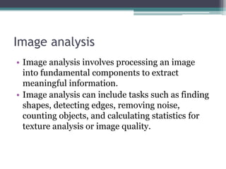
Retinal Image Analysis using Machine Learning and Deep.pptx
1. Retinal Image Analysis using
Machine Learning and Deep
Learning
Name : Deval Sachin Bhapkar
Roll No. 2218204
2. Introduction
• With the feasibility and development of deep learning methods, machines are now
able to interpret complex features in medical data, which leads to rapid
advancements in automation.
• Efforts have been made in ophthalmology to analyse retinal images and build
frameworks based on analysis for the identification of retinopathy and the
assessment of its severity.
• Using AI and Deep Learning, we can build Image analysis systems which can help
increase the accuracy and precision in detecting and treating various eye-related
diseases like Glaucoma, Diabetic retinopathy and various other diseases, thus helping
in proper treatment at the right time.
3. Benefits of using AI in Ophthalmology
• AI has tremendous potential to help manage patients in a world of limited
resources.
• AI can help in terms of time efficiency .It can save time as it can
automatically detect diseases at the right time, and the patient can be given
a treatment earlier, which can help in reducing complexities.
• AI also helps in terms of cost. Its expensive to hire people to read medical
images . We have limited resources, and we want to use our resources as
effectively as possible. AI can get accurate readings in a cost-efficient way.
• The next potential advantage of AI is accuracy . There is a lot of evidence
that AI can grade images as accurately-and potentially more accurately-
than human graders , and do it more quickly and cost-efficiently.
4. Image analysis
• Image analysis involves processing an image
into fundamental components to extract
meaningful information.
• Image analysis can include tasks such as finding
shapes, detecting edges, removing noise,
counting objects, and calculating statistics for
texture analysis or image quality.
5. Objectives
• The objective of this project is to see various
deep learning and image analysis techniques
used on retinal image data to increase accuracy
in prediction of eye related diseases.
6. Sr. no Title Author Abstract Conclusion Citation
1 Application of generative
adversarial networks
(GAN) for
ophthalmology image
domains: a survey
Aram You,
Jin Kuk Kim,
Ik Hee Ryu &
Tae Keun Yoo
In this work, we
present a literature
review on the
application of GAN in
ophthalmology image
domains to discuss
important
contributions and to
identify potential
future research
directions
The use of GAN has
benefited the various
tasks in ophthalmology
image domains.
However, the proper
selection of the GAN
technique and
statistical modeling of
ocular imaging will
greatly improve the
performance of each
image analysis.
https://rdcu.be/c
RlAH
2 Artificial intelligence:
the unstoppable
revolution in
ophthalmology
DavidBenetMsca,O
scar J.Pellicer-
ValeroMscb
This paper presents a
review of the state of
the art of AI in the
field of
ophthalmology,
focusing on the
strengths and
weaknesses of current
systems, and defining
the vision that will
enable us to advance
scientifically in this
digital era.
AI may be the answer
to healthcare system
sustainability amidst
an aging world
population, the quick
developments of LMIC
countries, and a deadly
global pandemic.
https://doi.
org/10.1016
/j.survophth
al.2021.03.0
03
3 Assessment of image
quality on color fundus
retinal images using the
automatic retinal image
analysis
Chuying Shi,
Jack Lee,
Gechun Wang,
Xinyan Dou,
Fei Yuan & Benny
This study developed
an automatic retinal
image analysis (ARIA)
method, incorporating
transfer net ResNet50
Our ARIA approach
showed good
performance in
identifying eye-
abnormality-
https://rd
cu.be/cRl
BY
8. Generative Adversarial Networks(GAN)
• Given a training set, this technique learns to generate new data with the same
statistics as the training set. For example, a GAN trained on photographs can
generate new photographs that look at least superficially authentic to human
observers, having many realistic characteristics.
• The core idea of a GAN is based on the "indirect" training through the discriminator,
another neural network that is able to tell how much an input is "realistic", which
itself is also being updated dynamically. This means that the generator is not trained
to minimize the distance to a specific image, but rather to fool the discriminator. This
enables the model to learn in an unsupervised manner.
• Randomly generated images and original real retinal images are classified by the
discriminator and this result is back-propagated and reflected in the training of both
the generator and the discriminator. Finally, the desired outcome after training the
GAN is that the pixel distributions from the generated retinal images should
approximate the distribution of real original retinal images.
12. Conclusion
• We concluded that GANs can help in image
analysis on retinal images(By performing de-
noising, augmentation, segmentation, super
resolution and feature extraction) to detect and
classify various eye-related diseases, more
accurately and precisely.