
Recommended
More Related Content
Similar to Immunity and Microbiota Interactions
Similar to Immunity and Microbiota Interactions (20)
Recently uploaded
Recently uploaded (20)
Immunity and Microbiota Interactions
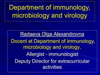
- 1. Department of immunology, microbiology and virology Radaeva Olga Alexandrovna Docent at Department of immunology, microbiology and virology. Allergist - immunologist Deputy Director for extracurricular activities
- 2. «Anatomical and chemical barriers. Communication between host immune system and commensal microbiota. Cells of the innate immunity. Phagocytosis»
- 3. Immunity – is a way of protection against genetically foreign substances (simple example: microorganism) that damage the organism. The main purpose is to maintain homeostasis.
- 4. Function of immunity Protection of "non-self" Tolerance of "non-self" this not damages protection of infectious diseases - the fetus in the uterus, - the сommensal microbiota Immunity controls homeostasis if damages
- 5. Types of immunity: 1. Innate immunity 2. Adaptive immunity
- 6. Innate immunity not only provides the early defense against infections but also instructs the adaptive immune system to respond to different microbes in ways that are effective for combating these microbes. Conversely, the adaptive immune response often uses mechanisms of innate immunity to eradicate infections. Thus, a constant bidirectional cross-talk occurs between innate immunity and adaptive immunity.
- 7. Mechanisms of innate immunity: 1/ Anatomical and chemical barriers (the skin and mucous membranes, body temperature, pH and various body secretions). 2/ Commensal microbiota (nonpathogenic bacteria colonize epithelial surfaces). 3/ Cellular elements (neutrophils, eosinophils, basophils, mast cells, monocytes/ macrophages, dendritic cells, NK cells, and NK T cells). 4/Humoral elements (the complement system, the interferon system, LPS binding protein,
- 8. Anatomical and chemical mechanisms of innate immunity : 1. The skin and mucous membranes (mechanical barrier). 2. PH and various body secretions. 3. Body temperature.
- 9. Anatomic and physiologic barriers provide the crucial first line of defense against pathogens. The Skin. Some of the reasons skin is a barrier to infection include: - the outer layer is relatively dry and is dead, acting as a mechanical barrier - secretions from sweat glands inhibit growth (high in salt; contains lysozyme which breaks down cell walls by attacking peptidoglycan's sugar links, antimicrobial peptides) - sebum (sē-bum) from oil glands is a chemical barrier, coats skin/hair oil, sebum lowers the pH between 4-6.
- 10. Mucous Membranes - Mucous Membranes line the body cavities that are open to the exterior and act as a mechanical barrier to infection. - They have secretions (mucus, antimicrobial peptides) that act as nonspecific mechanical and chemical barriers to infection. - Certain mucous membranes have specialized defenses -trachea have cilia. - They have two layers, an outer epithelium and an inner connective tissue layer. - The thin living epithelial layer provides a less efficient barrier than the skin, with the respiratory and reproductive systems being common entry sites for pathogens.
- 11. • nose hairs that filter out dust and microbes • mucous that traps many microbes (mechanical barrier) • mucous that contains antimicrobial peptides and antibodies (chemical barriers) • the ciliary escalator (ciliated cells which propel microbes up toward the mouth)when you cough • gastric juice kills many bacteria and inactivates many toxins (chemical barrier). The Respiratory System and its Defenses:
- 12. Other Defenses and Barriers: • A mechanical barrier to infection includes the flow of urine through the urinary system which sweeps bacteria out. • Vaginal secretions are both mechanical & chemical barriers. • Blood contains transferrins, proteins which bind iron. By reducing the amount of available iron these chemical barriers inhibit the growth of bacteria in the blood. • Tears, saliva, and nasal mucus contain lysosyme (an enzyme that degrades peptidoglycan). • Tears are a mechanical barrier that wash the eyes upon blinking. • Gastric juices contain digestive enzymes and acids.
- 14. Commensal microbiota. • These nonpathogenic microorganisms are found on the skin, in the mouth and in the reproductive and gastrointestinal tract. • The gastrointestinal tract contains many billions of bacteria that have a symbiotic relationship. • Imbalance of the commensal microbiota, called dysbiosis. • The relationship between the host immune system and the commensal microbiota is highly complex because these microbes possess many of the same traits as pathogenic microorganisms.
- 15. Hence, the immune system must coexist with commensal microbiota and maintain a state of tolerance to self and innocuous environmental, yet be able to vigorously repel invasion by a wide spectrum of pathogens.
- 16. The presence of a functional barrier, with normal amounts of PRRs with mucus, and secreted IgA, promotes intestinal homeostasis with the microbiota. The microbiota is segregated away from the immunity, and the intestinal immune system directs a largely tolerant response to the resident commensals. A breach in the epithelial barrier by the bacterial agents, pathogens and other human diseases (cholecystitis, pancreatitis, endocrine pathology) can lead to contact receptors and microbiota. Immunity attacks and kills сommensal microbiota. Functional barrier and barrier defect (the intestines)
- 17. Commensal microbiota Normal microbiota does not damage and does not activate the epithelium (only PAMPs)). Normal microbiota has no contact with the pattern recognition receptors The epithelium synthesizes anti-inflammatory cytokines Toleration Pathogens Pathogens damage and activate the epithelium (PAMPs + DAMPs) Pathogens have contact with the pattern recognition receptors The epithelium synthesizes inflammatory cytokines Elimination The more important issue of how the immune system can distinguish between beneficial and pathogenic microbes is still largely a mystery. Innate barriers ensure a tolerant response to the microbiota.
- 18. The Microbiota serves many functions. • Competition for adherence to the epithelia and mucosal surfaces as well as acidification of the microenvironment to prevent attachment pathogens • Consuming nutrients that would otherwise be available to pathogens. • Secretion of anti-microbial peptides • Development of gut-associated lymphoid tissues (GALT) • Normal flora of the intestines improve our overall health by producing several types of vitamins. While epithelial cells are often viewed as the first line of defense against pathogenic insult, the commensal microbiota thriving on epithelial surfaces serves as an invisible barrier to invading pathogens.
- 19. Development of gut-associated lymphoid tissues (GALT) M cells (or microfold cells) are cells found in the follicle-associated epithelium of the Peyer's patch as well as in bronchus-associated lymphoid tissue (BALT). They transport organisms and particles from the gut lumen to immune cells across the epithelial barrier, and thus are important in stimulating mucosal immunity.
- 20. Functional barrier (the intestines) M cells Cellular elements Anti-inflammatory Cytokines Mucus PRR
- 21. The relationship between the host immune system and the commensal microbiota is highly complex because these microbes possess many of the same traits as pathogenic microorganisms. Epithelium Intestines A principal function of the microbiota is to protect the intestine against colonization by exogenous pathogens and potentially harmful indigenous microorganisms via several mechanisms, which include direct competition for limited nutrients and the modulation of host immune responses.
- 22. The immune system must coexist with commensal microbiota and maintain a state of tolerance to self and innocuous environmental, yet be able to vigorously repel invasion by a wide spectrum of pathogens. Epithelium Intestines
- 23. Cells of the innate immunity. radaevamed@mail.ru
- 24. Cells of the innate immunity. • Dendritic cell • Natural helper (NH) Lymphocytes of innate immunity • Natural killer (NK) Epithelial cells Mast cells Innate immunity protection is a task performed by cells of both hematopoietic and non hematopoietic origin. Hematopoietic cells involved in innate immunity responses include neutrophils, eosinophils, basophil, monocytes-macrophages, dendritic cells, mast cells, lymphocytes of innate immunity. Cells of the innate immune have PRRs (except for lymphocytes of innate immunity (NK, NH)).
- 25. Myelopoiesis. Hematopoietic stem cells have the ability to differentiate into many types of blood cells. The two types of circulating phagocytes (neutrophils and monocytes), are blood cells that are recruited to sites of infection, where they recognize and ingest microbes for intracellular killing. Development of granulocytes and monocytes. The total body neutrophil content can be divided conceptually into the following 3 compartments: the bone marrow, the blood, and the tissues. After differentiation in the bone marrow, neutrophils are released into the peripheral blood and circulate for 7 to 10 hours before migrating into the tissues, where they have a life
- 28. Neutrophils IL-23 IL-17 G-CSF BLOOD INFLAMMATION НОРМА в крови 2 – 7,5 ·10 в 9 /л Other mediators of inflammation stimulate bone marrow release and promote margination and adhesion of neutrophils to vascular endothelium at the site of inflammation. The time for production of new cells from the bone marrow is 4 to 6 days Neutrophils are white blood cells that move from the blood into the cells to kill invading bacteria and fungi. If neutrophil levels become too high, neutrophilia results.
- 29. Neutrophils • Neutrophils are the first inflammatory cells to enter damaged tissue from the blood after tissue damage has being caused. They are the predominant cell 4 - 6 hours after the beginning of an inflammatory reaction; at 12 hours there are also substantial numbers of macrophages and at 24 hours there are equal numbers of neutrophils and macrophages. • The main function of neutrophils is the protection of the body from the local bacterial infections.
- 30. Microorganism resident macrophages IL1β, TNF-α Neutrophil's chemotaxis Lamellipodia Neutrophils and monocytes migrate to extravascular sites of infection by binding to endothelial adhesion molecules and in response to chemoattractants that are produced on encounter with microbes in tissue. Leukocyte migration from the blood into tissues is a multistep process that consists of initial loose attachment of the leukocytes to endothelial cells (rolling), followed by firm adhesion (adhesion) and transmigration through the endothelium (diapedesis). If an infectious microbe breaches an epithelium and enters the subepithelial tissue, resident macrophages recognize the microbe and respond by producing cytokines (described in more detail later).
- 31. Neutrophils have three methods for directly attacking microorganisms: • phagocytosis (ingestion), • release of soluble antimicrobials (including granule proteins) • generation of neutrophil extracellular traps (NETs). Neutrophils undergo apoptosis after the destruction of bacterial cells. Receptors for the recognition and absorption by macrophages appears on the membranes of neutrophils. The macrophage then has the task of clearing both the dead pathogens and the dead neutrophils.
- 33. Monocytes • Маke up about 5% to 10% of white blood cells and are a heterogeneous group of cells that migrate into tissues and differentiate into a diverse array of tissue-resident phagocytic cells, including macrophages and dendritic cells. • The mononuclear phagocyte system (monocytes/ macrophages) is a complex system that can be viewed in different ways. Macrophages as recognized by Metchnikoff can be found in all tissues, and as circulating cells called blood monocytes. • They, too, ingest microbes in the blood and in tissues. Unlike neutrophils, monocytes that enter extravascular tissues survive in these sites for long periods; in the tissues, these monocytes differentiate into cells called macrophages
- 34. Blood monocytes and tissue macrophages are two stages of the same cell lineage. Resident macrophages are found in connective tissues and in every organ in the body
- 35. Macrophages functions: • 1) phagocytosis of «non-self»; • 2) phagocytosis of dead, dying, and aging cell ( are also implicated in the removal of cell debris and apoptotic cell). Macrophages clear apoptotic cells using scavenger receptors and the vitronectin receptor). • 3) pinocytosis macromolecules; • 4) regulating repair and regeneration; • 5) presentation of antigens (macrophages present antigen in the pe-riphery to activated T cells. • 6) secretion of biologically active substances (activated macrophages secrete cytokines, enzymes, complement system molecules, and procoagulant).
- 36. Macrophages Tissue- resident macrophages Inflammatory macrophages 1) Regulating repair and regeneration; 2) Detoxification; 3) Removal of dead tissue cells - the main function !!!!! 4) The cleansing blood of degrading material, fibrin clots, the bad cholesterol, iron. The macrophage then has the task of clearing both the dead pathogens and the dead neutrophils. - If inflammatory macrophages recognise damaged "non-self" (a greater percentage of PAMPs), they will synthesize inflammatory cytokines. - If inflammatory macrophages recognise damaged "self" (a greater percentage of DAMPs), they will synthesize antiinflammatory cytokines.
- 37. Functions of tissue-resident macrophages: 1) Regulating repair and regeneration; 2) Detoxification; 3) Removal of dead tissue cells - the main function !!!!! 4) The cleansing blood of degrading material, fibrin clots, the bad cholesterol, iron.
- 39. Receptors on macrophage membrane: • CD 14 – is receptor for a variety of bacterial products (LPS); • Scavenger receptors participate in the removal of many foreign substances and waste materials in the living body by extensive ligand specificity and a variety of receptor molecules; • Complement receptor – CR3; • Fc gamma receptor I (FcγRI) – high affinity Rc for “tails” (FC-fragment) of IgG. • MHC I, MHC II.
- 40. Phagocytosis • Phagocytosis - the process by which a cell, such as a white blood cell, ingests microorganisms, other cells, and foreign particles. • In an organism's immune system, phagocytosis is a major mechanism used to remove pathogens (it is especially important for defense against extracellular microbes) and cell debris.
- 41. Elie (Illya) Metchnikoff (also spelled Mechnikov) (1845–1916) History of immunology Elie Mechnikov was a Russian zoologist best known for his pioneering research into the immune system. In particular, he is credited with the discovery of phagocytes (macrophages) in 1882, and his discovery turned out to be the major defence mechanism in innate immunity. He was awarded the 1908 Nobel Prize in Physiology or Medicine "in recognition of their work on immunity". In immunology he is given an epithet the "father of natural immunity or innate immunity"
- 42. 1) For example, when a macrophage ingests a pathogenic microorganism, the pathogen becomes trapped in a phagosome which then fuses with a lysosome to form a phagolysosome. 2) Within the phagolysosome, enzymes and toxic peroxides digest the pathogen. Bacteria, dead tissue cells, and small mineral particles are all examples of objects that may be phagocytised.
- 43. Phases of phagocytoses 1. Chemotaxis. 2. Adhesion (adherence) 3. Bacterium is ingested (formation of a phagocytic cup), forming phagosome 4. Phagosome fuses with lysosome. 5. Destruction of the Microbes 6. Discharge of waste materials (exocytosis). 4 5 6 MHC II + antigens On completion the membrane of the phagolysosome is disrupted. Antigenic fragments may become diverted to the acidic endosome compartment for interaction with MHC class II molecules and antigen presentation.
- 44. Microorganism resident macrophages IL1β, TNF-α Neutrophil's chemotaxis Lamellipodia Chemotaxis is the migration of polymorphonuclear leukocytes and macrophages toward higher concentrations of chemoattractants. Сhemoattractant concentration increases around microorganisms or damaged tissue.
- 45. Because of certain microbial defenses (production of M proteins or slippery capsules), adherence of the phagocyte cell membrane to the surface of the microbe may be difficult. Coating the surface of the microorganism with complement proteins and antibody proteins facilitates phagocytosis (process is called opsonization; proteins are called opsonins). Adhesion (adherence). Phagocyte cell Antibody Bacteria Receptor
- 46. • The phagocyte engulfs the microbe with its plasma membrane. The plasma membrane surrounds the microbe and pinches off around the microbe, forming a vesicle. актин Фагоцитарная чаша Formation of a phagocytic cup
- 47. Phagocytic cup
- 48. These signaling pathways trigger actin polymerization, resulting in membrane extensions around the microbe particles and their internalization, forming phagosomes
- 49. The formation of the phagosome
- 50. Phagosome fuses with lysosome. Phagosome fuses with lysosome.
- 51. Destruction of the Microbes The phagosomes then fuse with lysosomes and, in phagocytes, with preformed primary and secondary granules. The resulting phagolysosomes contain an arsenal of antimicrobial agents that then kill and degrade the internalized microbes. These agents include antimicrobial proteins and peptides (including defensins and cathelicidins), low pH, acid-activated hydrolytic enzymes (including lysozyme and proteases), and specialized molecules that mediate oxidative attack.
- 52. Degradation can be oxygen-dependent or oxygen-independent: • oxygen-dependent degradation depends on NADPH and the production of reactive oxygen species. Hydrogen peroxide and myeloperoxidase activate a halogenating system, which leads to the creation of hypochlorite and the destruction of bacteria. • Oxygen-independent degradation depends on the release of granules, containing proteolytic enzymes such as defensins, lysozyme, and cationic proteins. Other antimicrobial peptides are present in these granules, including lactoferrin
- 53. Discharge of waste materials (exocytosis). • On completion the membrane of the phagolysosome is disrupted. Antigenic fragments may become diverted to the acidic endosome compartment for interaction with MHC class II molecules and antigen presentation. The process induces secretion of toxic molecules and cytokines
- 54. Discharge of waste materials (exocytosis).
- 55. Example of a bacterium that resists digestion by a phagocyte. 1. Some intracellular pathogens, including Mycobacterium tuberculosis, have evolved strategies to inhibit phagosomal maturation and can survive and replicate within the immature phagosome. 2. Other pathogens, such as Escherichia coli and Neisseria meningitides, have developed mechanisms to fix, shed and/or degrade opsonins to prevent activation of the immune response and thus evade immune surveillance and phagocytosis.
- 58. • Thank you for attention
- 59. Cells of the innate immunity: Basophils, mast cells and eosinophils. Dendritic cells. Nature killer. Humoral elements. The Compliment System
- 60. Basophils, mast cells and eosinophils • are critical to our response to parasites, particularly helminths (worms). These cells also play an important role in the development of allergies. • Basophils, mast cells and eosinophils have receptors for Ig (immunoglobulin) E. • Ig E is essential for combating large parasitic worms. • sIgE (antibodies) are responsible for the symptoms experienced in allergic reactions
- 61. Eosinophils Eosinophil granulocytes, usually called eosinophils are white blood cells and one of the immune system components responsible for combating multicellular parasites They usually bind to an antibody-coated parasite through surface Fc receptors and release the contents of their granules (degranulate) onto the parasite surface. The granules contain peroxides and a toxin, major basic protein, which kill the parasite. The count can increase with allergic disorders, during parasitic infections Eosinophils are formed exclusively in the bone marrow where they spend about 8 days in the process of maturation before moving into the blood vessels. They travel through the vessels for 8 to 12 hours before they finally arrive at destination tissues, where they remain for 1 to 2 weeks.
- 62. Basophils and mast cells Basophil granulocytes, mostly referred to as basophils, are the least common of the granulocytes, representing about 0.01% to 0.3% of circulating white blood cells. Basophils contain large cytoplasmic granules that obscure the cell nucleus under the microscope when stained. However, when unstained, the nucleus is visible and it usually has two lobes. The mast cell, another granulocyte, is similar in appearance and function. Both cell types store histamine, a chemical that is secreted by the cells when stimulated. However, they arise from different cell lines, and mast cells usually do not circulate in the blood stream, but instead are located in connective tissue. Like all circulating granulocytes, basophils can be recruited out of the blood into a tissue when needed
- 63. Some distinctive and common characteristics of basophils and mast cells The stimulus for mast cell or basophil degranulation is often an allergen. Mediators such as histamine, released by degranulation, cause the adverse symptoms of allergy, but, on the positive side, also play a role in immunity against parasites by enhancing acute inflammation.
- 64. Basophils, mast cells and eosinophils • are critical to our response to parasites, particularly helminths (worms). • These cells also play an important role in the development of allergies (a type I hypersensitivity reactions). • Basophils, mast cells and eosinophils have receptors for Ig E.
- 65. Dendritic cells ❑ - Dendritic cells (DCs) are so called because of their many surface membrane folds that are similar in appearance to dendrites of the nervous system. ❑ - DCs are considered cellular bridges between the innate and adaptive immune systems.
- 66. • Dendritic cells make contact with a pathogen at the site of infection. They recognise pathogen-associated molecular patterns (microorganisms). • They are highly phagocytic, take up microbes, process the foreign microbial antigens, and become mature APCs carrying the processed antigen on their surface with specialized MHC molecules. • Dendritic cells communicate this encounter to T lymphocytes in the lymph node (“antigen presentation”)
- 67. Types of dendritic cells Most classical DCs derive from one of two bone marrow precursors: • a myeloid progenitor (DC1) that gives rise to myeloid DCs (localization – diffuse –epidermis, mucosae, thymus, and T cell areas of secondary lymphoid organs and tissues); • a lymphoid progenitor (DC2) that develops into plasmacytoid DCs (T cell areas of secondary lymphoid organs and tissues).
- 68. • Dendritic cells are generally viewed as the most effective of the APCs. Because these cells constitutively express a MHC class I, II molecules. • The immune system typically uses different pathways to eliminate intracellular and extracellular antigens.
- 69. As a rule, endogenous antigens (those generated within the cell) are processed in the cytosolic or endogenous pathway and presented on the membrane with class I MHC molecules. If a virus had infected the APC (dendritic cell), viral peptides would also be presented, allowing the immune system to recognize and kill the infected cell. The endogenous pathway is used to present cellular peptide fragments on the cell surface on MHC class I molecules.
- 70. • Exogenous antigens (those taken up from the extracellular environment by endocytosis) are typically processed in the exogenous pathway and presented on the membrane with class II MHC molecules. • The exogenous pathway is utilised by specialised antigen presenting cells to present peptides derived from proteins that the cell has endocytosed. The peptides are presented on MHC class II molecules. Proteins are endocytosed and degraded by acid-dependent proteases in endosomes.
- 71. • Remarkably, DCs are able to present internalized antigens to T cells via MHC class I as well as class II molecules, in a phenomenon called cross- presentation. • In Cross-presentation, peptides derived from extracellular virus are presented in the context of MHC class I and class II . The cell starts off with the exogenous pathways but diverts the antigens (cytosolic diversion) to the endogenous pathway.
- 72. APCs are a heterogeneous population of cells that are impotent in innate immunity and play a pivotal role in the induction of functional activity of T helper cells.
- 74. Natural killer (NK) cells • They are derived from the bone marrow and morphologically have the appearance of large granular lymphocytes. • CD16 and CD56 are important markers of NK cells
- 75. Natural killer (NK) cells
- 76. Natural killer (NK) cells The function of NK cells is to recognize and kill through apoptosis: • •virus-infected cells; • certain tumor cells.
- 77. • Activation of NK cells leads to lysis of target cells, which is mediated by perforin and granzymes, and cytolytic granule mediated cell apoptosis . • Upon release in close proximity to a cell slated for killing, perforin forms pores in the cell membrane of the target cell, creating an aqueous channel through which the granzymes and associated molecules can enter, inducing either apoptosis or osmotic cell lysis.
- 78. Лектор: Радаева О.А. Humoral elements. Molecules of the innate immune system
- 79. Molecules of the innate immune system 1. The complement system 2. The interferons system 3. Antimicrobial peptides (Lysozyme, β-lysin, Lactoferri, Collectins, Defensins) 4. Acute phase proteins (pentraxins, fibronectin, haptoglobin)
- 80. The complement system Jules Bordet(1870-1961) Paul Ehrlich (1854-1915)
- 81. The complement system is a proteins, that widely distributed throughout body fluids and tissues without adverse effect. At sites of infection, however, they are activated locally and trigger a series of potent inflammatory events. The complement system activates through a triggered- enzyme cascade. In such a cascade, an active complement enzyme generated by cleavage of its zymogen precursor then cleaves its substrate, another complement zymogen, to its active enzymatic form. This in turn cleaves and activates the next zymogen in the complement pathway. Over 30 proteins (nine major components - abbreviated as C') and protein fragments make up the complement system. Complement components constitute approximately 15% of the globulin protein fraction in plasma, and their combined concentration can be as high as 3 mg/ml.
- 82. The functions of the complement system include: 1. Cell Lysis – rupturing membranes of foreign cells; 2. Opsonisation – enhancing phagocytosis of antigens; 3. Chemotaxis – attracting macrophages and neutrophils; 4. The triggering and amplification of inflammatory reactions
- 83. • Nine major components: the first component of the pathway, C1, is a complex molecule comprising a large recognition unit termed C1q and two molecules each of C1r and C1s, the enzymatic units of the complex С2, C3, C4, C5 split into two parts (C2a/C2b, C3a/ C3b, C4a/C4b, C5a/C5b). As a rule, the larger fragment of a cleaved complement component is designated with the suffix “b,” and the smaller with the suffix “a.” However, there is one exception to this rule: the larger, enzymatically active form of the C2 component is named C2a. • the components C6, C7, C8, and C9 not disintegrate when activated (membrane attack proteins). The proteins of the membrane attack complex (MAC) insert into the cell membranes of invading microorganisms and punch holes that result in lysis of the pathogen. The complement components of the MAC are C5b, C6, C7, C8, and multiple copies of C9.
- 84. Cytolytic function of the complement system
- 85. The complement cascade may be activated by any of three pathways
- 86. The Classical pathway • The classical pathway links to the adaptive immune system. The classical pathway is activated by antibody bound to antigen
- 87. С1qrs
- 88. C1qrs BACTERIA
- 90. С3 конвертаза мембранная С4bC2b С4bC2bС3b С5 конвертаза
- 91. The membrane attack complex • Membrane-bound C5bC6C7 recruits C8 from the plasma, and, finally, multiple copies of C9 are incorporated in the complex to form the MAC.
- 92. • The membrane attack pathway results in the formation of a transmembrane pore. • The pore has an inner diameter approaching 10 nm: • allowing the free flow of solutes and electrolytes across the cell membrane; • because of the high internal osmotic pressure, causing the cell to swell and sometimes burst.
- 93. Видео
- 94. The lectin pathway The lectin pathway differs from the classical pathway only in the initial recognition and activation steps. The C1 complex is replaced by a structurally similar multimolecular complex, comprising the C1q-like recognition unit mannan-binding lectin (MBL)
- 95. The alternative pathway Initiation of the alternative pathway of complement activation is independent of antibody-antigen interactions. С3 C3b C3a Spontaneous hydrolysis of soluble C3 to C3b Фактор В С3-convertase – С3bВ
- 96. The membrane attack complex • Membrane-bound C5bC6C7 recruits C8 from the plasma, and, finally, multiple copies of C9 are incorporated in the complex to form the MAC.
- 98. Membrane-binding components or opsonins The term opsonisation refers to the capacity of antibodies and com-plement components (as well as other proteins) to coat dangerous antigens that can then be recognised by Fc receptors (for antibodies) or complement receptors (for complement components) on phagocytic cells. Binding of complement-coated antigen by phagocytic cells results in complement re- ceptor-mediated phagocytosis and antigen destruction. С3b СR 1 Фактор I For C3 and C4, the larger fragments, C3b and C4b, serve as opso-nins, enhancing phagocytosis by binding to microbial cells and serving as binding tags for phagocytic cells bearing receptors for C3b or C4b.
- 99. • Initiation of (acute) inflammation by direct activation of mast cells (C3a, C5a). • Chemotaxis. C3a and C5a are structurally similar, small proteins that promote inflammation and serve as chemoattractants for certain classes of leukocytes
- 100. The interferons system • IFNs belong to the large class of proteins known as cytokines, molecules used for communication between cells to trigger the defenses of the immune system.
- 101. Human interferons have been classified into three major types Types: • Interferon type I: IFN α, IFN β, IFN ω, IFN ε, IFN κ; • Interferon type II (IFN-γ in humans); • The recently classified type III interferon group consists of three IFN-λ (lambda) molecules called IFN-λ1, IFN-λ2 and IFN-λ3.
- 103. Common characteristic: Like all cytokines, the ability of a cell to respond to IFN is completely dependent on the presence of its cognate receptor on the surface of the target cell. Human IFN valid only on human cells (species specificity). IFN inhibits the proliferation of various viruses in the cells (universal mechanism of action).
- 104. Interferon type I: IFN α, IFN β Основные клетки-продуценты ИФН I: • The main producer cells IFN I: • 1) plasmacytoid dendritic cells; • 2) monocytes / macrophages; • 3) epithelial cells; • 4) fibroblasts (IFNβ, IFNα); • 5) virus-infected nucleated cells Peak production of type I interferons observed 6-12 h after virus infection.
- 105. Functions: I типа: Antiviral function. 1 A virus is a small infectious agent that replicates only inside the living cells of other organisms. 2 A virus-infected cell releases viral particles that can infect nearby cells. However, the infected cell can prepare neighboring cells against a potential infection by the virus by releasing interferons.
- 106. 1. Interferon binds to the cell receptor. 2. The signal is transmitted to the nucleus. Protein synthesis in the cell is blocked. 3. The virus penetrates the cell but can not replicate. Interferon does not kill the virus. 4. Inteferon blocks viral replication in human cells.
- 109. If antiviral immunity is working correctly, a viral infection last for ten days.
- 110. • Immunomodulatory function. Another function of interferons is to upregulate MHC I and MHC II, and increase immunoproteasome activity. • Antitumor functions of type I interferons. IFN-α can act indirectly on the tumor by inhibiting angiogenesis which is induced by the tumors and is required to promote their growth and metastasis.
- 111. Interferon type II (IFN-γ in humans). The main producer cells: NK, macrophages and dendritic cells; cytotoxic CD8 + T lymphocytes, ThI lymphocytes. Synthesis of IFNγ reaches its maximum 48-72 hours after stimulation of cells. Note: the antiproliferation and antiviral activities are weaker than those of IFN alpha and IFN beta. IFN gamma is the most potent of the three at macrophage activation and in inducing class II MHC expression
- 112. - IFN-gamma enhances the microbicidal function of macrophages through formation of nitric oxide and reactive oxygen intermediates (ROI) - IFN-gamma stimulates the expression of class I and class II MHC molecules and co-stimulatory molecules on antigen presenting cells - IFN-gamma promotes the differentiation of naive helper T cells into Th1 cells - IFN-gamma activates polymorphonuclear leukocytes (PMN) and cytotoxic T cells and increases the cytotoxicity of NK cells.
- 113. 1. Russian healthcare promotes the use of interferons and interferon inductors as a prophylaxis and treatment of influenza and other viral infections. 2. Interferons can selectively inhibit viral replication process without compromising cell metabolism. Genetically engineered recombinant compounds of interferon (Grippferon, Viferon, Reaferon) are widely used in medical practice. 3. Interferon therapy is used (in combination with chemotherapy and radiation) as a treatment for some cancers. Interferons used therapeutically are manufactured using recombinant DNA technology.
- 114. Бактерицидные пептиды • Lysozyme (Lysozyme, also known as muramidase, are glycoside hydrolases (enzyme)) Lysozyme is abundant in a number of secretions, such as blood, lymph, tears, saliva, human milk, and mucus. It is also present in cytoplasmic granules of the macrophages and the polymorphonuclear neutrophils. Ly- sozyme is not detected in the cerebrospinal fluid and the anterior chamber. Macrophages and neutrophils synthesize lysozyme. The enzyme functions by attacking peptidoglycans (found in the cell walls of bacteria). Lysozyme kills bacteria, especially Gram-positive.
- 115. Acute phase proteins and their functions. These molecules include: • pentraxins (C-reactive protein (CRP), serum amyloid protein A (SAA)); • complement components; • opsonic proteins such as mannose-binding protein (MBP), metal-binding proteins and protease inhibitors; • haptoglobin, hemopexin, ceruloplasmin; • Blood coagulation factors (fibrinogen, prothrombin, factor VIII, and plasminogen).
- 116. CRP • CRP was so named because it was first identified as a substance in the serum of patients with acute inflammation that reacted with the C-polysaccharide of Pneumococcus. CRP binds to the phosphocholine expressed on the surface of dead or dying cells and some bacteria. This activates the complement system, promoting phagocytosis by macrophages, which clears necrotic and apoptotic cells and bacteria. • Diagnostic use. CRP is used mainly as a marker of inflammation.
- 117. Pattern recognition receptors (PRRs) • are proteins expressed by cells of the innate immune system to identify pathogen-associated molecular patterns (PAMPs), which are associated with microbial pathogens or cellular stress, as well as damage-associated molecular patterns (DAMPs), which are associated with cell components released during cell damage.
- 118. PRR types and localization • Secreted (soluble) PRRs. • Membrane-bound PRRs - Endocytic PRRs - Signaling PRRs • Cytoplasmic PRRs
- 119. Secreted (soluble) PRRs. • A number of PRRs do not remain associated with the cell that produces them. • One very important collectin is mannan- binding lectin (MBL), a major PRR of the innate immune system. MBL predominantly recognizes certain sugar groups on the surface of microorganisms but also binds phospholipids. Once bound to the ligands MBL and initiate the lectin pathway of complement activation.
- 120. Membrane-bound PRRs. Endocytic PRRs • Endocytic PRRs - promote the attachment, engulfment and destruction of microorganisms by phagocytes, without relaying an intracellular signal. These PRRs recognize carbohydrates and include mannose receptors of macrophages, glucan receptors present on all phagocytes and scavenger receptors that recognize charged ligands, are found on all phagocytes and mediate removal of apoptotic cells.
- 121. Example The mannose receptor present on the surface of macrophages. The receptor recognises terminal mannose, N-acetylglucosamine residues on glycans attached to proteins found on the surface of some microorganisms. Binding to the receptor causes phagocytosis and destruction of the foreign cell.
- 122. Membrane-bound PRRs. Signaling PRRs Signaling PRRs include the large families of membrane-bound Toll- like receptors. Signaling PRRs recognize evolutionarily conserved structures on pathogens, PAMPs, and trigger innate immune responses, including production of pro- inflammatory cytokines and type I IFN. To date, 10 TLRs have been identified in humans, and they each recognize distinct PAMPs derived from various microbial pathogens, including viruses, bacteria, fungi, and protozoa.
- 123. Overview of innate immune signalling pathways and components. BACTERIA VIRUS Antibacterial protection IL-1, pro- inflammatory cytokines Antiviral protection Type I IFN (IFN-α, IFN-β)
- 126. Cytokines ❑ Cytokines are a large, diverse family of small proteins ❑ Cytokines are mediators of intercellular interaction ❑ Cytokines are produced by a broad range of cells, including immune cells like macrophages, B lymphocytes, T lymphocytes and mast cells, as well as endothelial cells, fibroblasts, and various stromal cells; a given cytokine may be produced by more than one type of cells
- 127. Nomenclature
- 128. This action may occur in • an autocrine (acts on same cell), • paracrine (acts on nearby cell) • or endocrine (acts on distant cell; not the normal manner for cytokine responses) manner. Receptor engagement triggers intracellular signalling cascades leading to altered gene expression in the target cell, which lead to a biological effect. Differentiation, proliferation and activation of the target cell are all effects which can be detected after cytokine stimulation.
- 129. Biological functions of cytokines: • 1. Control of the embryonic development • 2. Regulation a number of physiological functions • 3. Regulation of the body's defenses at the local and systemic levels. • 4. Regulation of development of allergic, autoimmune and other immune-pathological processes. • 5. Participation in the regulation of tumor cell transformation and tumor progression. • 6. Regulation of tissue regeneration.
- 130. Cytokines exhibit the attributes of • pleiotropy, • redundancy, • synergism, • antagonism, and • cascade induction, • short lived, which permit them to regulate cellular activity in a coordinated, interactive way
- 131. Cytokines • A cytokine that induces different biological effects depending on the nature of the target cells is said to have a pleiotropic action, whereas two or more cytokines that mediate similar functions are said to be redundant. • Cytokine synergy occurs when the combined effect of two cytokines on cellular activity is greater than the additive effects of the individual cytokines. • In some cases, the effects of one cytokine inhibit or antagonize the effects of another. • Cascade induction occurs when the action of one cytokine on a target cell induces that cell to produce one or more additional cytokines.
- 132. Proinflammatory or anti-inflammatory cytokines • A proinflammatory cytokine is a cytokine that promotes systemic inflammation. Interleukin (IL)-1 and tumor necrosis factor (TNF) are proinflammatory cytokines, and when they are administered to humans, they produce fever, inflammation, tissue destruction, and, in some cases, shock and death. • The anti-inflammatory cytokines are a series of immunoregulatory molecules that control the proinflammatory cytokine response • The net effect of an inflammatory response is determined by the balance between pro-inflammatory and anti-inflammatory cytokines.