Clinical Cancer Research Publication

Purpose: An inherited mutation in KRAS (LCS6-variant or rs61764370) results in altered control of the KRAS oncogene. We studied this biomarker’s correlation to anti-EGFR monoclonal antibody (mAb) therapy response in patients with metastatic colorectal cancer. Experimental Design: LCS6-variant and KRAS/BRAF mutational status was determined in 512 patients with metastatic colorectal cancer treated with salvage anti-EGFR mAb therapy, and findings correlated with outcome. Reporters were tested in colon cancer cell lines to evaluate the differential response of the LCS6- variant allele to therapy exposure. Results: In this study, 21.2% (109 of 512) of patients with metastatic colorectal cancer had the LCS6- variant (TG/GG), which was found twice as frequently in the BRAF-mutated versus the wild-type (WT) group (P = 0.03). LCS6-variant patients had significantly longer progression- free survival (PFS) with anti-EGFR mAb monotherapy treatment in the whole cohort (16.85 vs. 7.85 weeks; P = 0.019) and in the double WT (KRAS and BRAF) patient population (18 vs. 10.4 weeks; P = 0.039). Combination therapy (mAbs plus chemotherapy) led to improved PFS and overall survival (OS) for nonvariant patients, and brought their outcome to levels comparable with LCS6-variant patients receiving anti-EGFR mAb monotherapy. Combination therapy did not lead to improved PFS or OS for LCS6-variant patients. Cell line studies confirmed a unique response of the LCS6-variant allele to both anti-EGFR mAb monotherapy and chemotherapy. Conclusions: LCS6-variant patients with metastatic colorectal cancer have an excellent response to anti-EGFR mAb monotherapy, without any benefit from the addition of chemotherapy. These findings further confirm the importance of thismutation as a biomarker of anti-EGFR mAb response in patients with metastatic colorectal cancer, and warrant further prospective confirmation.

Recommended
Recommended
More Related Content
What's hot
What's hot (20)
Viewers also liked
Viewers also liked (18)
Similar to Clinical Cancer Research Publication
Similar to Clinical Cancer Research Publication (20)
Recently uploaded
Recently uploaded (20)
Clinical Cancer Research Publication
- 1. Personalized Medicine and Imaging A let-7 microRNA-Binding Site Polymorphism in KRAS Predicts Improved Outcome in Patients with Metastatic Colorectal Cancer Treated with Salvage Cetuximab/ Panitumumab Monotherapy Zenia Saridaki1,2, Joanne B. Weidhaas3, Heinz-Josef Lenz4, Pierre Laurent-Puig5, Bart Jacobs2, Jef De Schutter2, Wendy De Roock6, David W. Salzman3, Wu Zhang4, Dongyun Yang7, Camilla Pilati8, Olivier Bouche9, Hubert Piessevaux10, and Sabine Tejpar2 Abstract Purpose: An inherited mutation in KRAS (LCS6-variant or rs61764370) results in altered control of the KRAS oncogene.Westudied this biomarker’s correlation to anti-EGFR monoclonal antibody (mAb) therapy response in patients with metastatic colorectal cancer. Experimental Design: LCS6-variant and KRAS/BRAF mutational status was determined in 512 patients with metastatic colorectal cancer treated with salvage anti-EGFR mAb therapy, and findings correlated with outcome. Reporters were tested in colon cancer cell lines to evaluate the differential response of the LCS6- variant allele to therapy exposure. Results: In this study, 21.2% (109 of 512) of patients with metastatic colorectal cancer had the LCS6- variant (TG/GG), which was found twice as frequently in the BRAF-mutated versus the wild-type (WT) group (P ¼ 0.03). LCS6-variant patients had significantly longer progression- free survival (PFS) with anti-EGFR mAb monotherapy treatment in the whole cohort (16.85 vs. 7.85 weeks; P ¼ 0.019) and in the double WT (KRAS and BRAF) patient population (18 vs. 10.4 weeks; P ¼ 0.039). Combination therapy (mAbs plus chemotherapy) led to improved PFS and overall survival (OS) for nonvariant patients, and brought their outcome to levels comparable with LCS6-variant patients receiving anti-EGFR mAb monotherapy. Com-bination therapy did not lead to improved PFS or OS for LCS6-variant patients. Cell line studies confirmed a unique response of the LCS6-variant allele to both anti-EGFR mAb monotherapy and chemotherapy. Conclusions: LCS6-variant patients with metastatic colorectal cancer have an excellent response to anti-EGFR mAb monotherapy, without any benefit from the addition of chemotherapy. These findings further confirm the importance of thismutation as a biomarker of anti-EGFRmAbresponse in patients with metastatic colorectal cancer, and warrant further prospective confirmation. Clin Cancer Res; 20(17); 4499–510. 2014 AACR. Introduction In the western world, colorectal cancer remains a major public health problem, with an estimated 136,830 new cases and 50,310 deaths estimated to occur in 2014 in the United States alone (1). The incorporation of two mAbs targeting EGFR (anti-EGFR mAbs), cetuximab and panitu-mumab, in metastatic colorectal cancer clinical practice either given as monotherapy or in combination with che-motherapy, has proved to provide a modest clinical benefit in pretreated patients (2–5). Nevertheless, their efficacy is restricted to a subset of patients, as nonrandomized retro-spective studies (6–9), retrospective analysis of prospective randomized trials (10–13), a summary of the above men-tioned publications, and a grand European consortium study (14) have demonstrated. For example, the presence of tumor-acquired KRAS mutations are predictive of resis-tance to anti-EGFR mAb therapy and are associated with worse prognosis and shorter survival. 1Laboratory of Tumor Cell Biology School of Medicine, University of Crete, Heraklion, Greece. 2Laboratory of Molecular Digestive Oncology, Depart-ment of Oncology, Katholieke Universiteit Leuven, Leuven, Belgium. 3Department of Therapeutic Radiology, Yale School of Medicine, New Haven, Connecticut. 4Department of Medical Oncology, Norris Compre-hensive Cancer Center, University of Southern California, Los Angeles, California. 5UMR-S775 INSERM Laboratory, Descartes University Medical School, Paris, France. 6Oncology Unit, Ziekenhuis Oost-Limburg, Genk, Belgium. 7Department of Preventive Medicine, Norris Comprehensive Cancer Center, University of Southern California, Los Angeles, California. 8Faculte deMedecine, Universite Paris Descartes, Paris, France. 9Service d'Hepato Gastroenterologie et de Cancerologie Digestive, CHU Robert Debre, Reims, Champagne Ardenne, France. 10Cliniques Universitaires Saint-Luc, Universite Catholique de Louvain, Brussels, Belgium. Note: Supplementary data for this article are available at Clinical Cancer Research Online (http://clincancerres.aacrjournals.org/). Z. Saridaki and J.B. Weidhaas contributed equally to this article. Corresponding Author: Joanne B. Weidhaas, Yale School of Medicine, 333 Cedar Street, New Haven, CT 06510. Phone: 203-737-4267; Fax: 203- 785-6309; E-mail: joanne.weidhaas@yale.edu doi: 10.1158/1078-0432.CCR-14-0348 2014 American Association for Cancer Research. Clinical Cancer Research www.aacrjournals.org 4499
- 2. Saridaki et al. Although tumor-acquired KRAS mutational status testing is now mandatory for the initiation of anti-EGFR mAb treatment, approximately 50% to 65% of the patients with metastatic colorectal cancer with KRAS wild-type (WT) tumors still derive no benefit from these treatments, imply-ing that additional genetic determinants of resistance, or perhaps sensitivity, exist (7, 14–16). Mounting evidence from retrospective studies suggest that the BRAF V600E mutation also confers resistant to anti-EGFR mAbs (14, 17–20), and, although not entirely clear yet, it also seems that PIK3CA-mutant tumors derive no or little benefit from anti-EGFR mAb treatment (14, 21–23). miRNAs, discovered almost 20 years ago (24), are an abundant class of highly conserved, endogenous, noncod-ing small RNA molecules, 18 to 25 nucleotides in length, which negatively regulate gene expression by binding to partially complementary sites in the 30-untranslated region (UTR) of their target mRNAs (25, 26). The binding of miRNAs to their target mRNAs is critical for the regulation of mRNA levels and subsequent protein expression, and this regulation can be affected by SNPs or mutations in miRNA target sites in the 30-UTR of genes. Such 30-UTR variants appear to play an important role in human dis-eases like cancer (or conferring an increased risk for certain diseases, such as cancer; refs. 27, 28). A rapidly accelerating area of research is the systematic genomic evaluation of these sites, which are emerging as potential powerful biomarkers in the growing area of personalized medicine (29, 30). Such variants seem to affect not only gene expression but also tumor biology, drug response, and drug resistance (31–33), likely due to the critical role of miRNAs in managing the response to cytotoxic cancer therapy (33). The lethal-7 (let-7) family of miRNAs was among the first discovered, and their differential expression has been found in a number of cancers (34). The KRAS oncogene is a validated direct target of the let-7 miRNA family, as let-7 induces KRAS downregulation upon binding to the 30-UTR of the KRAS mRNA (34, 35). Recently, a functional variant was identified in a let-7 complementary site (LCS6) in the KRAS 30-UTR mRNA (36). This variant (rs61764370) con-sists of a T toGbase substitution and has been found to alter the binding capability of mature let-7 to the KRAS mRNA, resulting in both increased expression of the KRAS onco-genic protein and its downstream pathways (37), as well as altered let-7 miRNA levels in vivo, possibly due to a negative feedback loop. Consistent with the oncogenic nature of KRAS, the variant G allele has been shown to confer an increased risk of non–small cell lung cancer in moderate smokers (36), triple-negative breast cancer (37), and, in a subset of women, ovarian cancer (38, 39). More recently, significantly worse survival and platinum resistance was found in patients with ovarian cancer with the Gallele (40). These findings indicate the functional and clinical signifi-cance of the KRAS 30-UTR LCS6-variant. In the metastatic colorectal cancer anti-EGFR mAb ther-apy setting to date, the KRAS LCS6-variant has been eval-uated in four studies with selected populations and with contradicting (conflicting) results (30, 41–43). Graziano and colleagues (41) found within a KRAS and BRAF WT patients’ population treated with salvage irinotecan–cetux-imab combination therapy that LCS6-variant carriers had a statistically significant worse progression-free survival (PFS) and overall survival (OS). In Sebio and colleagues (43), again patients with metastatic colorectal cancer treated primarily with irinotecan and cetuximab mAb were signif-icantly more likely to be nonresponders (P ¼ 0.004); however, in this study the KRAS LCS6-variant did not predict a different outcome in a cohort of people treated with non–mAb-based treatment, suggesting that the pre-dictive properties of this mutation were primarily for cetux-imab. In contrast to these studies, in a study where patients were given salvage cetuximab monotherapy (30), KRAS LCS6-variant carriers exhibited a longer PFS and OS, and had a better objective response rate (ORR). In addition, in the most recent study (42) of 180 patients with metastatic colorectal cancer receiving Nordic FLOX (bolus 5-fluoro-uracil/ folinic acid and oxaliplatin) versus 355 patients receiving Nordic FLOX þ cetuximab in the NORDIC-VII trial (NCT00145314), there were no significant differences in outcome for KRAS LCS6-variant patients. However, in fact, the addition of cetuximab seemed to improve response rate for LCS6-variant carriers more than nonvariant carriers (from35%to57%vs.44%to 47%); however, the difference was not statistically significant (interaction P¼0.16). These conflicting results have led investigators to question if different chemotherapy agents given in addition to cetux-imab could impact the response to cetuximab for KRAS LCS6-variant patients (44). Here, our goal was to clarify the role of this mutation as a biomarker of cetuximab response by evaluating the KRAS Translational Relevance The KRAS-variant (LCS6-variant or rs61764370) represents a new type of germ-line mutation, which is located in the nonprotein coding region (the 30-untrans-lated region), of the KRAS oncogene. This mutation disrupts binding of the let-7 miRNA to KRAS and leads to KRAS and downstream cellular pathway signaling disruption, oncogenesis, and altered tumor biology. The KRAS-variant has been found to be a strong biomarker of treatment response in many cancers, yet seems to act fundamentally differently from tumor-acquired KRAS mutations. Here, we show that this mutation predicts a positive response to anti-EGFR monoclonal antibody therapy in a large cohort of patients with metastatic colorectal cancer, without any benefit of additional chemotherapy for these patients. Cell line reporter stud-ies also shed light on the functional biology of this mutation. These findings bring this powerful mutation one step closer to helping direct therapy for patients with metastatic colorectal cancer, allowing improved out-come and personalized treatment. 4500 Clin Cancer Res; 20(17) September 1, 2014 Clinical Cancer Research
- 3. LCS6-variant along with other molecular markers (KRAS and BRAF) in a series of 512 patients with metastatic colorectal cancer who underwent either salvage anti-EGFR mAb monotherapy or mAbs in combination with chemo-therapy. Weshow in our patient cohort as well as in cell lines that the KRAS LCS6-variant allele predicts a positive response to mAb monotherapy, without any additional benefit of cytotoxic chemotherapy. Materials and Methods Patients’ characteristics A total of 559 patients with metastatic colorectal cancer, 300 treated in the University Hospital of Leuven with anti- EGFR mAb monotherapy or mAb in combination with chemotherapy, as well as 148 patients from the Universite Paris Descartes treated with cetuximab-based salvage com-bination chemotherapy (14), and 111 previously published patients with metastatic colorectal cancer (30) treated with cetuximab monotherapy after failing fluoropyrimidine, iri-notecan, and oxaliplatin containing regimens (30, 45), had tumor tissue available and amenable for analysis of the KRAS LCS6-variant. The mutational status of the KRAS and BRAF genes in the above mentioned patients’ populations has been previously published (14, 30). KRAS LCS6-variant status was successfully determined in 512 of the 559 patients with metastatic colorectal cancer tested. Molecular characteristics were correlated with ORR, PFS, and OS. Genetic analyses Formalin-fixed, paraffin-embedded normal tissue from the patients’ specimens was macroscopically dissected using a scalpel blade andDNAwas isolated as previously described (14, 30). DNA was amplified using, as previously described (25), a custom-made Taqman genotyping assay (Applied Biosystems) designed specifically to identify the T or variant G allele of the KRAS-variant (rs61764370) with the forward primer: 50-GCCAGGCTGGTCTCGAA-30, reverse primer: 50-CTGAATAAATGAGTTCTGCAAAACAGGT T-30, VIC reporter probe: 50-CTCAAGTGATTCACCCAC-30, andFAM reporter probe: 50-CAAGTGATTCACCCAC- 30. The KRAS and BRAF mutational status was determined as previously described (14, 30). Luciferase reporter analysis KRAS 30-UTR reporter constructs were created by ampli-fying the entire KRAS 30-UTR from cal27 cells (heterozygous for the LCS6-variant) using the following primers: Forward: CGTATGACTCGAGATACAATTTGTACTTTTTTC-TTAAGGCATAC. Reverse: ATGAGCGGCCGCTAGGAGTAGTACAGTTCATG-ACAAAAA. The amplicons were subcloned into the Xho1 and Not1 sites in the 30-UTR of the Renilla luciferase gene of the psiCHECK2 dual-luciferase vector (Promega). Reporters with the LCS6-variant (G allele) and without the variant (T allele) were confirmed by sequencing. The KRAS-Variant Predicts Cetuximab Response in Colon Cancer Dual-luciferase reporters (50 ng) containing either the KRAS 30-UTR T allele or G allele were transfected into HCT- 116 cells (grown at log phase), in a 24-well plate using Lipofectamine 2000 according to the manufacturer’s pro-tocol. After a 16-hour incubation, the cells were lysed and lysates were analyzed for dual-luciferase activities by quan-titative titration using the Dual-Luciferase Reporter Assay System (Promega) according to the manufacturer’s proto-col. Renilla luciferase was normalized to firefly luciferase. The mean and SD of 4 independent experiments preformed in duplicate were graphed. The P value was calculated by the Students t test. For drug response analysis, dual-luciferase reporters (500 ng) were transfected into HCT-116 cells (grown at log phase) in a 6-mm plate using Lipofectamine 2000 according to the manufacturer’s protocol. Following a 6- hour transfection, the cells were trypsonized and replated at a density of 5.0 104 cells per well in a 24-well plate along with the indicated amount of each drug. Following a 16- hour incubation, the cells were lysed and lysates were analyzed for dual-luciferase activities as described above. The mean and SD of independent experiments preformed in duplicate were graphed. Statistical analyses The distribution of genotypes was in Hardy–Weinberg equilibrium, with a P value of 0.8. Because of the low frequency of homozygotes for the variant allele, patient samples that were either heterozygous (TG) or homozy-gous (GG) for the variant allele were considered positive for the KRAS LCS6-variant and entered the analyses as one group. PFS and OS were measured as previously described (14, 30). The two-tailed Fisher exact test was used to compare proportions between nonvariant (46) TT patients and LCS-variant (TG and GG) patients. PFS and OS were esti-mated by the Kaplan–Meier method, and their association with genotypes was tested with the log-rank test. The asso-ciation of genotypes with objective response was deter-mined by contingency table and the Fisher exact test. To fully explore the possible influence of the KRAS LCS6- variant, analysis was performed in the whole metastatic colorectal cancer population, in the patients harboring no tumor-acquired mutations in the KRAS and BRAF genes (double WT population) and in the KRAS-mutated popu-lation. The level of significance was set at a two-sided P value of 0.05. All statistical tests were performed using the statistical package SPSS version 13. Results KRAS LCS6-variant in the entire patient cohort In the 512 patients with metastatic colorectal cancer, 102 (19.9%) patients were heterozygous for the KRAS LCS6- variant and 7 (1.3%) patients homozygous for the variant, thus 109 (21.2%) had the KRAS LCS6-variant overall. KRAS tumor-acquired mutations in codons 12, 13, and 61 were found in 184 patients (33%) and the BRAF V600E mutation in 29 patients (5.3%). All patients received www.aacrjournals.org Clin Cancer Res; 20(17) September 1, 2014 4501
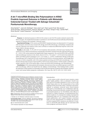
- 4. Saridaki et al. anti-EGFR mAb-based salvage treatment. There were no statistically significant differences found between KRAS LCS6-variant and nonvariant carriers for sex or age at diagnosis. The characteristics of the 559 patients have been previously published (14, 30) and are also presented here in Supplementary Table S1. As shown in Table 1, the distribution of the KRAS LCS6- variant genotypes was different among patients harboring tumor-acquired KRAS and BRAF mutations. Specifically, although the percentage of patients with the KRAS LCS6- variant was the same in KRAS WT and mutant groups (20% in each), variant patients were found twice as frequently in the BRAF V600E-mutant group versus in the BRAF-WT group (20%), resulting in a statistically significant differ-ence (P ¼ 0.030). Outcome and survival analysis in the entire patient cohort In the cohort as a whole, there were no significant differences in median PFS or OS between the nonvariant patients and the LCS6-variant patients (Supplementary Fig. S1A and S1B). Similarly, there were no differences in PFS or OS in the double (KRAS and BRAF) WT or in the KRAS-mutated patients’ cohorts comparing LCS6-variant and nonvariant patients. Finally, there were no significant cor-relations about response (n ¼ 483) and skin rash (n ¼ 359) with the LCS6-variant and nonvariant patients in the whole and in the double WT patients’ cohorts (Supplementary Table S3). In the cohort as a whole however, in univariate analysis, tumor-acquired KRAS mutations [hazard ratio (HR), 1.688; P, 0.0001; 95%confidence interval, CI, 1.395–2.042], BRAF mutations (HR, 2.206; P, 0.0001; 95% CI, 1.501–3.243), and type of treatment (HR, 1.748; P, 0.0001; 95% CI, 1.450–2.108) were correlated with PFS and OS. Multivar-iate analysis revealed that the above factors have an inde-pendent association with decreased PFS and OS (Supple-mentary Table S4), and thus were incorporated into the below analysis. PFS with monotherapy versus combination treatment Next, we separately analyzed patients that received mAb monotherapy versus mAb combination therapy. From 501 patients with known treatment, 160 (32%) received anti- EGFR mAbs as monotherapy and 341 (68%) were treated with multiple chemotherapy combinations plus EGFRmAb therapy. Of the monotherapy patients, 32 (20%) had the LCS6-variant, and of the combination treatment patients, 75 (22%) had the LCS6-variant (NS). There were no sig-nificant differences in tumor-acquired KRAS and BRAF mutations between patients that received anti-EGFR mAb monotherapy versus combination therapy (Supplementary Table S2). The median PFS of the whole monotherapy-treated patients’ population was 10.43 weeks (95% CI, 7.73– 13.12 weeks). There was a statistically significant benefit of monotherapy for the LCS6-variant patients versus non-variant patients, with a PFS of 16.86 weeks (95% CI, 10.2– 23.51 weeks) versus 7.85 weeks (95% CI, 3.897–11.817 weeks; Fig. 1A; P ¼ 0.019, log-rank test). The median PFS of the whole combination therapy patients’ population was 18 weeks (95% CI, 15.87–20.12 weeks), and there was no statistically significant difference observed between the LCS6-variant versus nonvariant patients [18 weeks (95% CI, 9.97–26.02 weeks) vs. 18.43 weeks (95% CI, 16.16– 20.69 weeks); Fig. 1B; P ¼ 0.760, log-rank test]. There was strong evidence for an interaction effect for PFS (P¼0.051). Interestingly, there was no improved PFS for LCS6-var-iant patients that received mAb therapy [16.86 weeks (95% CI, 8.55–25.18 weeks)] versus combination therapy [18 weeks (95% CI, 13.37–22.64 weeks); Fig. 1C; P ¼ 0.291, log-rank test]. In contrast, there was a significant benefit in PFS with the addition of chemotherapy for nonvariant patients (P 0.0001, log-rank test), 7.86 weeks for mono-therapy (95% CI, 3.9–11.82 weeks) versus 19.29 weeks for combination therapy (95% CI, 17–21.58 weeks; Fig. 1D). Of note, there was no significant difference in median PFS for monotherapy-treated LCS6-variant patients versus com-bination- treated nonvariant patients. In the double (KRAS and BRAF) WT patients’ popula-tion, the median PFS of the monotherapy-treated patients was 12 weeks (95% CI, 8.38–15.61 weeks), and again a statistically significant difference was observed between nonvariant patients and LCS6-variant patients [10.43 weeks (95% CI, 6.74–14.11 weeks) vs. 18 weeks (95% CI, 5.16–30.83 weeks); Fig. 2A; P ¼ 0.039, log-rank test]. The PFS for the combination therapy–treated patients was 28.71 weeks (95% CI, 24.98–32.43 weeks), and no statis-tically significant difference (P ¼ 0.39, log-rank test) was observed between the nonvariant patients and LCS6-var-iant patients [28.3 weeks (95% CI, 24.15–32.45 weeks) vs. Table 1. Distribution of the KRAS 30-UTR LCS6 genotypes according to mutation, clinical, and demographic data in the cohort of patients with metastatic colorectal cancer TG TT or GG N (%) N (%) P value Age (median, min–max) 61 (22–89) 61 (37–80) 0.654 Sex Male 229 (57.4) 59 (54.6) 0.662 Female 170 (42.6) 49 (45.4) KRAS status Mutant 138 (36.3) 36 (34.6) 0.818 WT 242 (63.7) 68 (65.4) BRAF status Mutant 17 (4.3) 11 (10.2) 0.030 WT 379 (95.7) 97 (89.8) Treatment Monotherapy 128 (32.1) 32 (29.6) 0.726 Combination 271 (67.9) 76 (70.4) 4502 Clin Cancer Res; 20(17) September 1, 2014 Clinical Cancer Research
- 5. The KRAS-Variant Predicts Cetuximab Response in Colon Cancer A B PFS and LCS6 SNP genotype in all monotherapy patients TG or GG (n = 32) TT (n = 128) TG or GG-censored TT-censored PFS according to type of therapy in all LCS6 SNP carriers 0.00 20.00 40.00 60.00 80.00 100.00 120.00 28.85 weeks (95% CI, 14.82–42.87 weeks); Fig. 2B]. Here, again, there was no significant improvement (P ¼ 0.061, log-rank test) in PFS for LCS6-variant patients that received mAb monotherapy [18 weeks (95% CI, 5.1–30.8 weeks)] versus combination therapy [28.85 weeks (95%CI, 14.83– 42.87 weeks); Fig. 2C], whereas there was improvement in PFS for nonvariant patients (P 0.0001, log-rank test) that received mAb monotherapy [10.43 weeks (95% CI, 6.75–14.15 weeks)] versus combination therapy [28 weeks (95% CI, 24.1–31.8 weeks); Fig. 2D]. Again, there was no significant difference in median PFS between LCS6- variant patients receiving mAb monotherapy and non-variant patients receiving combination therapy (18 vs. 28.8 weeks). OS analysis correlated with treatment The median OS of the whole monotherapy patients’ population was 33.14 weeks (95% CI: 26.70–39.57 weeks), and no statistically significant difference (P ¼ 0.139, log-rank test) was observed between the nonvariant patients PFS and LCS6 SNP genotype in all combination therapy patients TG or GG (n = 75) TT (n = 266) TG or GG-censored TT-censored PFS according to type of therapy in all non-LCS6 SNP carriers 0.00 20.00 40.00 60.00 80.00 100.00 120.00 and the LCS6-variant patients [28.85 weeks (95% CI, 22.53–35.18 weeks) vs. 45 weeks (95% CI, 35.02–54.97 weeks); Fig. 3A]. The median OS of the whole combination therapy patients’ population was 44 weeks (95% CI, 40.11– 47.88 weeks), and no statistically significant difference (P¼ 0.759, log-rank test) was observed between the nonvariant patients and the LCS6-variant patients [44 weeks (95% CI, 40.06–47.93 weeks) vs. 43 weeks (95% CI, 29.8–56.2 weeks); Fig. 3B]. For OS, the interaction term was clearly nonsignificant (P ¼ 0.248). There was no significant difference in OSfor LCS6-variant patients that received mAb monotherapy [45 weeks (95% CI, 35–55 weeks)] versus combination therapy [43 weeks (95% CI, 29.8–56.2 weeks); Fig. 3C; P ¼ 0.574, log-rank test], yet there was a significant benefit for OS with the addition of chemotherapy for nonvariant patients [mAb monotherapy 28.86 weeks (95% CI, 22.53–35.18 weeks) vs. combination therapy 44 weeks (95% CI, 40–47.93 weeks); P 0.0001, log-rank test; Fig. 3D]. Again, the difference in OS for LCS6-variant patients receiving 0.00 20.00 40.00 60.00 80.00 100.00 1.0 0.8 0.6 0.4 0.2 0.0 Cum survival Log-rank P = 0.019 PFS (weeks) 0.00 20.00 40.00 60.00 80.00 100.00 120.00 1.0 0.8 0.6 0.4 0.2 0.0 Log-rank P = 0.760 PFS (weeks) C PFS (weeks) 1.0 0.8 0.6 0.4 0.2 0.0 Monotherapy (n = 32) Combination (n = 75) Monotherapy-censored (n = 2) Combination therapy-censored (n = 11) Cum survival Log-rank P = 0.291 Cum survival D PFS (weeks) Cum survival 1.0 0.8 0.6 0.4 0.2 0.0 Monotherapy (n = 128) Combination (n = 266) Monotherapy-censored (n = 2) Combination therapy-censored (n = 30) Log-rank P 0.0001 Figure 1. LCS6-variant patients have improved PFS versus nonvariant patients in response to EGFR mAb monotherapy for all patients, with no benefit of additional chemotherapy. A, median PFS by KRAS LCS6 genotype in all patients treated with anti-EGFR mAb monotherapy as salvage treatment. B, median PFS according to the KRAS 30-UTR LCS6 SNP genotype status in all patients treated with anti-EGFR mAb-based combination chemotherapy as salvage treatment. C, median PFS according to type of therapy in all KRAS 30-UTR LCS6 SNP carriers. D, median PFS according to type of therapy in all non-KRAS 30-UTR LCS6 SNP carriers. www.aacrjournals.org Clin Cancer Res; 20(17) September 1, 2014 4503
- 6. Log-rank P = 0.039 PFS according to type of therapy in KRAS and BRAF WT LCS6 SNP carriers Monotherapy (n = 20) Combination (n = 38) Combination therapy -censored (n = 7) Saridaki et al. monotherapy and nonvariant patients receiving combina-tion therapy was nonsignificant. In the double (KRAS and BRAF) tumor WT patients’ population, the median OS of the monotherapy patients was 37 weeks (95% CI, 30.82–43.17 weeks). A trend toward a statistically significant difference was observed between the nonvariant patients [35.71 weeks (95% CI, 32.03–39.4 weeks)] and the LCS6-variant patients [55.43 weeks (95% CI, 36.98–73.87 weeks; Fig. 4A; P¼0.087, log-rank test]. In this population, the median OS of the combination therapy patients was 55 weeks (95% CI, 48.3–61.7 weeks), and there was no statistically significant difference (P ¼ 0.649, log-rank test) between the nonvariant patients [57 weeks (95% CI, 49.4–64.6 weeks)] and the LCS6-variant patients [54 weeks (95% CI, 45.46–62.53 weeks); Fig. 4B]. Again, there was no significant improvement (P ¼ 0.705, log-rank test) in OS for LCS6-variant patients that received mAb monotherapy [55.43 weeks (95% CI, 37–73.87 weeks)] versus combination therapy [54 weeks (95% CI, 45.47– 62.54 weeks); Fig. 4C], whereas there was improvement in OS for nonvariant patients (P 0.0001, log-rank test) that PFS and LCS6 SNP genotype in KRAS and BRAF WT combination therapy patients Monotherapy (n = 77) Combination (n =144) Combination therapy-censored (n =19) Log-rank P 0.0001 received mAb monotherapy [35.71 weeks (95% CI, 32– 39.4 weeks)] versus combination therapy [57 weeks (95% CI, 49.4–64.6 weeks); Fig. 4D]. Again, the difference in OS for LCS6 G variant patients receiving monotherapy and KRAS WT patients receiving combination therapy was nonsignificant. The LCS6-variant is not prognostic in KRAS- and BRAF-mutated patients In the KRAS- and BRAF-mutated patients’ population, no statistical significant differences in PFS or OS were observed in patients treated with either anti-EGFRmAbmonotherapy or with mAbs in combination with chemotherapy (data not shown). Median PFS times were identical between LCS6-variant and nonvariant patients, with no significant improvement (P ¼ 0.641, log-rank test) between PFS for LCS6-variant patients that received mAb monotherapy [6 weeks (95% CI, 0–13.25 weeks)] versus combination ther-apy [12 weeks (95% CI, 6.45–17.56 weeks); Supplementary Fig. S2A]. However, there was a significant improvement in PFS for nonvariant patients treated with combination A 0.00 20.00 40.00 60.00 80.00 1.0 0.8 0.6 0.4 0.2 0.0 PFS and LCS6 SNP genotype in KRAS and BRAF WT monotherapy patients PFS (weeks) TG or GG (n = 20) TT (n = 77) Cum survival B 0.00 20.00 40.00 60.00 80.00 100.00 120.00 1.0 0.8 0.6 0.4 0.2 0.0 Cum survival TG or GG (n = 38) TT (n = 144) TG or GG-censored TT-censored PFS (weeks) Log-rank P = 0.393 C 0.00 20.00 40.00 60.00 80.00 100.00 120.00 1.0 0.8 0.6 0.4 0.2 0.0 PFS (weeks) Cum survival Log-rank P = 0.096 D 0.00 20.00 40.00 60.00 80.00 100.00 120.00 Cum survival 1.0 0.8 0.6 0.4 0.2 0.0 PFS according to type of therapy in KRAS and BRAF WT non-LCS6 SNP carriers PFS (weeks) Figure 2. LCS6-variant patients have improved PFS versus nonvariant patients in response to EGFR mAb monotherapy for doubleWTpatients, with no benefit of additional chemotherapy. A, median PFS according to the KRAS 30-UTR LCS6 SNP genotype status in the double (KRAS and BRAF) WT patients' population treated with anti-EGFR mAb monotherapy as salvage treatment. B, median PFS according to the KRAS 30-UTR LCS6 SNP genotype status in the double (KRAS and BRAF) WT patients' population treated with anti-EGFR mAb-based combination chemotherapy as salvage treatment. C, median PFS according to type of therapy in the double (KRAS and BRAF) WT KRAS 30-UTR LCS6 SNP carriers. D, median PFS according to type of therapy in the double (KRAS and BRAF) WT non-KRAS 30-UTR LCS6 SNP carriers. 4504 Clin Cancer Res; 20(17) September 1, 2014 Clinical Cancer Research
- 7. The KRAS-Variant Predicts Cetuximab Response in Colon Cancer Figure 3. LCS6-variant patients do not have improved OS with the addition of chemotherapy for all patients. A, median OS according to the KRAS 30-UTR LCS6 SNP genotype status in all patients treated with anti-EGFR mAb monotherapy as salvage treatment. B, median OS according to the KRAS 30-UTR LCS6 SNP genotype status in all patients treated with anti-EGFR mAb-based combination chemotherapy as salvage treatment. C, median OS according to type of therapy in all KRAS 30-UTR LCS6 SNP carriers. D, median OS according to type of therapy in all non-KRAS 30-UTR LCS6 SNP carriers. therapy [P 0.0001, log-rank test, PFS for mAb monother-apy 6 weeks, (95% CI, 4.46–7.53 weeks) versus combina-tion therapy 12 weeks (95% CI, 9.72–14.28 weeks)] (Sup-plementary Fig. S2B). Likewise, for OS, there was no significant difference (P ¼ 0.303, log-rank test) in OS for LCS6-variant patients that received mAb monotherapy [28.43 weeks (95% CI, 9.47– 47.39 weeks)] versus combination therapy [23 weeks (95% CI, 10.8–35.19 weeks); Supplementary Fig. S2C], whereas there was a difference in OS for nonvariant patients (P ¼ 0.002, log-rank test) that received mAb monotherapy [21.29 weeks (95% CI, 15–27.55 weeks) versus combina-tion therapy [31 weeks (95% CI, 25.65–36.34 weeks); Supplementary Fig. S2D]. The LCS6-variant and response to mAb therapy From the whole population of 483 patients that were evaluable for both response and LCS6-variant genotyp-ing, 147 (30.4%) had received anti-EGFR mAb as mono-therapy and 336 (69.6%) with multiple chemotherapy combinations. In the monotherapy group, 123 (83.6%) patients were nonresponders [stable disease and progres-sive disease (PD)], and 24 (16.4%) were responders [partial response (PR) and complete response (CR)]. There were significantly more LCS6-variant patients in the monotherapy responders group versus the nonre-sponders group (11/24 vs. 19/123; Fisher exact test, P ¼ 0.002). In the combination with chemotherapy group, 252 (75%) patients were nonresponders (stable disease and PD) and 84 (25%) were responders (PR and CR). There was no statistically significant difference between the proportion of the nonvariant and LCS6- variant patients in these groups (Fisher exact test, P ¼ 1). In the 270 double (KRAS and BRAF) WT populations, 90 (33.3%) received anti-EGFR mAb as monotherapy and 180 (66.6%) with multiple chemotherapy combinations. In the www.aacrjournals.org Clin Cancer Res; 20(17) September 1, 2014 4505
- 8. OS and LCS6 SNP genotype in KRAS and BRAF WT monotherapy patients OS according to type of therapy in KRAS and BRAF WT LCS6 SNP carriers Monotherapy (n = 20) Combination (n = 37) Monotherapy-censored (n = 5) Combination therapy–censored (n = 5) Log-rank P = 0.705 Saridaki et al. monotherapy group, 71 (78.8%) patients were nonrespon-ders (stable disease and PD), and 19 (21.2%) were respon-ders (PR and CR). Again, there were significantly more LCS6-variant patients in the responders versus the nonre-sponders groups (9/19 vs. 11/71; Fisher exact test, P ¼ 0.010). In the combination with chemotherapy group, 102 (56.6%) patients were nonresponders (stable disease and PD) and 78 (43.4%) were responders (PR and CR). There was no statistically significant difference between the proportion of nonvariant and LCS6-variant patients in the groups (Fisher exact test, P ¼ 1). The effect of the LCS6-variant on gene expression upon therapy exposure Because patients with the LCS6-variant in this analysis were sensitive to EGFR mAb monotherapy, it suggested that they are not EGFR-independent, as tumor-acquired OS and LCS6 SNP genotype in KRAS and BRAF WT combination therapy patients TG or GG (n = 37) TT (n = 147) TG or GG-censored TT-censored Log-rank P = 0.649 OS according to type of therapy in KRAS and BRAF WT non-LCS6 SNP carriers Monotherapy (n = 77) Combination (n =147) Monotherapy-censored (n =11) Combination therapy–censored (n =18) Log-rank P 0.0001 KRAS mutant patients are (30). To better understand how these patients may respond differently to mAb monotherapy, we created dual-luciferase reporters con-taining the entire KRAS 30-UTR with the LCS6-variant (G allele) or without the variant (T allele). We used this system to test the hypothesis that mAb therapy and chemotherapy may differentially impact expression of KRAS for those with the LCS6-variant allele versus the nonvariant allele. We found, as has been previously reported in other cell lines (36), that the LCS6-variant allele (G allele reporter) displayed 1.8-fold higher expression at baseline in HCT-116 colon cancer cells when compared with the nonvariant allele (T allele reporter, Fig. 5A). This finding supports previous evidence that this is a functional mutation that permits KRAS overexpression in tumors. Next, we exposed these cells to cetuximab, and found that although there was A 0.00 50.00 100.00 150.00 1.0 0.8 0.6 0.4 0.2 0.0 Cum survival OS (weeks) TG or GG (n = 20) TT (n = 77) TG or GG-censored TT-censored Log-rank P = 0.087 B 0.00 50.00 100.00 150.00 200.00 250.00 300.00 1.0 0.8 0.6 0.4 0.2 0.0 Cum survival OS (weeks) C 0.00 50.00 100.00 150.00 200.00 250.00 300.00 1.0 0.8 0.6 0.4 0.2 0.0 Cum survival OS (weeks) D 0.00 50.00 100.00 150.00 200.00 250.00 1.0 0.8 0.6 0.4 0.2 0.0 Cum survival OS (weeks) Figure 4. LCS6-variant patients do not have improved OS with the addition of chemotherapy for double WT patients. A, median OS according to the KRAS 30-UTR LCS6 SNP genotype status in the double (KRAS and BRAF) WT patients' population treated with anti-EGFR mAb monotherapy as salvage treatment. B, medianOSaccording to theKRAS 30-UTR LCS6SNP genotype status in the double (KRAS and BRAF)WTpatients' population treated with anti- EGFR mAb-based combination chemotherapy as salvage treatment. C, median OS according to type of therapy in the double (KRAS and BRAF) WT KRAS 30-UTR LCS6 SNP carriers. D, median OS according to type of therapy in the double (KRAS and BRAF) WT non-KRAS 30-UTR LCS6 SNP carriers. 4506 Clin Cancer Res; 20(17) September 1, 2014 Clinical Cancer Research
- 9. no impact on KRAS expression in the nonvariant allele reporter system, there was a significant increase in the overexpression of KRAS for the LCS6-variant allele reporter system. We found similar increased overexpression of KRAS for the LCS6-variant allele with exposure to 5-fluorouracil. In contrast, we saw little to no change in expression of the LCS6-variant allele compared to the nonvariant allele with exposure to irinotecan (Fig. 5B). These results indicate that the LCS6-variant allele leads to KRAS protein overexpres-sion in response to specific chemotherapy treatments as well as mAb therapy, a finding not seen in the presence of the nonvariant allele. Discussion Here, we have shown a statistically significant improve-ment in median PFS for all LCS6-variant patients with metastatic colorectal cancer treated with anti-EGFR mAb monotherapy. This improved prognosis is not enhanced by the addition of chemotherapy, and in fact, LCS6-variant patients seemed to experience no benefit from the addition of chemotherapy to anti-EGFR mAb therapy. This finding is in contrast to nonvariant patients, who derived a significant benefit from the addition of chemotherapy to anti-EGFR mAbs across all cohorts, and only then achieved compara-ble outcomes as those of LCS6-variant patients. This clinical finding was supported by cell line studies indicating that the LCS6-variant allele responds differently than the nonvar-iant allele in response to chemotherapy and anti-EGFRmAb exposure, with increased expression and likely further dependence on the KRAS pathway (47). A different distribution of the LCS6 genotypes according to the KRAS and BRAF mutational status was observed in our population of patients with metastatic colorectal cancer The KRAS-Variant Predicts Cetuximab Response in Colon Cancer than that observed in prior reports. LCS6-variant patients were equally likely to have acquired KRAS tumor mutations as not, but, LCS6-variant patients were significantly more likely to be in the BRAF-mutated group. In a previously studied metastatic colorectal cancer population similar to ours, Graziano and colleagues (41) found an increased prevalence of the LCS6-variant in the KRAS mutant, but not in BRAF-mutant patients (41). Although one explana-tion for our different results could be that we used tumor DNAfor the majority of testing, this seems unlikely, since, it has previously been well documented that the genotype of normal and tumor tissue is the same in LCS6-variant patients (36). Another hypothesis could be that in the later stages of colorectal cancer carcinogenesis, the LCS6-variant allele mediates the selection of less differentiated and more aggressive clones that harbor BRAF mutations, and perhaps our cohort was more advanced. In addition, there could be a selective pressure to develop KRAS or BRAF mutations in the presence of the LCS6-variant, depending on exposure to specific therapies, and prior therapy likely differed between our two studies. The finding that this single base pair change in the 30-UTR of KRAS leads to a significant difference in both baseline expression as well as response to chemotherapy in a lucif-erase reporter construct is intriguing. Although tumor-acquired KRAS mutations are always turned on, these cell line reporter studies further indicate how fundamentally different this mutation is than a simple tumor-acquired KRAS mutation. By their nature, miRNA-binding disrupting mutations, such as the KRAS LCS6-variant, are dependent on trans-activating factors, such as miRNAs, that change in response to stress. It has been know for several years that miRNAs are used to dynamically regulate the response to cytotoxic cancer therapy (33). It is perhaps not surprising 12 10 8 6 4 2 0 4 3.5 3 2.5 2 1.5 1 0.5 Normalized luciferase (Renilla/firefly) Relative expression of G allele (compared with T allele) P = 0.000018 Cetuximab A B 5FU Irinotecan T allele (WT) G allele (MT) Untreated 0.5 1 2.5 5 KRAS 3¢UTR Reporter [compound] (mmol/L) Figure 5. The KRAS LCS6-variant causes overexpression of a KRAS reporter. A, HCT-116 colon cancer cells were transfected with dual-luciferase reporters harboring either the full-length KRAS 30-UTR T allele or G allele (LCS6-variant), as indicated. Dual-luciferase activities were measured, and Renilla was normalized to firefly. Results are graphed as the mean andSDof the mean of four independent experiments performed in duplicate. The Pvalue was calculated using the Student t test. B, the KRAS LCS6-variant exhibits altered gene expression in response to anticancer agents. HCT-116 colon cancer cells transfected with either the KRAS LCS6-variant or nonvariant 30-UTR reporters were exposed to various concentrations of anticancer compounds (as indicated). The results are expressed as the relative expression of the LCS-variant versus the nonvariant allele. Graphed is the mean andSDof themean for two experiments performed in duplicate. www.aacrjournals.org Clin Cancer Res; 20(17) September 1, 2014 4507
- 10. therefore that a mutation such as the LCS6-variant would be predictive of cancer treatment response, as cancer treat-ments will lead to changes in the very factors that regulate the mutation, and subsequent downstream gene and path-way expression. However, further molecular studies of the exact mechanisms by which this mutation alters response to EGFR mAb treatment are still required in tissue and animal models. Recently, two large studies of patients with colon cancer investigating outcome found that the LCS6-variant allele predicted a good prognosis, especially when in combina-tion with tumor-acquired mutations in KRAS, in both early- (48) and late-stage (49) patients. These authors hypothe-sized that at least in early-stage colon cancer, the LCS6- variant plus KRAS mutations could lead to too much KRAS and tumor cell senescence. On the basis of our cell line data, indicating that anti-EGFR mAb monotherapy leads to sig-nificantly higher KRAS expression, as does 5FU, but not irinotecan, we hypothesize that this may be a viable expla-nation of the very favorable anti-EGFR mAb monotherapy response in advanced KRAS LSC6-variant patients as well. It does further support the hypothesis that the combination of therapy delivered with anti-EGFR mAb monotherapy is critical, as there seems to be no benefit of additional chemotherapy in our study, and in fact chemotherapy could possibly be a detriment to patients with LCS6-variant met-astatic colorectal cancer. As is also true for other cancers, an important step in the development of colorectal cancer seems to be the deregu-lation of miRNAs. Over the past few years, miRNAs have been brought to the central stage of molecular oncology and have substantially changed the way we view and understand gene regulation (50). The KRAS LCS6-variant was the first mutation in a miRNA-binding site to be implicated in cancer risk, and although it certainly will not be the last (36), it seems to also play a significant predictive role that could guide therapy decisions. Our findings here suggest that patients carrying the LCS6-variant are biologically different than nonvariant patients, have a higher probabil-ity of benefit from anti-EGFR mAb monotherapy, and deserve prospective clinical studies to determine what, if anything, they should receive in addition to cetuximab treatment in the metastatic colorectal cancer setting. Disclosure of Potential Conflicts of Interest J.B. Weidhaas has ownership interest (including patents) in and is a consultant/advisory board member for Mira Dx. H.-J. Lenz is a consultant/ advisory board member for Bristol-Myers Squibb and Merck. P. Laurent-Puig is a consultant/advisory board member for Amgen and Merck Serono. O. Bouche reports receiving speakers bureau honoraria from Amgen and is a consultant/advisory board member for Merck Sereno. S. Tejpar is a consul-tant/ advisory board member for Merck Serono. No potential conflicts of interest were disclosed by the other authors. Authors' Contributions Conception and design: Z. Saridaki, J.B. Weidhaas, H.-J. Lenz, P. Laurent- Puig, D.W. Salzman, S. Tejpar Development of methodology: Z. Saridaki, J.B. Weidhaas, B. Jacobs, D.W. Salzman, H. Piessevaux, S. Tejpar Acquisition of data (provided animals, acquired and managed patients, provided facilities, etc.): Z. Saridaki, H.-J. Lenz, P. Laurent-Puig, W. De Roock, D.W. Salzman, W. Zhang, C. Pilati, O. Bouche, S. Tejpar Analysis and interpretation of data (e.g., statistical analysis, biosta-tistics, computational analysis): Z. Saridaki, J.B. Weidhaas, H.-J. Lenz, B. Jacobs, D.W. Salzman, D. Yang, C. Pilati, H. Piessevaux, S. Tejpar Writing, review, and/or revision of the manuscript: Z. Saridaki, J.B. Weidhaas, H.-J. Lenz, P. Laurent-Puig, B. Jacobs, D. Yang, C. Pilati, O. Bouche, H. Piessevaux, S. Tejpar Administrative, technical, or material support (i.e., reporting or orga-nizing data, constructing databases): Z. Saridaki, H.-J. Lenz, J. De Schut-ter, W. Zhang, S. Tejpar Study supervision: Z. Saridaki, S. Tejpar Acknowledgments Z. Saridaki was a recipient of a research fellowship from the Hellenic Society of Medical Oncology (Hesmo). Grant Support J.B. Weidhaas was supported by two R01 grants: CA131301-04 and CA157749-01A1. The costs of publication of this article were defrayed in part by the payment of page charges. This article must therefore be hereby marked advertisement in accordance with 18 U.S.C. Section 1734 solely to indicate this fact. Received February 13, 2014; revised May 9, 2014; accepted June 16, 2014; published online September 2, 2014. References 1. Siegel R, Desantis C, Jemal A. Cancer statistics, 2014. CA Cancer J Clin 2014;64:104–17. 2. Cunningham D, Humblet Y, Siena S, Khayat D, Bleiberg H, Santoro A, et al. Cetuximab monotherapy and cetuximab plus irinotecan in irino-tecan- refractory metastatic colorectal cancer. N Engl J Med 2004; 351:337–45. 3. Saltz LB, Cox JV, Blanke C, Rosen LS, Fehrenbacher L, Moore MJ, et al. Irinotecan plus fluorouracil and leucovorin for metastatic colo-rectal cancer. Irinotecan Study Group.NEngl JMed 2000;343:905–14. 4. Saltz LB, Meropol NJ, Loehrer PJ, Needle MN, Kopit J, Mayer RJ. Phase II trial of cetuximab in patients with refractory colorectal cancer that expresses the epidermal growth factor receptor. J Clin Oncol 2004;22:1201–8. 5. Van Cutsem E, Peeters M, Siena S, Humblet Y, Hendlisz A, Neyns B, et al. Open-label phase III trial of panitumumab plus best supportive care compared with best supportive care alone in patients with chemotherapy-refractory metastatic colorectal cancer. J Clin Oncol 2007;25:1658–64. 6. Amado RG, Wolf M, Peeters M, Van Cutsem E, Siena S, Freeman DJ, et al. Wild-type KRAS is required for panitumumab efficacy in patients with metastatic colorectal cancer. J Clin Oncol 2008;26: 1626–34. 7. De Roock W, Piessevaux H, De Schutter J, Janssens M, De Hertogh G, Personeni N, et al. KRAS wild-type state predicts survival and is associated to early radiological response in metastatic colorectal cancer treated with cetuximab. Ann Oncol 2008;19:508–15. 8. Lievre A, Bachet J-B, Boige V, Cayre A, Le Corre D, Buc E, et al. KRAS mutations as an independent prognostic factor in patients with advanced colorectal cancer treated with cetuximab. J Clin Oncol 2008;26:374–9. 9. Sartore-Bianchi A, Moroni M, Veronese S, Carnaghi C, Bajetta E, Luppi G, et al. Epidermal growth factor receptor gene copy number and clinical outcome of metastatic colorectal cancer treated with panitu-mumab. J Clin Oncol 2007;25:3238–45. 10. Bokemeyer C, Bondarenko I, Makhson A, Hartmann JT, Aparicio J, de Braud F, et al. Fluorouracil, leucovorin, and oxaliplatin with and without Saridaki et al. 4508 Clin Cancer Res; 20(17) September 1, 2014 Clinical Cancer Research
- 11. cetuximab in the first-line treatment of metastatic colorectal cancer. J Clin Oncol 2009;27:663–71. 11. Douillard J-Y, Siena S, Cassidy J, Tabernero J, Burkes R, Barugel M, et al. Randomized, phase III trial of panitumumab with infusional fluorouracil, leucovorin, and oxaliplatin (FOLFOX4) versus FOLFOX4 alone as first-line treatment in patients with previously untreated metastatic colorectal cancer: the PRIME study. J Clin Oncol 2010; 28:4697–705. 12. Karapetis CS, Khambata-Ford S, Jonker DJ, O'Callaghan CJ, Tu D, Tebbutt NC, et al. K-ras mutations and benefit from cetuximab in advanced colorectal cancer. N Engl J Med 2008;359:1757–65. 13. Van Cutsem E, Kohne C-H, Hitre E, Zaluski J, Chang Chien C-R, Makhson A, et al. Cetuximab and chemotherapy as initial treatment for metastatic colorectal cancer. N Engl J Med 2009;360:1408–17. 14. De Roock W, Claes B, Bernasconi D, De Schutter J, Biesmans B, Fountzilas G, et al. Effects of KRAS, BRAF, NRAS, and PIK3CA mutations on the efficacy of cetuximab plus chemotherapy in chemo-therapy- refractory metastatic colorectal cancer: a retrospective con-sortium analysis. Lancet Oncol 2010;11:753–62. 15. Allegra CJ, Jessup JM, Somerfield MR, Hamilton SR, Hammond EH, Hayes DF, et al. American Society of Clinical Oncology provisional clinical opinion: testing for KRAS gene mutations in patients with metastatic colorectal carcinoma to predict response to anti-epidermal growth factor receptor monoclonal antibody therapy. J Clin Oncol 2009;27:2091–6. 16. De Roock W, De Vriendt V, Normanno N, Ciardiello F, Tejpar S. KRAS, BRAF, PIK3CA, and PTEN mutations: implications for targeted ther-apies in metastatic colorectal cancer. Lancet Oncol 2011;12:594–603. 17. Di Nicolantonio F, Martini M, Molinari F, Sartore-Bianchi A, Arena S, Saletti P, et al. Wild-type BRAF is required for response to panitumu-mab or cetuximab in metastatic colorectal cancer. J Clin Oncol 2008;26:5705–12. 18. Laurent-Puig P, Cayre A, Manceau G, Buc E, Bachet J-B, Lecomte T, et al. Analysis of PTEN, BRAF, and EGFR status in determining benefit from cetuximab therapy in wild-type KRAS metastatic colon cancer. J Clin Oncol 2009;27:5924–30. 19. Saridaki Z, Tzardi M, Papadaki C, Sfakianaki M, Pega F, Kalikaki A, et al. Impact of KRAS, BRAF, PIK3CA mutations, PTEN, AREG, EREG expression and skin rash in /¼ 2 line cetuximab-based therapy of colorectal cancer patients. PLoS One 2011;6:e15980. 20. Souglakos J, Philips J, Wang R, Marwah S, Silver M, Tzardi M, et al. Prognostic and predictive value of common mutations for treatment response and survival in patients with metastatic colorectal cancer. Br J Cancer 2009;101:465–72. 21. Ogino S, Nosho K, Kirkner GJ, Shima K, Irahara N, Kure S, et al. PIK3CA mutation is associated with poor prognosis among patients with curatively resected colon cancer. J Clin Oncol 2009;27: 1477–84. 22. Prenen H, De Schutter J, Jacobs B, De Roock W, Biesmans B, Claes B, et al. PIK3CA mutations are not a major determinant of resistance to the epidermal growth factor receptor inhibitor cetuximab in metastatic colorectal cancer. Clin Cancer Res 2009;15:3184–8. 23. Sartore-Bianchi A, Martini M, Molinari F, Veronese S, Nichelatti M, Artale S, et al. PIK3CA mutations in colorectal cancer are associated with clinical resistance to EGFR-targeted monoclonal antibodies. Cancer Res 2009;69:1851–7. 24. Lee RC, Feinbaum RL, Ambros V. The C. elegans heterochronic gene lin-4 encodes small RNAs with antisense complementarity to lin-14. Cell 1993;75:843–54. 25. Hollestelle A, Pelletier C, Hooning M, Crepin E, Schutte M, Look M, et al. Prevalence of the variant allele rs61764370 TG in the 30UTR of KRAS among Dutch BRCA1, BRCA2 and non-BRCA1/BRCA2 breast cancer families. Breast Cancer Res Treat 2011;128:79–84. 26. Reddy SDN, Gajula RP, Pakala SB, Kumar R. MicroRNAs and cancer therapy: the next wave or here to stay? Cancer Biol Ther 2010;9: 479–82. 27. Faber C, Kirchner T, Hlubek F. The impact of microRNAs on colorectal cancer. Virchows Arch 2009;454:359–67. 28. Landi D, Gemignani F, Naccarati A, Pardini B, Vodicka P, Vodick-ova L, et al. Polymorphisms within micro-RNA-binding sites The KRAS-Variant Predicts Cetuximab Response in Colon Cancer and risk of sporadic colorectal cancer. Carcinogenesis 2008;29: 579–84. 29. Chen K, Song F, Calin GA, Wei Q, Hao X, Zhang W. Polymorphisms in microRNA targets: a gold mine for molecular epidemiology. Carcino-genesis 2008;29:1306–11. 30. Zhang W, Winder T, Ning Y, Pohl A, Yang D, Kahn M, et al. A let-7 microRNA-binding site polymorphism in 30-untranslated region of KRAS gene predicts response in wild-type KRAS patients with met-astatic colorectal cancer treated with cetuximab monotherapy. Ann Oncol 2011;22:104–9. 31. Mishra PJ, Bertino JR. MicroRNA polymorphisms: the future of phar-macogenomics, molecular epidemiology and individualized medicine. Pharmacogenomics 2009;10:399–416. 32. Sethupathy P, Collins FS. MicroRNA target site polymorphisms and human disease. Trends Genet 2008;24:489–97. 33. Weidhaas JB, Babar I, Nallur SM, Trang P, Roush S, Boehm M, et al. MicroRNAs as potential agents to alter resistance to cytotoxic anti-cancer therapy. Cancer Res 2007;67:11111–6. 34. Jerome T, Laurie P, Louis B, Pierre C. Enjoy the silence: the Story of let-7 MicroRNA and Cancer. Curr Genomics 2007;8: 229–33. 35. Johnson SM, Grosshans H, Shingara J, Byrom M, Jarvis R, Cheng A, et al. RAS is regulated by the let-7 microRNA family. Cell 2005; 120:635–47. 36. Chin LJ, Ratner E, Leng S, Zhai R, Nallur S, Babar I, et al. A SNP in a let- 7 microRNA complementary site in the KRAS 30 untranslated region increases non-small cell lung cancer risk. Cancer Res 2008;68: 8535–40. 37. Paranjape T, Heneghan H, Lindner R, Keane FK, Hoffman A, Hollestelle A, et al. A 30-untranslated region KRAS variant and triple-negative breast cancer: a case-control and genetic analysis. Lancet Oncol 2011;12:377–86. 38. Pharoah PD, Palmieri RT, Ramus SJ, Gayther SA, Andrulis IL, Anton- Culver H, et al. The role of KRAS rs61764370 in invasive epithelial ovarian cancer: implications for clinical testing. Clin Cancer Res 2011;17:3742–50. 39. Ratner E, Lu L, Boeke M, Barnett R, Nallur S, Chin LJ, et al. A KRAS-variant in ovarian cancer acts as a genetic marker of cancer risk. Cancer Res 2010;70:6509–15. 40. Ratner ES, Keane FK, Lindner R, Tassi RA, Paranjape T, Glasgow M, et al. A KRAS variant is a biomarker of poor outcome, platinum chemotherapy resistance and a potential target for therapy in ovarian cancer. Oncogene 2012;31:4559–66. 41. Graziano F, Canestrari E, Loupakis F, Ruzzo A, Galluccio N, Santini D, et al. Genetic modulation of the Let-7 microRNA binding to KRAS 30- untranslated region and survival of metastatic colorectal cancer patients treated with salvage cetuximab-irinotecan. Pharmacoge-nomics J 2010;10:458–64. 42. Kjersem JB, Ikdahl T, Guren T, Skovlund E, Sorbye H, Hamfjord J, et al. Let-7 miRNA-binding site polymorphism in the KRAS 30UTR; colorectal cancer screening population prevalence and influence on clinical outcome in patients with metastatic colorectal cancer trea-ted with 5-fluorouracil and oxaliplatin þ/ cetuximab. BMC Cancer 2012;12:534. 43. Sebio A, Pare L, Paez D, Salazar J, Gonzalez A, Sala N, et al. The LCS6 polymorphism in the binding site of let-7 microRNA to the KRAS 30- untranslated region: its role in the efficacy of anti-EGFR-based therapy in metastatic colorectal cancer patients. Pharmacogenet Genomics 2013;23:142–7. 44. Winder T, Zhang W, Khoueiry AE, Yang D, Pohl A, Lurje G, et al. Association of a germ-line variant in the K-ras 30 untranslated region with response and progression-free survival in patients with mCRC treated with single-agent cetuximab (IMCL-0144) or in combination with cetuximab (EPIC) independent of K-ras mutation status. J Clin Oncol 27:15s, 2009 (suppl; abstr 4061). 45. Lenz HJ, Van Cutsem E, Khambata-Ford S, Mayer RJ, Gold P, Stella P, et al. Multicenter phase II and translational study of cetuximab in metastatic colorectal carcinoma refractory to irino-tecan, oxaliplatin, and fluoropyrimidines. J Clin Oncol 2006;24: 4914–21. www.aacrjournals.org Clin Cancer Res; 20(17) September 1, 2014 4509
- 12. Saridaki et al. 46. Thwaites SE, Gurung B, Yao J, Kable K, Robertson P, Ryan BJ, et al. Excellent outcomes of simultaneous pancreas kidney transplantation in patients from rural and urban Australia: a national service experi-ence. Transplantation 2012;94:1230–5. 47. Paranjape T, Slack FJ, Weidhaas JB. MicroRNAs: tools for cancer diagnostics. Gut 2009;58:1546–54. 48. Smits KM, Paranjape T, Nallur S, Wouters KAD, Weijenberg MP, Schouten LJ, et al. A let-7 microRNA SNP in the KRAS 30UTR is prognostic in early-stage colorectal cancer. Clin Cancer Res 2011; 17:7723–31. 49. Ryan BM, Robles AI, Harris CC. KRAS-LCS6 genotype as a prog-nostic marker in early-stage CRC–letter. Clin Cancer Res 2012; 18:3487–8. 50. Slaby O, Svoboda M, Michalek J, Vyzula R. MicroRNAs in colorectal cancer: translation of molecular biology into clinical application. Mol Cancer 2009;8:102–7. 4510 Clin Cancer Res; 20(17) September 1, 2014 Clinical Cancer Research