1. Comparison of Superoxide Dismutase Mimetics Made
With PABA and Bacitracin Ligands
William Magilton , Aura Enache, April Viola, Catherine Reis, David N. Juboor, Fred Bicknese, Sergio Davila-Martinez, Piper Gauthier, Sana Nadeem, Darryl Peters, Northampton Community College
Abstract
Superoxide dismutases (SODs) are the primary defense against oxidative stress
during the conversion of diatomic oxygen into water (Fridovich, 1997, p. 18516).
This experiment synthesized compounds with SOD-like behavior and compared
those having a 4-Aminobenzoic acid ligand with those containing a bacitracin
ligand to see which better displayed SOD activity. The benefit of SOD-like
compounds would be in their medicinal purposes. Each compound contained a
metal cation bound to one or two of four chosen compounds previously recorded
as displaying SOD-like activity when bound to a metal cation. SOD activity was
determined by an enzymatic assay which can be monitored using
Ultraviolet/Visible Spectroscopy (UV/Vis). The results showed similar SOD
activity for the 4-Aminobenzoic acid / salicylic acid / nicotinic acid (PABA/SA/NA)
and bacitracin complexes. However, only the bacitracin compounds showed
bacterial inhibition, suggesting that further experimentation with bacitracin-
metalloid compounds is a promising direction for future studies.
Introduction
Procedure
Melting Point
Test
• Done to
analyze the
purity of the
complex
• Compared to
melting point
of ligands
Infrared
Spectroscopy
Test
• Identifies the
movement and
vibration of the
bonds in
complex and
ligands
Solubility
Test
• Determined
the best
solvent to use
for Ultraviolet-
visible light
spectroscopy
and enzymatic
assay
Enzymatic
Assay
• Tests the
effectiveness
of the
complexes
• Ultraviolet
Visible
spectroscopy
measured the
absorption of
the complex at
Physical Properties
Assay
Solutions
Loading
of
samples
in
cuvettes
Assay Results
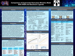
Bacterial Inhibition
TSA agar plates were inoculated with Staphylococcus epidermidis. Sterile disks
loaded with 1-2 drops of each synthesized metalloid complex were placed on the
agar, one disk/complex per labeled quadrant. Agar plates were then incubated at
37oC for 24 hours. Quadrants containing disks loaded with metallo-bacitracin
complexes exhibit zones of clearing indicating bacterial inhibition while all other
complexes do not. (See images below.)
Conclusion
Works Cited
Fridovich, I. (1997). Superoxide anion radial (O.-2), superoxide dismutases, and
rellated matters. The Journal of Biological Chemistry, 272(30), 18515-
18517.doi:10.1074/jbc.272.30.18515
Fridovich, I. (2012). Oxygen: how do we stand it?. Medical Principles and
Practice (International Journal of the Kuwait University Health Science
Synthesis of Complexes by Reflux
1mmol of the first ligand was dissolved in 30 mL of methanol and placed in the
reflux system. 1mmol of the chosen metal chloride was also dissolved in 2 mL of
methanol and added to the system drop-wise. After mixture was heated and
stirred for 1-3 hours,1 mmol of the second ligand was dissolved in 2 mL of
methanol and added drop-wise. If a second ligand was added, the mixture was
refluxed for another hour. After refluxing, the mixture was left to cool at room
temperature. Product was collected by vacuum filtration, rinsed with cold
methanol and dried in a desiccator for one week.
23.0 mL purified water
25.0 mL of 216mM potassium
phosphate buffer
1.0 mL of 10.7mM EDTA
1.0 mL of 1.1mM Cytochrome c
50 mL of 0.108 mM Xanthine
• Second the Xanthine
oxidase and bovine
erythrocyte superoxide
dismutase solutions
were made and kept on
ice until use
Blank
2.8mL of
cocktail
0.2 mL of
purified water
Uninhibited
2.8mL of cocktail
0.1 mL of purified
water
0.1 mL of
Xanthine
Oxidase
Superoxide
2.8 mL cocktail
0.1 mL of bovine
erythrocyte
superoxide dismutase
0.1 mL of Xanthine
oxidase
Complex
2.8 mL of cocktail
0.1 mL of complex
0.1 mL of
Xanthine oxidase
An enzymatic assay was used to test the complexes for SOD-like
activity. The test is based on the ability of SODs to inhibit the
reduction of Cytochrome C3+ to Cytochrome C2+ (2) by neutralizing
the superoxide radical (3). The inhibition of this reduction reaction due
to the SOD can be monitored using Ultraviolet/Visible Spectroscopy
which detects Cytochrome C at 550nm.
1. Xanthine + O2 + H2O XOD→ Uric Acid + O2 •- + H+
2. Cytochrome 3+ c + O2 •- → Cytochrome 2+ c + O2
3. 2 O2 •- + 2H+ SOD→ O2 + H2O2
• First the reagent cocktail was prepared with all the components
for Reaction 1 and 2 except the catalyst Xanthine oxidase:
Inhibition was determined by recording absorption over time at 550nm
Compound Color Melting Point
Range
Solubility
PABA-Zn2+-
Nicotinic Acid
Aquamarine
crystals
295.2 °C to
295.8°C
Methanol
PABA-Fe2+-
Salicylic Acid
Dark purple crystals >300°C Methanol, Acetonitrile
Ethyl Alcohol, Ethyl Acetate
PABA-Cu2+ Brown crystals >300°C
PABA-Cu2+-
Nicotinic Acid
Aquamarine
crystals
286 °C to
287.7°C
Methanol, Acetonitrile
Ethanol, Ethyl Acetate
PABA-CuCl2-
Salicylic Acid
Olive brown crystals 211.2°C to
213.7°C
Methanol
PABA-Co2+-
Nicotinic Acid
Purple crystals >300°C Methanol
Ethanol (slightly)
PABA-Ni2+-
Salicylic Acid
Green crystals >300°C Methanol
Ethyl alcohol
PABA-Ni2+-
Nicotinic Acid
Light green crystals >300°C Methanol
Ethanol (slightly)
Bacitracin-
CuCl2
Crystals changed
from blue to brown
to a final color of
dark green
221.3°C to
229.3°C
Methanol
Bacitracin-
CoCl2
Dark metallic purple
crystals
297.4°C Methanol
Bacitracin-Ni2+ Light frog-green
crystals
236.9°C to
296.9°C
Methanol
Ethyl acetate (slightly)
Bacitracin-Zn2+ Yellow crystals 255°C Methanol
Ethyl acetate (slightly)
Ethyl alcohol (slightly)
Fig. 2: Clockwise from
top left –
Zn/Bacitracin; Water
(control);
PABA/Ni/Nicotinic
Acid; Cu/Bacitracin
Fig. 3: Clockwise from
top left – PABA/Cu;
PABA/Fe/Salicylic Acid;
PABA/Cu/Salicylic Acid;
PABA/Ni/Salicylic Acid
Fig. 1: Clockwise from top
left – Ni/Bacitracin;
PABA/Cu/Nicotinic Acid;
PABA/Co/Nicotinic Acid;
PABA/Zn/Nicotinic Acid
Based on the results shown, a multitude of aspects can be confirmed concerning the
ability of the synthesized complexes to mimic superoxide dismutase (SOD). Firstly,
all complexes were synthesized successfully based on the recorded tight melting
points and the observed shifting of peaks on the IR spectra for each complex. From
the results of the assays, it can be concluded that all complexes inhibited the
formation of the superoxide radical via an SOD-like process, as expected from most
benzoic acids. One complex, consisting of PABA/Fe2+/SA inhibited the formation of
the superoxide radical by nearly 85%. Two other complexes, PABA/Cu2+ and
PABA/Cu2+/NA, both did a fairly good job, with percent inhibition as high as 78%
and 57%, respectively. The bacterial inhibition assay, though, is what exhibited a
marked difference between the PABA and bacitracin complexes. Complexes
synthesized with bacitracin inhibited growth of bacteria, which is logical since the
medical application of bactitracin is as an antibiotic. However, the PABA complexes
promoted the growth of bacteria. These results also make sense due to the natural
function of PABA in life processes. Similar experimentation will continue in the future
Superoxide dismutase is an enzyme found in living cells which reduces the
toxicity of diatomic oxygen necessary for aerobic/biological processes (Fridovich,
2012, p. 131). Atmospheric oxygen contains two unpaired electrons with parallel
spin states. The metabolism of aerobic organisms generates reactive
intermediates during the reduction of atmospheric oxygen, O2, such as
superoxide radicals (O2-), hydrogen peroxide (H2O2), and hydroxyl radicals
(HO-) (Fridovich, 2012, p. 132). These reactive intermediates are called free
radicals due to having an unpaired valence electron, which makes them high in
energy and therefore unstable. This instability can damage all cellular
macromolecules, and unless opposed by cellular defenses, such as superoxide
dismutase, aerobic life would be inconceivable. Superoxide dismutase catalyzes
the conversion of oxygen radicals into hydrogen peroxide and oxygen, thereby
reducing the concentration of free radicals which are able to cause cellular
damage (Fridovich, 2012, p. 133). With many medical conditions tracing their
origins back to damage caused at the cellular level, superoxide dismutase
presents a promising possibility for a wide range of medical applications
Bacterial Inhibition