CYTOO Stories Mitochondria
•
1 like•342 views

Researchers from Angers University Hospital, led by Arnaud Chevrollier, have recently published an innovative study about mitochondrial dynamics, conducted on CYTOO micropatterns. Their technique allows standardized quantitative analysis of mitochondrial networks and provides new insight into mitochondrial dysfunction. The method has strong potential in defining new diagnostic criteria for neurodegenerative disorders, cardiomyopathies, metabolic syndrome, cancer, and obesity.

Recommended
Recommended
More Related Content
What's hot
What's hot (20)
Similar to CYTOO Stories Mitochondria
Similar to CYTOO Stories Mitochondria (20)
More from CYTOO
Recently uploaded
Recently uploaded (20)
CYTOO Stories Mitochondria
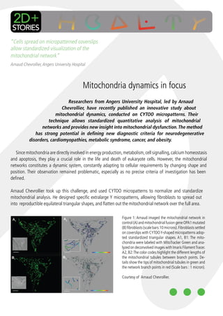
- 1. “Cells spread on micropatterned coverslips allow standardized visualization of the mitochondrial network” Arnaud Chevrollier, Angers University Hospital Mitochondria dynamics in focus Researchers from Angers University Hospital, led by Arnaud Chevrollier, have recently published an innovative study about mitochondrial dynamics, conducted on CYTOO micropatterns. Their technique allows standardized quantitative analysis of mitochondrial networks and provides new insight into mitochondrial dysfunction. The method has strong potential in defining new diagnostic criteria for neurodegenerative disorders, cardiomyopathies, metabolic syndrome, cancer, and obesity. Since mitochondria are directly involved in energy production, metabolism, cell signalling, calcium homeostasis and apoptosis, they play a crucial role in the life and death of eukaryote cells. However, the mitochondrial networks constitutes a dynamic system, constantly adapting to cellular requirements by changing shape and position. Their observation remained problematic, especially as no precise criteria of investigation has been defined. Arnaud Chevrollier took up this challenge, and used CYTOO micropatterns to normalize and standardize mitochondrial analysis. He designed specific extralarge Y micropatterns, allowing fibroblasts to spread out into reproductible equilateral triangular shapes, and flatten out the mitochondrial network over the full area. Figure 1: Arnaud imaged the mitochondrial network in control (A) and mitochondrial fusion gene OPA1 mutated (B) fibroblasts (scale bars:10 microns). Fibroblasts settled on coverslips with CYTOO Y-shaped micropatterns adop- ted standardized triangular shapes. A1, B1: The mito- chondria were labeled with MitoTracker Green and ana- lyzed on deconvolved images with Imaris Filament Tracer. A2, B2: The color codes highlight the different lengths of the mitochondrial tubules between branch points. De- tails show the tips of mitochondrial tubules in green and the network branch points in red (Scale bars : 1 micron). Courtesy of Arnaud Chevrollier.
- 2. Innovation in Cell-based Assays Contact us to discuss your application contact@cytoo.com +33 438 88 47 05 Observing mitochondrial dysfunction www.cytoo.com Cell Architects 7 parvis Louis Néel, BHT, Arnaud explored the mitochondrial dynamics of skin BP 50 - 38040 Grenoble fibroblasts, carrying the OPA1 and MFN2 gene mutations, FRANCE which are known to be involved in mitochondrial fusion, and in severe pathologies like Autosomal Dominant Optic Atrophy and Inc Harvard Square Charcot Marie Tooth neuropathy. Combining the normalization induced by One Mifflin Place micropatterning and Imaris Filament Tracer to automatically detect mitochondrial Suite 400 Cambridge, filaments in three dimensions, he analyzed mitochondrial parameters in single cells and MA 02138, USA cell populations. He examined the total mitochondrial volume per cell, the connectivity of the +1 617 674 7711 mitochondrial network, the number of mitochondrial branch points, and general mitochondrial organization. This complete exploration led him to conclude that the volume and cellular distribution of mitochondria is affected by mutations in the OPA1 and MFN2 genes. He also discovered that the volume of mitochondrial network is closely adapted to that of the cell, which means that the mitochondrial volume, rather than being genetically determined, may depend on the size and shape adopted by the cell according to its specific environment and anchoring points, making the use of micropatterns particularly relevant to remove biais present in classical 2D culture. “The role of mitochondrial division and its potential as a therapeutic target for neurodegeneration, call for the development of our approach in high throughput drug screening” finally concluded Arnaud Chevrollier. Michel Bornens, CSO at CYTOO, commented this article: “Arnaud Chevrollier’s work is amazing. He is the first one who succeeded in establishing a standardized method to observe what used to be hidden! Combining micropatterns and Imaris technology led him to discover that mitochondrial organization depends on the adhesion geometry of the extracellular matrix, which is crucial for cell polarity. He also pinpointed a third region in mitochondrial networks, associated with the microtubule organizing center. This unknown region could constitute a specific mitochondrial structure, involved in anchoring the network and promoting the biogenesis and distribution of mitochondria all though the cytoplasm. Chevrollier’s research could represent the first step in defining better diagnostic criteria for mitochondrial diseases and bring new understanding in their mechanisms.” Figure 2: visualization of the mitochondrial network from human skin fibroblasts. Magnification 630 X. Red: mitotracker M7510 / Green: Phalloidin / Blue: Hoechst Courtesy of the lab of Drs Donna McPhie and Bruce Cohen, Harvard Medical School, MA, USA. Further reading: - Chevrollier A, Cassereau J, Ferré M, et al. Standardized mitochondrial analysis gives new insights into mitochondrial dynamics and OPA1 viewbox.fr function. Int. J. Biochem. Cell Biol. 2012;44(6):980–988. - Bitplane Case Study. Automated Analysis of Dynamic Mitochondrial Networks. 2012.