SlideShare a Scribd company logo
1 of 144
CAMPBELL BIOLOGY IN FOCUS
© 2014 Pearson Education, Inc.
Urry • Cain • Wasserman • Minorsky • Jackson • Reece
Lecture Presentations by
Kathleen Fitzpatrick and Nicole Tunbridge
25
The Origin and
Diversification
of Eukaryotes
Overview: Shape Changers
 Protist is the informal name of the diverse group
of mostly unicellular eukaryotes
 Some protists, like the ciliate Didinium, are able
to perform dramatic shape changes due to the
structural complexity of their cells
© 2014 Pearson Education, Inc.
Figure 25.1
Concept 25.1: Eukaryotes arose by endosymbiosis
more than 1.8 billion years ago
 Early eukaryotes were unicellular
 Eukaryotic cells have organelles and are structurally
more complex than prokaryotic cells
 A well-developed cytoskeleton enables eukaryotic
cells to have asymmetrical forms and to change
shape
© 2014 Pearson Education, Inc.
The Fossil Record of Early Eukaryotes
 Chemical evidence for the presence of eukaryotes
dates back to 2.7 billion years ago
 The earliest fossils of eukaryotic cells are 1.8
billion years old
© 2014 Pearson Education, Inc.
 Initial diversification of eukaryotes occurred 1.8 to 1.3
billion years ago
 Novel features of eukaryotes, including complex
multicellularity, sexual life cycles, and photosynthesis,
arose 1.3 billion to 635 million years ago
 The first large, multicellular eukaryotes represented
by the Ediacaran biota evolved 635 to 535 million
years ago
© 2014 Pearson Education, Inc.
Figure 25.2
4.5 bya 3.5 1.52.5 500 mya
1.8 bya 1.5 bya 1.3 bya 1.2 bya
20 µm
20µm
25 µm 25 µm
(a) A 1.8-billion-
year-old fossil
eukaryote
(b) Tappania, a
1.5-billion-year-old
fossil that may
represent an early
alga or fungus
(c) Bangiomorpha,
an ancient red
alga
20µm
(d) Proterocladus,
classified as a
green alga
(e) Bonneia, a
vase-shaped
eukaryote
(f) An early
member of
the Ediacaran
biota
(g) Spriggina
floundersi,
an early
animal with
many body
segments
750 mya 635 mya 600 mya
550
mya
535
mya
1 cm
0.5 cm
© 2014 Pearson Education, Inc.
Figure 25.2a
4.5 bya 3.5 1.52.5 500 mya
1.8 bya 1.5 bya
20 µm
20µm
(a) A 1.8-billion-
year-old fossil
eukaryote
(b) Tappania, a
1.5-billion-year-old
fossil that may
represent an early
alga or fungus
Initial diversification
© 2014 Pearson Education, Inc.
Figure 25.2aa
20 µm
(a) A 1.8-billion-year-old
fossil eukaryote
© 2014 Pearson Education, Inc.
Figure 25.2ab
(b) Tappania, a 1.5-billion-year-old
fossil that may represent an
early alga or fungus
20µm
© 2014 Pearson Education, Inc.
Figure 25.2b
4.5 bya 3.5 1.52.5 500 mya
Appearance of novel features
1.3 bya 1.2 bya 750 mya
20µm
25 µm 25 µm
(c) Bangiomorpha,
an ancient red
alga
(d) Proterocladus,
classified as a
green alga
(e) Bonneia, a
vase-shaped
eukaryote
© 2014 Pearson Education, Inc.
Figure 25.2ba
25 µm
(c) Bangiomorpha, an
ancient red alga
© 2014 Pearson Education, Inc.
Figure 25.2bb
25 µm
(d) Proterocladus,
classified as a
green alga
© 2014 Pearson Education, Inc.
Figure 25.2bc
20µm
(e) Bonneia, a vase-shaped
eukaryote
© 2014 Pearson Education, Inc.
Figure 25.2c
4.5 bya 3.5 1.52.5 500 mya
Rise of large eukaryotes
635 mya 600 mya
550
mya
535
mya
1 cm
0.5 cm
(f) An early
member of
the Ediacaran
biota
(g) Spriggina
floundersi,
an early
animal with
many body
segments
© 2014 Pearson Education, Inc.
Figure 25.2ca
1 cm
(f) An early member of
the Ediacaran biota
© 2014 Pearson Education, Inc.
Figure 25.2cb
0.5 cm
(g) Spriggina floundersi,
an early animal with
many body segments
 Eukaryotic cells exhibited greater structural
complexity than prokaryotic cells as early as 1.5
billion years ago
 The earliest multicellular eukaryotic fossils are of red
algae, which date back to 1.2 billion years ago, but
large, multicellular eukaryotes did not arise until 635
million years ago
 Severe ice ages from 750 to 580 million years ago
may have hindered the rise of large eukaryotes
© 2014 Pearson Education, Inc.
Endosymbiosis in Eukaryotic Evolution
 DNA sequence data indicate that eukaryotes are
“combination” organisms
 Eukaryotic genes and characteristics show evidence
of both archaeal and bacterial origins
 Evidence of mixed origins may be a consequence
of endosymbiosis, a symbiotic relationship in which
one organism lives inside the body or cell of another
organism
© 2014 Pearson Education, Inc.
Table 25.1
Origin of Mitochondria and Plastids
 Endosymbiont theory proposes that mitochondria
and plastids were formerly small prokaryotes that
began living within larger cells
 An endosymbiont is a cell that lives within a host cell
 Prokaryote ancestors to mitochondria and plastids
probably entered the host cell as undigested prey or
internal parasites
© 2014 Pearson Education, Inc.
 The relationship between endosymbiont and host
cells was mutually beneficial
 Anaerobic host cells benefited from endosymbionts’
ability to take advantage of an increasingly aerobic
world
 Heterotrophic host cells benefited from the nutrients
produced by photosynthetic endosymbionts
 In the process of becoming more interdependent, the
host and endosymbionts would have become a
single organism
© 2014 Pearson Education, Inc.
 Serial endosymbiosis supposes that mitochondria
evolved before plastids through a sequence of
endosymbiotic events
© 2014 Pearson Education, Inc.
Figure 25.3
Cytoplasm
DNA
Nucleus
Engulfing
of aerobic
bacterium
Engulfing
of photo-
synthetic
bacterium
Mitochondrion
Mito-
chondrion
Plastid
Plasma
membrane
Endoplasmic
reticulum
Nuclear
envelope
Ancestral
prokaryote
Ancestral
heterotrophic
eukaryote
Ancestral
photosynthetic
eukaryote
 Key evidence supporting an endosymbiotic origin of
mitochondria and plastids:
 Inner membranes are similar to plasma membranes of
prokaryotes
 Division is similar in these organelles and some
prokaryotes
 DNA structure is similar to that of prokaryotes
 These organelles transcribe and translate their own
DNA
 Their ribosomes are more similar to prokaryotic than
eukaryotic ribosomes
© 2014 Pearson Education, Inc.
 DNA sequence analysis indicates that mitochondria
arose from an alpha proteobacterium
 Eukaryotic mitochondria descended from a single
common ancestor
 Plastids arose from an engulfed cyanobacterium
 Some photosynthetic protists may have been
engulfed to become endosymbionts themselves
© 2014 Pearson Education, Inc.
 The ancestral host cell may have been an archaean
or a “protoeukaryote,” from a lineage related to, but
diverged from, archaeal ancestors
© 2014 Pearson Education, Inc.
Plastid Evolution: A Closer Look
 There is now considerable evidence that much
protist diversity has its origins in endosymbiosis
 Mitochondria arose first through descent from a
bacterium that was engulfed by a cell from an
archaeal lineage
 The plastid lineage evolved later from a
photosynthetic cyanobacterium that was engulfed
by a heterotrophic eukaryote
© 2014 Pearson Education, Inc.
Figure 25.4
Cyano-
bacterium
Membranes
are represented
as dark lines in
the cell.
Red alga
Primary
endo-
symbiosis
1 2
3
Nucleus
Heterotrophic
eukaryote
One of these
membranes
was lost in
red and
green algal
descendants.
Green
alga
Secondary
endo-
symbiosis
Secondary
endo-
symbiosis
Plastid
Euglenids
Chlorarachniophytes
Stramenopiles
Plastid
Secondary
endo-
symbiosis
Dinoflagellates
© 2014 Pearson Education, Inc.
Figure 25.4a
Cyano-
bacterium
Membranes
are represented
as dark lines in
the cell. Red alga
Primary
endosymbiosis
1 2 3
Nucleus
Heterotrophic
eukaryote
One of these
membranes
was lost in
red and
green algal
descendants.
Green alga
© 2014 Pearson Education, Inc.
Figure 25.4b
Secondary endosymbiosis
Red alga
Plastid
Dinoflagellates
Stramenopiles
© 2014 Pearson Education, Inc.
Figure 25.4c
Secondary endosymbiosis
Plastid
Euglenids
Green
alga
Chlorarachniophytes
 The plastid-bearing lineage of protists evolved into
red and green algae
 Like cyanobacteria, plastids of red algae and green
algae have two membranes
 Transport proteins in the membranes of red and
green algae are homologous to those found in
cyanobacteria
 On several occasions during eukaryotic evolution,
red and green algae underwent secondary
endosymbiosis, in which they were ingested by a
heterotrophic eukaryote
© 2014 Pearson Education, Inc.
Concept 25.2: Multicellularity has originated
several times in eukaryotes
 The evolution of eukaryotic cells allowed for a
greater range of unicellular forms
 A second wave of diversification occurred when
multicellularity evolved and gave rise to algae,
plants, fungi, and animals
© 2014 Pearson Education, Inc.
Multicellular Colonies
 The first multicellular forms were colonies, collections
of cells that are connected to one another but show
little or no cellular differentiation
 Multicellular colonies consist of simple filaments,
balls, or cell sheets
 Colonial cells may be attached by shared cell walls
or, in cells that lack rigid walls, held together by
proteins that physically connect adjacent cells
© 2014 Pearson Education, Inc.
Figure 25.5
Pediastrum
Independent Origins of Complex Multicellularity
 Multicellular organisms with differentiated cells
originated multiple times over the course of
eukaryotic evolution
 Genetic and morphological evidence indicates that
lineages of red, green, and brown algae, plants,
fungi, and animals arose independently from different
single-celled ancestors
© 2014 Pearson Education, Inc.
 Volvox is a multicellular green algae that forms a
monophyletic group with a single-celled alga
(Chlamydomonas) and several colonial species
 Volvox and all colonial members of this group have
proteins homologous to those found in the cell wall
of Chlamydomonas
 Multicellularity in Volvox may have originated
through evolution of increasingly complex colonial
forms descended from a single-celled common
ancestor
© 2014 Pearson Education, Inc.
Video: Chlamydomonas
© 2014 Pearson Education, Inc.
Figure 25.6
Flagellum
Cytoplasm
Outer cell wall
Inner cell wall
Outer cell wall
Cytoplasm
Extracellular matrix (ECM)
Chlamydomonas
Gonium
Pandorina
Volvox
 Only a few novel genes account for morphological
differences between Volvox and Chlamydomonas
 The transition to multicellularity may result from
changes in how existing genes are used rather
than the origin of large numbers of novel genes
© 2014 Pearson Education, Inc.
Steps in the Origin of Multicellular Animals
 Choanoflagellates are the closest living relatives of
animals
 The common ancestor of choanoflagellates and
living animals may have resembled present-day
choanoflagellates
© 2014 Pearson Education, Inc.
Figure 25.7
Individual
choanoflagellate
Choano-
flagellates
Other
animals
Collar cell
(choanocyte)
Sponges
Animals
OTHER
EUKARY-
OTES
 The origin of multicellularity in animals required
mechanisms for cells to adhere to each other and to
communicate with each other (cell signaling)
 Molecular similarities in domains of proteins
functioning in cell adherence (cadherins) and cell
signaling have been found between modern
choanoflagellates and representative animals
© 2014 Pearson Education, Inc.
Figure 25.8
Hydra
Mouse
“CCD” domain
Choano-
flagellate
Fruit
fly
 The transition to multicellularity in animals involved
new ways of using proteins encoded by genes
found in choanoflagellates rather than the evolution
of many novel genes
© 2014 Pearson Education, Inc.
Concept 25.3: Four “supergroups” of eukaryotes
have been proposed based on morphological and
molecular data
 Eukaryote diversity is influenced by their hybrid
origins and the independent origins of complex
multicellularity
 One hypothesis divides all eukaryotes into four
supergroups
© 2014 Pearson Education, Inc.
Four Supergroups of Eukaryotes
 It is no longer thought that amitochondriates (lacking
mitochondria) are the oldest lineage of eukaryotes
 Many have been shown to have mitochondria and
have been reclassified
 Our understanding of the relationships among protist
groups continues to change rapidly
 One hypothesis divides all eukaryotes (including
protists) into four supergroups
© 2014 Pearson Education, Inc.
Video: Amoeba Pseudopodia
Video: Volvox Colony
Video: Amoeba
Video: Volvox Female Spheroid
Video: Volvox Flagella
Video: Volvox Daughter
Video: Volvox Inversion 1
Video: Volvox Sperm
Video: Volvox Inversion 2
© 2014 Pearson Education, Inc.
Figure 25.9
Stramen-
opiles Diplomonads
Parabasalids
Euglenozoans
Excavata
Alveo-
lates
Rhiza-
rians
Diatoms
Brown algae
Dinoflagellates
Apicomplexans
Ciliates
Forams
Cercozoans
“SAR”clade
Green
algae
Amoebo-
zoans
Red algae
Chlorophytes
Charophytes
Land plants
Archaeplastida
Opisthokonts
Unikonta
Gymnamoebas
Slime molds
Nucleariids
Fungi
Choanoflagellates
Animals
100 µm
50 µm
“SAR” Clade
Excavata 5 µm Archaeplastida
20 µm
50 µm
Unikonta
100 µm
© 2014 Pearson Education, Inc.
Figure 25.9a
Stramen-
opiles
Diplomonads
Parabasalids
Euglenozoans
Excavata
Alveo-
lates
Rhiza-
rians
Diatoms
Brown algae
Dinoflagellates
Apicomplexans
Ciliates
Forams
Cercozoans
“SAR”clade
Green
algae
Amoebo-
zoans
Red algae
Chlorophytes
Charophytes
Land plants
Archaeplastida
Opisthokonts
Unikonta
Gymnamoebas
Slime molds
Nucleariids
Fungi
Choanoflagellates
Animals
© 2014 Pearson Education, Inc.
Figure 25.9aa
Stramen-
opiles
Diplomonads
Parabasalids
Euglenozoans
Excavata
Alveo-
lates
Rhiza-
rians
Diatoms
Brown algae
Dinoflagellates
Apicomplexans
Ciliates
Forams
Cercozoans
“SAR”clade
© 2014 Pearson Education, Inc.
Figure 25.9ab
Green
algae
Amoebo-
zoans
Red algae
Chlorophytes
Charophytes
Land plants
Archaeplastida
Opisthokonts
Unikonta
Gymnamoebas
Slime molds
Nucleariids
Fungi
Choanoflagellates
Animals
© 2014 Pearson Education, Inc.
Figure 25.9b
Excavata “SAR” Clade Archaeplastida
50 µm
50 µm
20 µm
5 µm
Unikonta
100 µm
100 µm
© 2014 Pearson Education, Inc.
Figure 25.9ba
Giardia intestinalis, a
diplomonad parasite
5 µm
© 2014 Pearson Education, Inc.
Figure 25.9bb
Diatom diversity
50 µm
© 2014 Pearson Education, Inc.
Figure 25.9bc
Globigerina, a rhizarian in the SAR supergroup
100 µm
© 2014 Pearson Education, Inc.
Figure 25.9bca
Globigerina, a rhizarian in the SAR supergroup
100 µm
© 2014 Pearson Education, Inc.
Figure 25.9bcb
© 2014 Pearson Education, Inc.
Figure 25.9bd
Volvox, a multicellular freshwater green
alga
20 µm
50 µm
© 2014 Pearson Education, Inc.
Figure 25.9bda
Volvox, a multicellular freshwater green
alga
50 µm
© 2014 Pearson Education, Inc.
Figure 25.9bdb
20 µm
© 2014 Pearson Education, Inc.
Figure 25.9be
100 µm
A unikont amoeba
Excavates
 The supergroup Excavata is characterized by its
cytoskeleton
 Some members have a feeding groove
 This monophyletic group includes the diplomonads,
parabasalids, and euglenozoans
© 2014 Pearson Education, Inc.
Figure 25.UN02
Diplomonads
Parabasalids
Euglenozoans
Excavata
SAR clade
Archaeplastida
Unikonta
Diplomonads and Parabasalids
 These two groups lack plastids and have modified
mitochondria, and most live in anaerobic
environments
 Diplomonads
 Have reduced mitochondria called mitosomes
 Derive energy from anaerobic biochemical pathways
 Are often parasites, for example, Giardia intestinalis
 Move using multiple flagella
© 2014 Pearson Education, Inc.
 Parabasalids
 Have reduced mitochondria called hydrogenosomes
that generate some energy anaerobically
 Include Trichomonas vaginalis, a sexually transmitted
parasite
© 2014 Pearson Education, Inc.
Figure 25.10
The parabasalid parasite, Trichomonas vaginalis
(colorized SEM)
Undulating
membrane
Flagella
5µm
Euglenozoans
 Euglenozoa is a diverse clade that includes
predatory heterotrophs, photosynthetic autotrophs,
and parasites
 The main feature distinguishing them as a clade is a
spiral or crystalline rod inside their flagella
 This clade includes the euglenids and kinetoplastids
© 2014 Pearson Education, Inc.
Video: Euglena Motion
Video: Euglena
© 2014 Pearson Education, Inc.
Figure 25.11
Euglenozoan flagellum
Ring of microtubules
(cross section)
Crystalline rod
(cross section)
8 µm
0.2 µm
Flagella
© 2014 Pearson Education, Inc.
Figure 25.11a
Ring of microtubules
(cross section)
Crystalline rod
(cross section)
0.2 µm
 Euglenids have one or two flagella that emerge from
a pocket at one end of the cell
 Some species can be both autotrophic and
heterotrophic
© 2014 Pearson Education, Inc.
 Kinetoplastids have a single mitochondrion with an
organized mass of DNA called a kinetoplast
 They include free-living consumers of prokaryotes
in aquatic ecosystems and parasites of animals,
plants, and other protists
 This group includes Trypanosoma, which causes
sleeping sickness in humans
© 2014 Pearson Education, Inc.
Figure 25.12
Trypanosoma, the kinetoplastid that causes sleeping
sickness
9 µm
The “SAR” Clade
 The “SAR” clade is a diverse monophyletic
supergroup named for the first letters of its three
major clades, stramenopiles, alveolates, and
rhizarians
© 2014 Pearson Education, Inc.
Figure 25.UN03
Archaeplastida
Unikonta
SARclade
Rhizarians
Alveolates
Stramenopiles
Excavata
Diatoms
Brown algae
Dinoflagellates
Apicomplexans
Ciliates
Forams
Cercozoans
Stramenopiles
 The subgroup stramenopiles includes the most
important photosynthetic organisms on Earth
 Diatoms and brown algae are members of the
stramenopiles clade
 Diatoms are highly diverse, unicellular algae with a
unique two-part, glass-like wall of silicon dioxide
© 2014 Pearson Education, Inc.
Video: Various Diatoms
Video: Diatoms Moving
© 2014 Pearson Education, Inc.
Figure 25.13
The diatom Triceratium morlandii
(colorized SEM)
40µm
 Brown algae are the largest and most complex
algae
 All are multicellular, and most are marine
 Brown algae include many species commonly called
“seaweeds”
© 2014 Pearson Education, Inc.
 Brown algal seaweeds have plantlike structures: the
rootlike holdfast, which anchors the alga, and a
stemlike stipe, which supports the leaflike blades
 Similarities between algae and plants are examples
of analogous structures
© 2014 Pearson Education, Inc.
Figure 25.14
Blade
Stipe
Holdfast
Alveolates
 The alveolates are a subgroup of the SAR clade
that have membrane-bounded sacs (alveoli) just
under the plasma membrane
 Dinoflagellates and ciliates are members of the
alveolata clade
© 2014 Pearson Education, Inc.
Figure 25.15
Flagellum Alveoli
Alveolate
0.2µm
© 2014 Pearson Education, Inc.
Figure 25.15a
Flagellum Alveoli
0.2µm
 Dinoflagellates have two flagella, and each cell is
reinforced by cellulose plates
 They are abundant components of both marine and
freshwater phytoplankton
 They are a diverse group of aquatic phototrophs,
mixotrophs, and heterotrophs
 Toxic “red tides” are caused by dinoflagellate
blooms
© 2014 Pearson Education, Inc.
Video: Dinoflagellate
© 2014 Pearson Education, Inc.
Figure 25.16
Flagella
Pfiesteria shumwayae, a dinoflagellate
3µm
 Ciliates, a large varied group of protists, are named
for their use of cilia to move and feed
 Most ciliates are predators of bacteria or small
protists
© 2014 Pearson Education, Inc.
Video: Paramecium Cilia
Video: Paramecium Vacuole
Video: Vorticella Cilia
Video: Vorticella Habitat
Video: Vorticella Detail
© 2014 Pearson Education, Inc.
Figure 25.17
Contractile
vacuole
Cilia
Oral groove
Cell mouth
Micronucleus
Food
vacuolesMacronucleus
50 µm
© 2014 Pearson Education, Inc.
Figure 25.17a
50 µm
 Many species of rhizarians are amoebas
 Amoebas move and feed by pseudopodia; some
but not all belong to the clade Rhizaria
 Forams and cercozoans are members of the
rhizarian clade
Rhizarians
© 2014 Pearson Education, Inc.
 Foraminiferans, or forams, are named for porous
shells, called tests
 Pseudopodia extend through the pores in the test
 Many forams have endosymbiotic algae
 Forams include both marine and freshwater species
© 2014 Pearson Education, Inc.
 Cercozoans include amoeboid and flagellated
protists with threadlike pseudopodia
 They are common in marine, freshwater, and soil
ecosystems
 Most are heterotrophs, including parasites and
predators
© 2014 Pearson Education, Inc.
 Paulinella chromatophora is an autotroph with a
unique photosynthetic structure called a
chromatophore
 This structure evolved from a different
cyanobacterium than the plastids of other
photosynthetic eukaryotes
© 2014 Pearson Education, Inc.
Figure 25.18
5 µm
Chromatophore
Archaeplastids
 Plastids arose when a heterotrophic protist acquired
a cyanobacterial endosymbiont
 The photosynthetic descendants of this ancient
protist evolved into red algae and green algae
 Land plants are descended from the green algae
 Archaeplastida is the supergroup that includes red
algae, green algae, and land plants
© 2014 Pearson Education, Inc.
Figure 25.UN04
Archaeplastida
Unikonta
SAR clade
Excavata
Chlorophytes
Charophytes
Red algae
Green algae
Land plants
Red Algae
 Red algae are reddish in color due to an accessory
pigment called phycoerythrin, which masks the
green of chlorophyll
 The color varies from greenish-red in shallow water
to dark red or almost black in deep water
 Red algae are usually multicellular; the largest are
seaweeds
 Red algae are the most abundant large algae in
coastal waters of the tropics
 Red algae reproduce sexually
© 2014 Pearson Education, Inc.
Figure 25.19
Bonnemaisonia
hamifera
8 mm
Red algae
Nori
© 2014 Pearson Education, Inc.
Figure 25.19a
Bonnemaisonia
hamifera 8 mm
© 2014 Pearson Education, Inc.
Figure 25.19b
Nori
© 2014 Pearson Education, Inc.
Figure 25.19c
Green Algae
 Green algae are named for their grass-green
chloroplasts
 Plants are descended from the green algae
 Green algae are a paraphyletic group
 The two main groups are charophytes and
chlorophytes
 Charophytes are most closely related to land plants
© 2014 Pearson Education, Inc.
 Most chlorophytes live in fresh water, although many
are marine and some are terrestrial
 Nearly all species of chlorophytes reproduce
sexually
 Some unicellular chlorophytes are free-living while
others live symbiotically within other eukaryotes
 Larger size and greater complexity are found in
multicellular species including Volvox and Ulva
© 2014 Pearson Education, Inc.
Figure 25.20
(a) Ulva, or sea
lettuce
(b) Caulerpa, an
intertidal
chlorophyte
Multicellular chlorophytes
2 cm
© 2014 Pearson Education, Inc.
Figure 25.20a
(a) Ulva, or sea lettuce
2 cm
© 2014 Pearson Education, Inc.
Figure 25.20b
(b) Caulerpa, an intertidal chlorophyte
Unikonts
 The supergroup Unikonta includes animals, fungi,
and some protists
 This group includes two clades: the amoebozoans
and the opisthokonts (animals, fungi, and related
protists)
 The root of the eukaryotic tree remains controversial
 It is unclear whether unikonts separated from other
eukaryotes relatively early or late
© 2014 Pearson Education, Inc.
Figure 25.UN05
Unikonta
Archaeplastida
SAR clade
Excavata
Gymnamoebas
Slime molds
Nucleariids
Fungi
Animals
Choanoflagellates
© 2014 Pearson Education, Inc.
Figure 25.21
Results
Common
ancestor
of all
eukaryotes
Choanoflagellates
Animals
Fungi
Amoebozoans
Unikonta
Excavata
Diplomonads
SAR clade
Euglenozoans
Stramenopiles
Alveolates
Rhizarians
Plants
Green algae
Red algae
Archaeplastida
DHFR-TS
gene
fusion
Amoebozoans
 Amoebozoans are amoebas that have lobe- or
tube-shaped, rather than threadlike, pseudopodia
 Most amoebozoans are free-living, but those that
belong to the genus Entamoeba are parasites
© 2014 Pearson Education, Inc.
 Slime molds are free-living amoebozoans that
were once thought to be fungi
 DNA sequence analyses indicate that slime molds
belong in the clade Amoebozoa
© 2014 Pearson Education, Inc.
 Dictyostelium is an example of a cellular slime mold
 Cellular slime molds consist of solitary cells that feed
individually but can aggregate to form a fruiting body
© 2014 Pearson Education, Inc.
Figure 25.22
Spores
(n)
Fruiting
bodies (n)
Emerging
amoeba
(n)
Solitary amoebas
(feeding stage) (n)
ASEXUAL
REPRODUCTION
SEXUAL
REPRODUCTION
MEIOSIS
FERTILIZATION
Zygote
(2n)
Amoebas
(n)
Aggregated
amoebas
600 µm
Migrating
aggregate
200 µm
Key
Haploid (n)
Diploid (2n)
© 2014 Pearson Education, Inc.
Figure 25.22a-1
Solitary amoebas
(feeding stage) (n)
SEXUAL
REPRODUCTION
FERTILIZATION
Zygote
(2n)
Key
Haploid (n)
Diploid (2n)
© 2014 Pearson Education, Inc.
Figure 25.22a-2
Solitary amoebas
(feeding stage) (n)
SEXUAL
REPRODUCTION
MEIOSIS
FERTILIZATION
Zygote
(2n)
Amoebas
(n)
Key
Haploid (n)
Diploid (2n)
© 2014 Pearson Education, Inc.
Figure 25.22a-3
Solitary amoebas
(feeding stage) (n)
ASEXUAL
REPRODUCTION
SEXUAL
REPRODUCTION
MEIOSIS
FERTILIZATION
Zygote
(2n)
Amoebas
(n)
Aggregated
amoebas
Key
Haploid (n)
Diploid (2n)
© 2014 Pearson Education, Inc.
Figure 25.22a-4
Fruiting
bodies (n)
Solitary amoebas
(feeding stage) (n)
ASEXUAL
REPRODUCTION
SEXUAL
REPRODUCTION
MEIOSIS
FERTILIZATION
Zygote
(2n)
Amoebas
(n)
Aggregated
amoebas
Migrating
aggregate
Key
Haploid (n)
Diploid (2n)
© 2014 Pearson Education, Inc.
Figure 25.22a-5
Spores
(n)
Fruiting
bodies (n)
Emerging
amoeba
(n)
Solitary amoebas
(feeding stage) (n)
ASEXUAL
REPRODUCTION
SEXUAL
REPRODUCTION
MEIOSIS
FERTILIZATION
Zygote
(2n)
Amoebas
(n)
Aggregated
amoebas
Migrating
aggregate
Key
Haploid (n)
Diploid (2n)
© 2014 Pearson Education, Inc.
Figure 25.22b
200 µm
© 2014 Pearson Education, Inc.
Figure 25.22c
600 µm
Opisthokonts
 Opisthokonts include animals, fungi, and several
groups of protists
© 2014 Pearson Education, Inc.
Concept 25.4: Single-celled eukaryotes play key
roles in ecological communities and affect
human health
 The majority of the eukaryotic lineages are
composed of protists
 Protists exhibit a wide range of structural and
functional diversity
© 2014 Pearson Education, Inc.
Structural and Functional Diversity in Protists
 Single-celled protists can be very complex, as all
biological functions are carried out by organelles in
each individual cell
© 2014 Pearson Education, Inc.
 Protists are found in diverse aquatic environments
 Some protists reproduce asexually, while others
reproduce sexually, or by the sexual processes of
meiosis and fertilization
© 2014 Pearson Education, Inc.
 Protists show a wide range of nutritional diversity,
including
 Photoautotrophs, which contain chloroplasts
 Heterotrophs, which absorb organic molecules or
ingest larger food particles
 Mixotrophs, which combine photosynthesis and
heterotrophic nutrition
© 2014 Pearson Education, Inc.
Photosynthetic Protists
 Many protists are important producers that obtain
energy from the sun
 In aquatic environments, photosynthetic protists and
prokaryotes are the main producers
 In aquatic environments, photosynthetic protists are
limited by nutrients
 These populations can explode when limiting
nutrients are added
© 2014 Pearson Education, Inc.
Figure 25.23
Other
consumers
Herbivorous
plankton
Carnivorous
plankton
Protistan
producers
Prokaryotic
producers
 Diatoms are a major component of the phytoplankton
 After a diatom population has bloomed, many dead
individuals fall to the ocean floor undecomposed
 This removes carbon dioxide from the atmosphere
and “pumps” it to the ocean floor
© 2014 Pearson Education, Inc.
 The biomass and growth of photosynthetic protists
and prokaryotes have declined as sea surface
temperature has increased
 Warmer surface temperatures may prevent nutrient
upwelling, a process critical to the growth of marine
producers
© 2014 Pearson Education, Inc.
 If sea surface temperature continues to warm due
to global warming, this could have large effects on
 Marine ecosystems
 Fishery yields
 The global carbon cycle
© 2014 Pearson Education, Inc.
Figure 25.24
NI
SI
S SP
SA
EA
NA
AEPNP
Sea-surface temperature (SST) change (°C)
−2.50 −1.25 0.00 1.25 2.50
(a) (b)
Southern (S)
South Pacific (SP)
Equatorial Pacific (EP)
North Pacific (NP)
South Indian (SI)
North Indian (NI)
South Atlantic (SA)
Equatorial Atlantic (EA)
North Atlantic (NA)
Arctic (A)
Chlorophyll change (mg/m2
• yr)
−0.02 −0.01 0.00 0.01 0.02
© 2014 Pearson Education, Inc.
Figure 25.24a
Sea-surface temperature (SST) change (°C)
−2.50 −1.25 0.00 1.25 2.50
SA
SP
EA
NA
AEPNP
NI
SI
S
(a)
© 2014 Pearson Education, Inc.
Figure 25.24b
−0.02 −0.01 0.00 0.01 0.02
Southern (S)
South Pacific (SP)
Equatorial Pacific (EP)
North Pacific (NP)
South Indian (SI)
North Indian (NI)
South Atlantic (SA)
Equatorial Atlantic (EA)
North Atlantic (NA)
Arctic (A)
Chlorophyll change (mg/m2
• yr)
(b)
Symbiotic Protists
 Some protist symbionts benefit their hosts
 Dinoflagellates nourish coral polyps that build reefs
 Wood-digesting protists digest cellulose in the gut of
termites
© 2014 Pearson Education, Inc.
10µm
Figure 25.25
 Some protists are parasitic
 Plasmodium causes malaria
 Pfiesteria shumwayae is a dinoflagellate that
causes fish kills
 Phytophthora ramorum causes sudden oak death
 Phytophthora infestans causes potato late blight
© 2014 Pearson Education, Inc.
Effects on Human Health
 Protists that cause infectious disease can pose
major challenges to human immune systems and
public health
 Trypanosoma is an excavate that causes sleeping
sickness in humans
© 2014 Pearson Education, Inc.
 Trypanosomes evade immune responses by
switching surface proteins
 A cell produces millions of copies of a single protein
 The new generation produces millions of copies of a
different protein
 These frequent changes prevent the host from
developing immunity
© 2014 Pearson Education, Inc.
 Entamoebas are amoebozoan parasites of vertebrates
and some invertebrates
 Entamoeba histolytica causes amebic dysentery, the
third-leading cause of human death due to eukaryotic
parasites
© 2014 Pearson Education, Inc.
 Apicomplexans are alveolate parasites of
animals, and some cause serious human diseases
 Most have sexual and asexual stages that require
two or more different host species for completion
© 2014 Pearson Education, Inc.
 The apicomplexan Plasmodium is the parasite that
causes malaria
 Plasmodium requires both mosquitoes and humans
to complete its life cycle
 Approximately 900,000 people die each year from
malaria
 Efforts are ongoing to develop vaccines that target
this pathogen
© 2014 Pearson Education, Inc.
Figure 25.26
0.5 µm
Apex
Red blood
cell
Merozoite
Liver
Liver
cell
Red
blood
cells
Merozoite
(n)
Zygote
(2n)
Oocyst
MEIOSIS
Sporozoites
(n)
Inside mosquito Inside human
FERTILIZATION
Gametes
Gametocytes
(n)
Key
Haploid (n)
Diploid (2n)
© 2014 Pearson Education, Inc.
Figure 25.26a
0.5 µm
Apex
Red blood
cell
Merozoite
© 2014 Pearson Education, Inc.
Figure 25.UN01
© 2014 Pearson Education, Inc.
Figure 25.UN06

More Related Content

What's hot

Moneran Kingdom
Moneran KingdomMoneran Kingdom
Moneran KingdomECSD
 
Classification of microorganisms lecture 2
Classification of microorganisms lecture 2Classification of microorganisms lecture 2
Classification of microorganisms lecture 2Ahmad Farid Azizi
 
structure & classification of microbes
structure & classification of microbesstructure & classification of microbes
structure & classification of microbesKHyati CHaudhari
 
BIS2C: Lecture 28: Lophotrochozoans
BIS2C: Lecture 28: LophotrochozoansBIS2C: Lecture 28: Lophotrochozoans
BIS2C: Lecture 28: LophotrochozoansJonathan Eisen
 
Viruses, bacteria, protists and fungi
Viruses, bacteria, protists and fungiViruses, bacteria, protists and fungi
Viruses, bacteria, protists and fungiSian Ferguson
 
Introduction to viruses
Introduction to virusesIntroduction to viruses
Introduction to virusesAreesha Ahmad
 
Biology in Focus - Chapter 29
Biology in Focus - Chapter 29Biology in Focus - Chapter 29
Biology in Focus - Chapter 29mpattani
 
BIS2C. Biodiversity and the Tree of Life. 2014. L15. Fungi
BIS2C. Biodiversity and the Tree of Life. 2014. L15. FungiBIS2C. Biodiversity and the Tree of Life. 2014. L15. Fungi
BIS2C. Biodiversity and the Tree of Life. 2014. L15. FungiJonathan Eisen
 
Ch 1 homework
Ch 1 homeworkCh 1 homework
Ch 1 homeworkkaro5858
 
Microbial Interactrion
Microbial InteractrionMicrobial Interactrion
Microbial InteractrionYOGESHHK
 
Bohomolets Microbiology Lecture#1
Bohomolets Microbiology Lecture#1Bohomolets Microbiology Lecture#1
Bohomolets Microbiology Lecture#1Dr. Rubz
 

What's hot (18)

Classification of microorganisms
Classification of microorganismsClassification of microorganisms
Classification of microorganisms
 
Moneran Kingdom
Moneran KingdomMoneran Kingdom
Moneran Kingdom
 
Classification of microorganisms lecture 2
Classification of microorganisms lecture 2Classification of microorganisms lecture 2
Classification of microorganisms lecture 2
 
Evolution
EvolutionEvolution
Evolution
 
structure & classification of microbes
structure & classification of microbesstructure & classification of microbes
structure & classification of microbes
 
BIS2C: Lecture 28: Lophotrochozoans
BIS2C: Lecture 28: LophotrochozoansBIS2C: Lecture 28: Lophotrochozoans
BIS2C: Lecture 28: Lophotrochozoans
 
Viruses, bacteria, protists and fungi
Viruses, bacteria, protists and fungiViruses, bacteria, protists and fungi
Viruses, bacteria, protists and fungi
 
Introduction to viruses
Introduction to virusesIntroduction to viruses
Introduction to viruses
 
Biology in Focus - Chapter 29
Biology in Focus - Chapter 29Biology in Focus - Chapter 29
Biology in Focus - Chapter 29
 
BIS2C. Biodiversity and the Tree of Life. 2014. L15. Fungi
BIS2C. Biodiversity and the Tree of Life. 2014. L15. FungiBIS2C. Biodiversity and the Tree of Life. 2014. L15. Fungi
BIS2C. Biodiversity and the Tree of Life. 2014. L15. Fungi
 
Ch 1 homework
Ch 1 homeworkCh 1 homework
Ch 1 homework
 
Microbial Interactrion
Microbial InteractrionMicrobial Interactrion
Microbial Interactrion
 
Bohomolets Microbiology Lecture#1
Bohomolets Microbiology Lecture#1Bohomolets Microbiology Lecture#1
Bohomolets Microbiology Lecture#1
 
Closer bacteria
Closer bacteriaCloser bacteria
Closer bacteria
 
Molecular Biology 1-1
Molecular Biology 1-1Molecular Biology 1-1
Molecular Biology 1-1
 
1290b 1 7
1290b 1 71290b 1 7
1290b 1 7
 
Virus
VirusVirus
Virus
 
2. B Bakteria
2. B Bakteria2. B Bakteria
2. B Bakteria
 

Similar to Origin and Diversification of Eukaryotes

Biology in Focus - Chapter 26
Biology in Focus - Chapter 26Biology in Focus - Chapter 26
Biology in Focus - Chapter 26mpattani
 
26lecturepresentation 160331224042
26lecturepresentation 16033122404226lecturepresentation 160331224042
26lecturepresentation 160331224042Cleophas Rwemera
 
Biology in Focus - Chapter 26
Biology in Focus - Chapter 26Biology in Focus - Chapter 26
Biology in Focus - Chapter 26mpattani
 
Chapter 28
Chapter 28Chapter 28
Chapter 28ktanaka2
 
evolution of eukaryotes.pptx
evolution of eukaryotes.pptxevolution of eukaryotes.pptx
evolution of eukaryotes.pptxAsnaaAasim
 
Biology in Focus - Chapter 27
Biology in Focus - Chapter 27Biology in Focus - Chapter 27
Biology in Focus - Chapter 27mpattani
 
27lecturepresentation 160421162913
27lecturepresentation 16042116291327lecturepresentation 160421162913
27lecturepresentation 160421162913Cleophas Rwemera
 
24lecturepresentation 160331123248
24lecturepresentation 16033112324824lecturepresentation 160331123248
24lecturepresentation 160331123248Cleophas Rwemera
 
Prokaryote Vs Eukaryotes Essay
Prokaryote Vs Eukaryotes EssayProkaryote Vs Eukaryotes Essay
Prokaryote Vs Eukaryotes EssayAlyssa Dennis
 
Biol108 chp10-pt2-ppt-spr12-120402132710-phpapp01
Biol108 chp10-pt2-ppt-spr12-120402132710-phpapp01Biol108 chp10-pt2-ppt-spr12-120402132710-phpapp01
Biol108 chp10-pt2-ppt-spr12-120402132710-phpapp01Cleophas Rwemera
 
INVERTEBRATES Phylum Porifera.pdf
INVERTEBRATES  Phylum Porifera.pdfINVERTEBRATES  Phylum Porifera.pdf
INVERTEBRATES Phylum Porifera.pdfNabeelTahir23
 
15 lecture presentation0 (1)
15 lecture presentation0 (1)15 lecture presentation0 (1)
15 lecture presentation0 (1)Uconn Stamford
 
2 the origin of life-2012
2 the origin of life-20122 the origin of life-2012
2 the origin of life-2012NELO TRAVER
 
Ch 20-22 lecture
Ch 20-22 lectureCh 20-22 lecture
Ch 20-22 lectureRoy Withers
 
Parfrey smbe euk_2013_final
Parfrey smbe euk_2013_finalParfrey smbe euk_2013_final
Parfrey smbe euk_2013_finalLaura_Parfrey
 
Chapter 28(2)
Chapter 28(2)Chapter 28(2)
Chapter 28(2)ktanaka2
 
Module 7 OverviewOrigin and Classification of LifeThe origin o.docx
Module 7 OverviewOrigin and Classification of LifeThe origin o.docxModule 7 OverviewOrigin and Classification of LifeThe origin o.docx
Module 7 OverviewOrigin and Classification of LifeThe origin o.docxmoirarandell
 

Similar to Origin and Diversification of Eukaryotes (20)

Biology in Focus - Chapter 26
Biology in Focus - Chapter 26Biology in Focus - Chapter 26
Biology in Focus - Chapter 26
 
26lecturepresentation 160331224042
26lecturepresentation 16033122404226lecturepresentation 160331224042
26lecturepresentation 160331224042
 
Biology in Focus - Chapter 26
Biology in Focus - Chapter 26Biology in Focus - Chapter 26
Biology in Focus - Chapter 26
 
Chapter 28
Chapter 28Chapter 28
Chapter 28
 
evolution of eukaryotes.pptx
evolution of eukaryotes.pptxevolution of eukaryotes.pptx
evolution of eukaryotes.pptx
 
Biology in Focus - Chapter 27
Biology in Focus - Chapter 27Biology in Focus - Chapter 27
Biology in Focus - Chapter 27
 
27lecturepresentation 160421162913
27lecturepresentation 16042116291327lecturepresentation 160421162913
27lecturepresentation 160421162913
 
Chapter 27
Chapter 27Chapter 27
Chapter 27
 
24lecturepresentation 160331123248
24lecturepresentation 16033112324824lecturepresentation 160331123248
24lecturepresentation 160331123248
 
Prokaryote Vs Eukaryotes Essay
Prokaryote Vs Eukaryotes EssayProkaryote Vs Eukaryotes Essay
Prokaryote Vs Eukaryotes Essay
 
Biol108 chp10-pt2-ppt-spr12-120402132710-phpapp01
Biol108 chp10-pt2-ppt-spr12-120402132710-phpapp01Biol108 chp10-pt2-ppt-spr12-120402132710-phpapp01
Biol108 chp10-pt2-ppt-spr12-120402132710-phpapp01
 
INVERTEBRATES Phylum Porifera.pdf
INVERTEBRATES  Phylum Porifera.pdfINVERTEBRATES  Phylum Porifera.pdf
INVERTEBRATES Phylum Porifera.pdf
 
5.4 5.5
5.4  5.55.4  5.5
5.4 5.5
 
5.4 5.5
5.4  5.55.4  5.5
5.4 5.5
 
15 lecture presentation0 (1)
15 lecture presentation0 (1)15 lecture presentation0 (1)
15 lecture presentation0 (1)
 
2 the origin of life-2012
2 the origin of life-20122 the origin of life-2012
2 the origin of life-2012
 
Ch 20-22 lecture
Ch 20-22 lectureCh 20-22 lecture
Ch 20-22 lecture
 
Parfrey smbe euk_2013_final
Parfrey smbe euk_2013_finalParfrey smbe euk_2013_final
Parfrey smbe euk_2013_final
 
Chapter 28(2)
Chapter 28(2)Chapter 28(2)
Chapter 28(2)
 
Module 7 OverviewOrigin and Classification of LifeThe origin o.docx
Module 7 OverviewOrigin and Classification of LifeThe origin o.docxModule 7 OverviewOrigin and Classification of LifeThe origin o.docx
Module 7 OverviewOrigin and Classification of LifeThe origin o.docx
 

More from Cleophas Rwemera

Chapter003 150907175411-lva1-app6891
Chapter003 150907175411-lva1-app6891Chapter003 150907175411-lva1-app6891
Chapter003 150907175411-lva1-app6891Cleophas Rwemera
 
Chapter002 150831173907-lva1-app6892
Chapter002 150831173907-lva1-app6892Chapter002 150831173907-lva1-app6892
Chapter002 150831173907-lva1-app6892Cleophas Rwemera
 
Chapter001 150823230128-lva1-app6892
Chapter001 150823230128-lva1-app6892Chapter001 150823230128-lva1-app6892
Chapter001 150823230128-lva1-app6892Cleophas Rwemera
 
Chapter25 cancer-140105085413-phpapp01
Chapter25 cancer-140105085413-phpapp01Chapter25 cancer-140105085413-phpapp01
Chapter25 cancer-140105085413-phpapp01Cleophas Rwemera
 
Chapter24 immunology-140105101108-phpapp02
Chapter24 immunology-140105101108-phpapp02Chapter24 immunology-140105101108-phpapp02
Chapter24 immunology-140105101108-phpapp02Cleophas Rwemera
 
Chapter23 nervecells-140105100942-phpapp02
Chapter23 nervecells-140105100942-phpapp02Chapter23 nervecells-140105100942-phpapp02
Chapter23 nervecells-140105100942-phpapp02Cleophas Rwemera
 
Chapter22 themolecularcellbiologyofdevelopment-140105100412-phpapp02
Chapter22 themolecularcellbiologyofdevelopment-140105100412-phpapp02Chapter22 themolecularcellbiologyofdevelopment-140105100412-phpapp02
Chapter22 themolecularcellbiologyofdevelopment-140105100412-phpapp02Cleophas Rwemera
 
Chapter21 cellbirthlineageanddeath-140105095914-phpapp02
Chapter21 cellbirthlineageanddeath-140105095914-phpapp02Chapter21 cellbirthlineageanddeath-140105095914-phpapp02
Chapter21 cellbirthlineageanddeath-140105095914-phpapp02Cleophas Rwemera
 
Chapter20 regulatingtheeukaryoticcellcycle-140105095738-phpapp01
Chapter20 regulatingtheeukaryoticcellcycle-140105095738-phpapp01Chapter20 regulatingtheeukaryoticcellcycle-140105095738-phpapp01
Chapter20 regulatingtheeukaryoticcellcycle-140105095738-phpapp01Cleophas Rwemera
 
Chapter19 integratingcellsintotissues-140105095535-phpapp02
Chapter19 integratingcellsintotissues-140105095535-phpapp02Chapter19 integratingcellsintotissues-140105095535-phpapp02
Chapter19 integratingcellsintotissues-140105095535-phpapp02Cleophas Rwemera
 
Chapter18 cellorganizationandmovementiimicrotubulesandintermediatefilaments-1...
Chapter18 cellorganizationandmovementiimicrotubulesandintermediatefilaments-1...Chapter18 cellorganizationandmovementiimicrotubulesandintermediatefilaments-1...
Chapter18 cellorganizationandmovementiimicrotubulesandintermediatefilaments-1...Cleophas Rwemera
 
Chapter17 cellorganizationandmovementimicrofilaments-140105094810-phpapp02
Chapter17 cellorganizationandmovementimicrofilaments-140105094810-phpapp02Chapter17 cellorganizationandmovementimicrofilaments-140105094810-phpapp02
Chapter17 cellorganizationandmovementimicrofilaments-140105094810-phpapp02Cleophas Rwemera
 
Chapter16 cellsignalingiisignalingpathwaysthatcontrolgeneactivity-14010509451...
Chapter16 cellsignalingiisignalingpathwaysthatcontrolgeneactivity-14010509451...Chapter16 cellsignalingiisignalingpathwaysthatcontrolgeneactivity-14010509451...
Chapter16 cellsignalingiisignalingpathwaysthatcontrolgeneactivity-14010509451...Cleophas Rwemera
 
Chapter15 cellsignalingisignaltransductionandshort-termcellularresponses-1401...
Chapter15 cellsignalingisignaltransductionandshort-termcellularresponses-1401...Chapter15 cellsignalingisignaltransductionandshort-termcellularresponses-1401...
Chapter15 cellsignalingisignaltransductionandshort-termcellularresponses-1401...Cleophas Rwemera
 
Chapter14 vesiculartrafficsecretionandendocytosis-140105094215-phpapp01
Chapter14 vesiculartrafficsecretionandendocytosis-140105094215-phpapp01Chapter14 vesiculartrafficsecretionandendocytosis-140105094215-phpapp01
Chapter14 vesiculartrafficsecretionandendocytosis-140105094215-phpapp01Cleophas Rwemera
 
Chapter13 movingproteinsintomembranesandorganelles-140105094005-phpapp01
Chapter13 movingproteinsintomembranesandorganelles-140105094005-phpapp01Chapter13 movingproteinsintomembranesandorganelles-140105094005-phpapp01
Chapter13 movingproteinsintomembranesandorganelles-140105094005-phpapp01Cleophas Rwemera
 
Chapter12 cellularenergetics-140105093734-phpapp01
Chapter12 cellularenergetics-140105093734-phpapp01Chapter12 cellularenergetics-140105093734-phpapp01
Chapter12 cellularenergetics-140105093734-phpapp01Cleophas Rwemera
 
Chapter11 transmembranetransportofionsandsmallmolecules-140105092904-phpapp02
Chapter11 transmembranetransportofionsandsmallmolecules-140105092904-phpapp02Chapter11 transmembranetransportofionsandsmallmolecules-140105092904-phpapp02
Chapter11 transmembranetransportofionsandsmallmolecules-140105092904-phpapp02Cleophas Rwemera
 
Chapter10 biomembranestructure-140105093829-phpapp02
Chapter10 biomembranestructure-140105093829-phpapp02Chapter10 biomembranestructure-140105093829-phpapp02
Chapter10 biomembranestructure-140105093829-phpapp02Cleophas Rwemera
 
Chapter9 visualizingfractionatingandculturingcells-140105092245-phpapp01
Chapter9 visualizingfractionatingandculturingcells-140105092245-phpapp01Chapter9 visualizingfractionatingandculturingcells-140105092245-phpapp01
Chapter9 visualizingfractionatingandculturingcells-140105092245-phpapp01Cleophas Rwemera
 

More from Cleophas Rwemera (20)

Chapter003 150907175411-lva1-app6891
Chapter003 150907175411-lva1-app6891Chapter003 150907175411-lva1-app6891
Chapter003 150907175411-lva1-app6891
 
Chapter002 150831173907-lva1-app6892
Chapter002 150831173907-lva1-app6892Chapter002 150831173907-lva1-app6892
Chapter002 150831173907-lva1-app6892
 
Chapter001 150823230128-lva1-app6892
Chapter001 150823230128-lva1-app6892Chapter001 150823230128-lva1-app6892
Chapter001 150823230128-lva1-app6892
 
Chapter25 cancer-140105085413-phpapp01
Chapter25 cancer-140105085413-phpapp01Chapter25 cancer-140105085413-phpapp01
Chapter25 cancer-140105085413-phpapp01
 
Chapter24 immunology-140105101108-phpapp02
Chapter24 immunology-140105101108-phpapp02Chapter24 immunology-140105101108-phpapp02
Chapter24 immunology-140105101108-phpapp02
 
Chapter23 nervecells-140105100942-phpapp02
Chapter23 nervecells-140105100942-phpapp02Chapter23 nervecells-140105100942-phpapp02
Chapter23 nervecells-140105100942-phpapp02
 
Chapter22 themolecularcellbiologyofdevelopment-140105100412-phpapp02
Chapter22 themolecularcellbiologyofdevelopment-140105100412-phpapp02Chapter22 themolecularcellbiologyofdevelopment-140105100412-phpapp02
Chapter22 themolecularcellbiologyofdevelopment-140105100412-phpapp02
 
Chapter21 cellbirthlineageanddeath-140105095914-phpapp02
Chapter21 cellbirthlineageanddeath-140105095914-phpapp02Chapter21 cellbirthlineageanddeath-140105095914-phpapp02
Chapter21 cellbirthlineageanddeath-140105095914-phpapp02
 
Chapter20 regulatingtheeukaryoticcellcycle-140105095738-phpapp01
Chapter20 regulatingtheeukaryoticcellcycle-140105095738-phpapp01Chapter20 regulatingtheeukaryoticcellcycle-140105095738-phpapp01
Chapter20 regulatingtheeukaryoticcellcycle-140105095738-phpapp01
 
Chapter19 integratingcellsintotissues-140105095535-phpapp02
Chapter19 integratingcellsintotissues-140105095535-phpapp02Chapter19 integratingcellsintotissues-140105095535-phpapp02
Chapter19 integratingcellsintotissues-140105095535-phpapp02
 
Chapter18 cellorganizationandmovementiimicrotubulesandintermediatefilaments-1...
Chapter18 cellorganizationandmovementiimicrotubulesandintermediatefilaments-1...Chapter18 cellorganizationandmovementiimicrotubulesandintermediatefilaments-1...
Chapter18 cellorganizationandmovementiimicrotubulesandintermediatefilaments-1...
 
Chapter17 cellorganizationandmovementimicrofilaments-140105094810-phpapp02
Chapter17 cellorganizationandmovementimicrofilaments-140105094810-phpapp02Chapter17 cellorganizationandmovementimicrofilaments-140105094810-phpapp02
Chapter17 cellorganizationandmovementimicrofilaments-140105094810-phpapp02
 
Chapter16 cellsignalingiisignalingpathwaysthatcontrolgeneactivity-14010509451...
Chapter16 cellsignalingiisignalingpathwaysthatcontrolgeneactivity-14010509451...Chapter16 cellsignalingiisignalingpathwaysthatcontrolgeneactivity-14010509451...
Chapter16 cellsignalingiisignalingpathwaysthatcontrolgeneactivity-14010509451...
 
Chapter15 cellsignalingisignaltransductionandshort-termcellularresponses-1401...
Chapter15 cellsignalingisignaltransductionandshort-termcellularresponses-1401...Chapter15 cellsignalingisignaltransductionandshort-termcellularresponses-1401...
Chapter15 cellsignalingisignaltransductionandshort-termcellularresponses-1401...
 
Chapter14 vesiculartrafficsecretionandendocytosis-140105094215-phpapp01
Chapter14 vesiculartrafficsecretionandendocytosis-140105094215-phpapp01Chapter14 vesiculartrafficsecretionandendocytosis-140105094215-phpapp01
Chapter14 vesiculartrafficsecretionandendocytosis-140105094215-phpapp01
 
Chapter13 movingproteinsintomembranesandorganelles-140105094005-phpapp01
Chapter13 movingproteinsintomembranesandorganelles-140105094005-phpapp01Chapter13 movingproteinsintomembranesandorganelles-140105094005-phpapp01
Chapter13 movingproteinsintomembranesandorganelles-140105094005-phpapp01
 
Chapter12 cellularenergetics-140105093734-phpapp01
Chapter12 cellularenergetics-140105093734-phpapp01Chapter12 cellularenergetics-140105093734-phpapp01
Chapter12 cellularenergetics-140105093734-phpapp01
 
Chapter11 transmembranetransportofionsandsmallmolecules-140105092904-phpapp02
Chapter11 transmembranetransportofionsandsmallmolecules-140105092904-phpapp02Chapter11 transmembranetransportofionsandsmallmolecules-140105092904-phpapp02
Chapter11 transmembranetransportofionsandsmallmolecules-140105092904-phpapp02
 
Chapter10 biomembranestructure-140105093829-phpapp02
Chapter10 biomembranestructure-140105093829-phpapp02Chapter10 biomembranestructure-140105093829-phpapp02
Chapter10 biomembranestructure-140105093829-phpapp02
 
Chapter9 visualizingfractionatingandculturingcells-140105092245-phpapp01
Chapter9 visualizingfractionatingandculturingcells-140105092245-phpapp01Chapter9 visualizingfractionatingandculturingcells-140105092245-phpapp01
Chapter9 visualizingfractionatingandculturingcells-140105092245-phpapp01
 

Recently uploaded

DATA STRUCTURE AND ALGORITHM for beginners
DATA STRUCTURE AND ALGORITHM for beginnersDATA STRUCTURE AND ALGORITHM for beginners
DATA STRUCTURE AND ALGORITHM for beginnersSabitha Banu
 
Types of Journalistic Writing Grade 8.pptx
Types of Journalistic Writing Grade 8.pptxTypes of Journalistic Writing Grade 8.pptx
Types of Journalistic Writing Grade 8.pptxEyham Joco
 
Full Stack Web Development Course for Beginners
Full Stack Web Development Course  for BeginnersFull Stack Web Development Course  for Beginners
Full Stack Web Development Course for BeginnersSabitha Banu
 
“Oh GOSH! Reflecting on Hackteria's Collaborative Practices in a Global Do-It...
“Oh GOSH! Reflecting on Hackteria's Collaborative Practices in a Global Do-It...“Oh GOSH! Reflecting on Hackteria's Collaborative Practices in a Global Do-It...
“Oh GOSH! Reflecting on Hackteria's Collaborative Practices in a Global Do-It...Marc Dusseiller Dusjagr
 
Hierarchy of management that covers different levels of management
Hierarchy of management that covers different levels of managementHierarchy of management that covers different levels of management
Hierarchy of management that covers different levels of managementmkooblal
 
Presiding Officer Training module 2024 lok sabha elections
Presiding Officer Training module 2024 lok sabha electionsPresiding Officer Training module 2024 lok sabha elections
Presiding Officer Training module 2024 lok sabha electionsanshu789521
 
Computed Fields and api Depends in the Odoo 17
Computed Fields and api Depends in the Odoo 17Computed Fields and api Depends in the Odoo 17
Computed Fields and api Depends in the Odoo 17Celine George
 
ECONOMIC CONTEXT - LONG FORM TV DRAMA - PPT
ECONOMIC CONTEXT - LONG FORM TV DRAMA - PPTECONOMIC CONTEXT - LONG FORM TV DRAMA - PPT
ECONOMIC CONTEXT - LONG FORM TV DRAMA - PPTiammrhaywood
 
Earth Day Presentation wow hello nice great
Earth Day Presentation wow hello nice greatEarth Day Presentation wow hello nice great
Earth Day Presentation wow hello nice greatYousafMalik24
 
Introduction to AI in Higher Education_draft.pptx
Introduction to AI in Higher Education_draft.pptxIntroduction to AI in Higher Education_draft.pptx
Introduction to AI in Higher Education_draft.pptxpboyjonauth
 
18-04-UA_REPORT_MEDIALITERAСY_INDEX-DM_23-1-final-eng.pdf
18-04-UA_REPORT_MEDIALITERAСY_INDEX-DM_23-1-final-eng.pdf18-04-UA_REPORT_MEDIALITERAСY_INDEX-DM_23-1-final-eng.pdf
18-04-UA_REPORT_MEDIALITERAСY_INDEX-DM_23-1-final-eng.pdfssuser54595a
 
Historical philosophical, theoretical, and legal foundations of special and i...
Historical philosophical, theoretical, and legal foundations of special and i...Historical philosophical, theoretical, and legal foundations of special and i...
Historical philosophical, theoretical, and legal foundations of special and i...jaredbarbolino94
 
Final demo Grade 9 for demo Plan dessert.pptx
Final demo Grade 9 for demo Plan dessert.pptxFinal demo Grade 9 for demo Plan dessert.pptx
Final demo Grade 9 for demo Plan dessert.pptxAvyJaneVismanos
 
Alper Gobel In Media Res Media Component
Alper Gobel In Media Res Media ComponentAlper Gobel In Media Res Media Component
Alper Gobel In Media Res Media ComponentInMediaRes1
 
Roles & Responsibilities in Pharmacovigilance
Roles & Responsibilities in PharmacovigilanceRoles & Responsibilities in Pharmacovigilance
Roles & Responsibilities in PharmacovigilanceSamikshaHamane
 
How to Configure Email Server in Odoo 17
How to Configure Email Server in Odoo 17How to Configure Email Server in Odoo 17
How to Configure Email Server in Odoo 17Celine George
 
MARGINALIZATION (Different learners in Marginalized Group
MARGINALIZATION (Different learners in Marginalized GroupMARGINALIZATION (Different learners in Marginalized Group
MARGINALIZATION (Different learners in Marginalized GroupJonathanParaisoCruz
 

Recently uploaded (20)

DATA STRUCTURE AND ALGORITHM for beginners
DATA STRUCTURE AND ALGORITHM for beginnersDATA STRUCTURE AND ALGORITHM for beginners
DATA STRUCTURE AND ALGORITHM for beginners
 
Types of Journalistic Writing Grade 8.pptx
Types of Journalistic Writing Grade 8.pptxTypes of Journalistic Writing Grade 8.pptx
Types of Journalistic Writing Grade 8.pptx
 
Full Stack Web Development Course for Beginners
Full Stack Web Development Course  for BeginnersFull Stack Web Development Course  for Beginners
Full Stack Web Development Course for Beginners
 
“Oh GOSH! Reflecting on Hackteria's Collaborative Practices in a Global Do-It...
“Oh GOSH! Reflecting on Hackteria's Collaborative Practices in a Global Do-It...“Oh GOSH! Reflecting on Hackteria's Collaborative Practices in a Global Do-It...
“Oh GOSH! Reflecting on Hackteria's Collaborative Practices in a Global Do-It...
 
Hierarchy of management that covers different levels of management
Hierarchy of management that covers different levels of managementHierarchy of management that covers different levels of management
Hierarchy of management that covers different levels of management
 
Presiding Officer Training module 2024 lok sabha elections
Presiding Officer Training module 2024 lok sabha electionsPresiding Officer Training module 2024 lok sabha elections
Presiding Officer Training module 2024 lok sabha elections
 
Computed Fields and api Depends in the Odoo 17
Computed Fields and api Depends in the Odoo 17Computed Fields and api Depends in the Odoo 17
Computed Fields and api Depends in the Odoo 17
 
ECONOMIC CONTEXT - LONG FORM TV DRAMA - PPT
ECONOMIC CONTEXT - LONG FORM TV DRAMA - PPTECONOMIC CONTEXT - LONG FORM TV DRAMA - PPT
ECONOMIC CONTEXT - LONG FORM TV DRAMA - PPT
 
Earth Day Presentation wow hello nice great
Earth Day Presentation wow hello nice greatEarth Day Presentation wow hello nice great
Earth Day Presentation wow hello nice great
 
Introduction to AI in Higher Education_draft.pptx
Introduction to AI in Higher Education_draft.pptxIntroduction to AI in Higher Education_draft.pptx
Introduction to AI in Higher Education_draft.pptx
 
18-04-UA_REPORT_MEDIALITERAСY_INDEX-DM_23-1-final-eng.pdf
18-04-UA_REPORT_MEDIALITERAСY_INDEX-DM_23-1-final-eng.pdf18-04-UA_REPORT_MEDIALITERAСY_INDEX-DM_23-1-final-eng.pdf
18-04-UA_REPORT_MEDIALITERAСY_INDEX-DM_23-1-final-eng.pdf
 
Historical philosophical, theoretical, and legal foundations of special and i...
Historical philosophical, theoretical, and legal foundations of special and i...Historical philosophical, theoretical, and legal foundations of special and i...
Historical philosophical, theoretical, and legal foundations of special and i...
 
Model Call Girl in Bikash Puri Delhi reach out to us at 🔝9953056974🔝
Model Call Girl in Bikash Puri  Delhi reach out to us at 🔝9953056974🔝Model Call Girl in Bikash Puri  Delhi reach out to us at 🔝9953056974🔝
Model Call Girl in Bikash Puri Delhi reach out to us at 🔝9953056974🔝
 
Final demo Grade 9 for demo Plan dessert.pptx
Final demo Grade 9 for demo Plan dessert.pptxFinal demo Grade 9 for demo Plan dessert.pptx
Final demo Grade 9 for demo Plan dessert.pptx
 
Alper Gobel In Media Res Media Component
Alper Gobel In Media Res Media ComponentAlper Gobel In Media Res Media Component
Alper Gobel In Media Res Media Component
 
Roles & Responsibilities in Pharmacovigilance
Roles & Responsibilities in PharmacovigilanceRoles & Responsibilities in Pharmacovigilance
Roles & Responsibilities in Pharmacovigilance
 
9953330565 Low Rate Call Girls In Rohini Delhi NCR
9953330565 Low Rate Call Girls In Rohini  Delhi NCR9953330565 Low Rate Call Girls In Rohini  Delhi NCR
9953330565 Low Rate Call Girls In Rohini Delhi NCR
 
TataKelola dan KamSiber Kecerdasan Buatan v022.pdf
TataKelola dan KamSiber Kecerdasan Buatan v022.pdfTataKelola dan KamSiber Kecerdasan Buatan v022.pdf
TataKelola dan KamSiber Kecerdasan Buatan v022.pdf
 
How to Configure Email Server in Odoo 17
How to Configure Email Server in Odoo 17How to Configure Email Server in Odoo 17
How to Configure Email Server in Odoo 17
 
MARGINALIZATION (Different learners in Marginalized Group
MARGINALIZATION (Different learners in Marginalized GroupMARGINALIZATION (Different learners in Marginalized Group
MARGINALIZATION (Different learners in Marginalized Group
 

Origin and Diversification of Eukaryotes

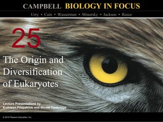
  • 1. CAMPBELL BIOLOGY IN FOCUS © 2014 Pearson Education, Inc. Urry • Cain • Wasserman • Minorsky • Jackson • Reece Lecture Presentations by Kathleen Fitzpatrick and Nicole Tunbridge 25 The Origin and Diversification of Eukaryotes
  • 2. Overview: Shape Changers  Protist is the informal name of the diverse group of mostly unicellular eukaryotes  Some protists, like the ciliate Didinium, are able to perform dramatic shape changes due to the structural complexity of their cells © 2014 Pearson Education, Inc.
  • 3. © 2014 Pearson Education, Inc. Figure 25.1
  • 4. Concept 25.1: Eukaryotes arose by endosymbiosis more than 1.8 billion years ago  Early eukaryotes were unicellular  Eukaryotic cells have organelles and are structurally more complex than prokaryotic cells  A well-developed cytoskeleton enables eukaryotic cells to have asymmetrical forms and to change shape © 2014 Pearson Education, Inc.
  • 5. The Fossil Record of Early Eukaryotes  Chemical evidence for the presence of eukaryotes dates back to 2.7 billion years ago  The earliest fossils of eukaryotic cells are 1.8 billion years old © 2014 Pearson Education, Inc.
  • 6.  Initial diversification of eukaryotes occurred 1.8 to 1.3 billion years ago  Novel features of eukaryotes, including complex multicellularity, sexual life cycles, and photosynthesis, arose 1.3 billion to 635 million years ago  The first large, multicellular eukaryotes represented by the Ediacaran biota evolved 635 to 535 million years ago © 2014 Pearson Education, Inc.
  • 7. © 2014 Pearson Education, Inc. Figure 25.2 4.5 bya 3.5 1.52.5 500 mya 1.8 bya 1.5 bya 1.3 bya 1.2 bya 20 µm 20µm 25 µm 25 µm (a) A 1.8-billion- year-old fossil eukaryote (b) Tappania, a 1.5-billion-year-old fossil that may represent an early alga or fungus (c) Bangiomorpha, an ancient red alga 20µm (d) Proterocladus, classified as a green alga (e) Bonneia, a vase-shaped eukaryote (f) An early member of the Ediacaran biota (g) Spriggina floundersi, an early animal with many body segments 750 mya 635 mya 600 mya 550 mya 535 mya 1 cm 0.5 cm
  • 8. © 2014 Pearson Education, Inc. Figure 25.2a 4.5 bya 3.5 1.52.5 500 mya 1.8 bya 1.5 bya 20 µm 20µm (a) A 1.8-billion- year-old fossil eukaryote (b) Tappania, a 1.5-billion-year-old fossil that may represent an early alga or fungus Initial diversification
  • 9. © 2014 Pearson Education, Inc. Figure 25.2aa 20 µm (a) A 1.8-billion-year-old fossil eukaryote
  • 10. © 2014 Pearson Education, Inc. Figure 25.2ab (b) Tappania, a 1.5-billion-year-old fossil that may represent an early alga or fungus 20µm
  • 11. © 2014 Pearson Education, Inc. Figure 25.2b 4.5 bya 3.5 1.52.5 500 mya Appearance of novel features 1.3 bya 1.2 bya 750 mya 20µm 25 µm 25 µm (c) Bangiomorpha, an ancient red alga (d) Proterocladus, classified as a green alga (e) Bonneia, a vase-shaped eukaryote
  • 12. © 2014 Pearson Education, Inc. Figure 25.2ba 25 µm (c) Bangiomorpha, an ancient red alga
  • 13. © 2014 Pearson Education, Inc. Figure 25.2bb 25 µm (d) Proterocladus, classified as a green alga
  • 14. © 2014 Pearson Education, Inc. Figure 25.2bc 20µm (e) Bonneia, a vase-shaped eukaryote
  • 15. © 2014 Pearson Education, Inc. Figure 25.2c 4.5 bya 3.5 1.52.5 500 mya Rise of large eukaryotes 635 mya 600 mya 550 mya 535 mya 1 cm 0.5 cm (f) An early member of the Ediacaran biota (g) Spriggina floundersi, an early animal with many body segments
  • 16. © 2014 Pearson Education, Inc. Figure 25.2ca 1 cm (f) An early member of the Ediacaran biota
  • 17. © 2014 Pearson Education, Inc. Figure 25.2cb 0.5 cm (g) Spriggina floundersi, an early animal with many body segments
  • 18.  Eukaryotic cells exhibited greater structural complexity than prokaryotic cells as early as 1.5 billion years ago  The earliest multicellular eukaryotic fossils are of red algae, which date back to 1.2 billion years ago, but large, multicellular eukaryotes did not arise until 635 million years ago  Severe ice ages from 750 to 580 million years ago may have hindered the rise of large eukaryotes © 2014 Pearson Education, Inc.
  • 19. Endosymbiosis in Eukaryotic Evolution  DNA sequence data indicate that eukaryotes are “combination” organisms  Eukaryotic genes and characteristics show evidence of both archaeal and bacterial origins  Evidence of mixed origins may be a consequence of endosymbiosis, a symbiotic relationship in which one organism lives inside the body or cell of another organism © 2014 Pearson Education, Inc.
  • 20. © 2014 Pearson Education, Inc. Table 25.1
  • 21. Origin of Mitochondria and Plastids  Endosymbiont theory proposes that mitochondria and plastids were formerly small prokaryotes that began living within larger cells  An endosymbiont is a cell that lives within a host cell  Prokaryote ancestors to mitochondria and plastids probably entered the host cell as undigested prey or internal parasites © 2014 Pearson Education, Inc.
  • 22.  The relationship between endosymbiont and host cells was mutually beneficial  Anaerobic host cells benefited from endosymbionts’ ability to take advantage of an increasingly aerobic world  Heterotrophic host cells benefited from the nutrients produced by photosynthetic endosymbionts  In the process of becoming more interdependent, the host and endosymbionts would have become a single organism © 2014 Pearson Education, Inc.
  • 23.  Serial endosymbiosis supposes that mitochondria evolved before plastids through a sequence of endosymbiotic events © 2014 Pearson Education, Inc.
  • 24. © 2014 Pearson Education, Inc. Figure 25.3 Cytoplasm DNA Nucleus Engulfing of aerobic bacterium Engulfing of photo- synthetic bacterium Mitochondrion Mito- chondrion Plastid Plasma membrane Endoplasmic reticulum Nuclear envelope Ancestral prokaryote Ancestral heterotrophic eukaryote Ancestral photosynthetic eukaryote
  • 25.  Key evidence supporting an endosymbiotic origin of mitochondria and plastids:  Inner membranes are similar to plasma membranes of prokaryotes  Division is similar in these organelles and some prokaryotes  DNA structure is similar to that of prokaryotes  These organelles transcribe and translate their own DNA  Their ribosomes are more similar to prokaryotic than eukaryotic ribosomes © 2014 Pearson Education, Inc.
  • 26.  DNA sequence analysis indicates that mitochondria arose from an alpha proteobacterium  Eukaryotic mitochondria descended from a single common ancestor  Plastids arose from an engulfed cyanobacterium  Some photosynthetic protists may have been engulfed to become endosymbionts themselves © 2014 Pearson Education, Inc.
  • 27.  The ancestral host cell may have been an archaean or a “protoeukaryote,” from a lineage related to, but diverged from, archaeal ancestors © 2014 Pearson Education, Inc.
  • 28. Plastid Evolution: A Closer Look  There is now considerable evidence that much protist diversity has its origins in endosymbiosis  Mitochondria arose first through descent from a bacterium that was engulfed by a cell from an archaeal lineage  The plastid lineage evolved later from a photosynthetic cyanobacterium that was engulfed by a heterotrophic eukaryote © 2014 Pearson Education, Inc.
  • 29. © 2014 Pearson Education, Inc. Figure 25.4 Cyano- bacterium Membranes are represented as dark lines in the cell. Red alga Primary endo- symbiosis 1 2 3 Nucleus Heterotrophic eukaryote One of these membranes was lost in red and green algal descendants. Green alga Secondary endo- symbiosis Secondary endo- symbiosis Plastid Euglenids Chlorarachniophytes Stramenopiles Plastid Secondary endo- symbiosis Dinoflagellates
  • 30. © 2014 Pearson Education, Inc. Figure 25.4a Cyano- bacterium Membranes are represented as dark lines in the cell. Red alga Primary endosymbiosis 1 2 3 Nucleus Heterotrophic eukaryote One of these membranes was lost in red and green algal descendants. Green alga
  • 31. © 2014 Pearson Education, Inc. Figure 25.4b Secondary endosymbiosis Red alga Plastid Dinoflagellates Stramenopiles
  • 32. © 2014 Pearson Education, Inc. Figure 25.4c Secondary endosymbiosis Plastid Euglenids Green alga Chlorarachniophytes
  • 33.  The plastid-bearing lineage of protists evolved into red and green algae  Like cyanobacteria, plastids of red algae and green algae have two membranes  Transport proteins in the membranes of red and green algae are homologous to those found in cyanobacteria  On several occasions during eukaryotic evolution, red and green algae underwent secondary endosymbiosis, in which they were ingested by a heterotrophic eukaryote © 2014 Pearson Education, Inc.
  • 34. Concept 25.2: Multicellularity has originated several times in eukaryotes  The evolution of eukaryotic cells allowed for a greater range of unicellular forms  A second wave of diversification occurred when multicellularity evolved and gave rise to algae, plants, fungi, and animals © 2014 Pearson Education, Inc.
  • 35. Multicellular Colonies  The first multicellular forms were colonies, collections of cells that are connected to one another but show little or no cellular differentiation  Multicellular colonies consist of simple filaments, balls, or cell sheets  Colonial cells may be attached by shared cell walls or, in cells that lack rigid walls, held together by proteins that physically connect adjacent cells © 2014 Pearson Education, Inc.
  • 36. © 2014 Pearson Education, Inc. Figure 25.5 Pediastrum
  • 37. Independent Origins of Complex Multicellularity  Multicellular organisms with differentiated cells originated multiple times over the course of eukaryotic evolution  Genetic and morphological evidence indicates that lineages of red, green, and brown algae, plants, fungi, and animals arose independently from different single-celled ancestors © 2014 Pearson Education, Inc.
  • 38.  Volvox is a multicellular green algae that forms a monophyletic group with a single-celled alga (Chlamydomonas) and several colonial species  Volvox and all colonial members of this group have proteins homologous to those found in the cell wall of Chlamydomonas  Multicellularity in Volvox may have originated through evolution of increasingly complex colonial forms descended from a single-celled common ancestor © 2014 Pearson Education, Inc. Video: Chlamydomonas
  • 39. © 2014 Pearson Education, Inc. Figure 25.6 Flagellum Cytoplasm Outer cell wall Inner cell wall Outer cell wall Cytoplasm Extracellular matrix (ECM) Chlamydomonas Gonium Pandorina Volvox
  • 40.  Only a few novel genes account for morphological differences between Volvox and Chlamydomonas  The transition to multicellularity may result from changes in how existing genes are used rather than the origin of large numbers of novel genes © 2014 Pearson Education, Inc.
  • 41. Steps in the Origin of Multicellular Animals  Choanoflagellates are the closest living relatives of animals  The common ancestor of choanoflagellates and living animals may have resembled present-day choanoflagellates © 2014 Pearson Education, Inc.
  • 42. © 2014 Pearson Education, Inc. Figure 25.7 Individual choanoflagellate Choano- flagellates Other animals Collar cell (choanocyte) Sponges Animals OTHER EUKARY- OTES
  • 43.  The origin of multicellularity in animals required mechanisms for cells to adhere to each other and to communicate with each other (cell signaling)  Molecular similarities in domains of proteins functioning in cell adherence (cadherins) and cell signaling have been found between modern choanoflagellates and representative animals © 2014 Pearson Education, Inc.
  • 44. © 2014 Pearson Education, Inc. Figure 25.8 Hydra Mouse “CCD” domain Choano- flagellate Fruit fly
  • 45.  The transition to multicellularity in animals involved new ways of using proteins encoded by genes found in choanoflagellates rather than the evolution of many novel genes © 2014 Pearson Education, Inc.
  • 46. Concept 25.3: Four “supergroups” of eukaryotes have been proposed based on morphological and molecular data  Eukaryote diversity is influenced by their hybrid origins and the independent origins of complex multicellularity  One hypothesis divides all eukaryotes into four supergroups © 2014 Pearson Education, Inc.
  • 47. Four Supergroups of Eukaryotes  It is no longer thought that amitochondriates (lacking mitochondria) are the oldest lineage of eukaryotes  Many have been shown to have mitochondria and have been reclassified  Our understanding of the relationships among protist groups continues to change rapidly  One hypothesis divides all eukaryotes (including protists) into four supergroups © 2014 Pearson Education, Inc.
  • 48. © 2014 Pearson Education, Inc. Video: Amoeba Pseudopodia Video: Volvox Colony Video: Amoeba Video: Volvox Female Spheroid Video: Volvox Flagella Video: Volvox Daughter Video: Volvox Inversion 1 Video: Volvox Sperm Video: Volvox Inversion 2
  • 49. © 2014 Pearson Education, Inc. Figure 25.9 Stramen- opiles Diplomonads Parabasalids Euglenozoans Excavata Alveo- lates Rhiza- rians Diatoms Brown algae Dinoflagellates Apicomplexans Ciliates Forams Cercozoans “SAR”clade Green algae Amoebo- zoans Red algae Chlorophytes Charophytes Land plants Archaeplastida Opisthokonts Unikonta Gymnamoebas Slime molds Nucleariids Fungi Choanoflagellates Animals 100 µm 50 µm “SAR” Clade Excavata 5 µm Archaeplastida 20 µm 50 µm Unikonta 100 µm
  • 50. © 2014 Pearson Education, Inc. Figure 25.9a Stramen- opiles Diplomonads Parabasalids Euglenozoans Excavata Alveo- lates Rhiza- rians Diatoms Brown algae Dinoflagellates Apicomplexans Ciliates Forams Cercozoans “SAR”clade Green algae Amoebo- zoans Red algae Chlorophytes Charophytes Land plants Archaeplastida Opisthokonts Unikonta Gymnamoebas Slime molds Nucleariids Fungi Choanoflagellates Animals
  • 51. © 2014 Pearson Education, Inc. Figure 25.9aa Stramen- opiles Diplomonads Parabasalids Euglenozoans Excavata Alveo- lates Rhiza- rians Diatoms Brown algae Dinoflagellates Apicomplexans Ciliates Forams Cercozoans “SAR”clade
  • 52. © 2014 Pearson Education, Inc. Figure 25.9ab Green algae Amoebo- zoans Red algae Chlorophytes Charophytes Land plants Archaeplastida Opisthokonts Unikonta Gymnamoebas Slime molds Nucleariids Fungi Choanoflagellates Animals
  • 53. © 2014 Pearson Education, Inc. Figure 25.9b Excavata “SAR” Clade Archaeplastida 50 µm 50 µm 20 µm 5 µm Unikonta 100 µm 100 µm
  • 54. © 2014 Pearson Education, Inc. Figure 25.9ba Giardia intestinalis, a diplomonad parasite 5 µm
  • 55. © 2014 Pearson Education, Inc. Figure 25.9bb Diatom diversity 50 µm
  • 56. © 2014 Pearson Education, Inc. Figure 25.9bc Globigerina, a rhizarian in the SAR supergroup 100 µm
  • 57. © 2014 Pearson Education, Inc. Figure 25.9bca Globigerina, a rhizarian in the SAR supergroup 100 µm
  • 58. © 2014 Pearson Education, Inc. Figure 25.9bcb
  • 59. © 2014 Pearson Education, Inc. Figure 25.9bd Volvox, a multicellular freshwater green alga 20 µm 50 µm
  • 60. © 2014 Pearson Education, Inc. Figure 25.9bda Volvox, a multicellular freshwater green alga 50 µm
  • 61. © 2014 Pearson Education, Inc. Figure 25.9bdb 20 µm
  • 62. © 2014 Pearson Education, Inc. Figure 25.9be 100 µm A unikont amoeba
  • 63. Excavates  The supergroup Excavata is characterized by its cytoskeleton  Some members have a feeding groove  This monophyletic group includes the diplomonads, parabasalids, and euglenozoans © 2014 Pearson Education, Inc.
  • 64. © 2014 Pearson Education, Inc. Figure 25.UN02 Diplomonads Parabasalids Euglenozoans Excavata SAR clade Archaeplastida Unikonta
  • 65. Diplomonads and Parabasalids  These two groups lack plastids and have modified mitochondria, and most live in anaerobic environments  Diplomonads  Have reduced mitochondria called mitosomes  Derive energy from anaerobic biochemical pathways  Are often parasites, for example, Giardia intestinalis  Move using multiple flagella © 2014 Pearson Education, Inc.
  • 66.  Parabasalids  Have reduced mitochondria called hydrogenosomes that generate some energy anaerobically  Include Trichomonas vaginalis, a sexually transmitted parasite © 2014 Pearson Education, Inc.
  • 67. © 2014 Pearson Education, Inc. Figure 25.10 The parabasalid parasite, Trichomonas vaginalis (colorized SEM) Undulating membrane Flagella 5µm
  • 68. Euglenozoans  Euglenozoa is a diverse clade that includes predatory heterotrophs, photosynthetic autotrophs, and parasites  The main feature distinguishing them as a clade is a spiral or crystalline rod inside their flagella  This clade includes the euglenids and kinetoplastids © 2014 Pearson Education, Inc. Video: Euglena Motion Video: Euglena
  • 69. © 2014 Pearson Education, Inc. Figure 25.11 Euglenozoan flagellum Ring of microtubules (cross section) Crystalline rod (cross section) 8 µm 0.2 µm Flagella
  • 70. © 2014 Pearson Education, Inc. Figure 25.11a Ring of microtubules (cross section) Crystalline rod (cross section) 0.2 µm
  • 71.  Euglenids have one or two flagella that emerge from a pocket at one end of the cell  Some species can be both autotrophic and heterotrophic © 2014 Pearson Education, Inc.
  • 72.  Kinetoplastids have a single mitochondrion with an organized mass of DNA called a kinetoplast  They include free-living consumers of prokaryotes in aquatic ecosystems and parasites of animals, plants, and other protists  This group includes Trypanosoma, which causes sleeping sickness in humans © 2014 Pearson Education, Inc.
  • 73. © 2014 Pearson Education, Inc. Figure 25.12 Trypanosoma, the kinetoplastid that causes sleeping sickness 9 µm
  • 74. The “SAR” Clade  The “SAR” clade is a diverse monophyletic supergroup named for the first letters of its three major clades, stramenopiles, alveolates, and rhizarians © 2014 Pearson Education, Inc.
  • 75. © 2014 Pearson Education, Inc. Figure 25.UN03 Archaeplastida Unikonta SARclade Rhizarians Alveolates Stramenopiles Excavata Diatoms Brown algae Dinoflagellates Apicomplexans Ciliates Forams Cercozoans
  • 76. Stramenopiles  The subgroup stramenopiles includes the most important photosynthetic organisms on Earth  Diatoms and brown algae are members of the stramenopiles clade  Diatoms are highly diverse, unicellular algae with a unique two-part, glass-like wall of silicon dioxide © 2014 Pearson Education, Inc. Video: Various Diatoms Video: Diatoms Moving
  • 77. © 2014 Pearson Education, Inc. Figure 25.13 The diatom Triceratium morlandii (colorized SEM) 40µm
  • 78.  Brown algae are the largest and most complex algae  All are multicellular, and most are marine  Brown algae include many species commonly called “seaweeds” © 2014 Pearson Education, Inc.
  • 79.  Brown algal seaweeds have plantlike structures: the rootlike holdfast, which anchors the alga, and a stemlike stipe, which supports the leaflike blades  Similarities between algae and plants are examples of analogous structures © 2014 Pearson Education, Inc.
  • 80. © 2014 Pearson Education, Inc. Figure 25.14 Blade Stipe Holdfast
  • 81. Alveolates  The alveolates are a subgroup of the SAR clade that have membrane-bounded sacs (alveoli) just under the plasma membrane  Dinoflagellates and ciliates are members of the alveolata clade © 2014 Pearson Education, Inc.
  • 82. © 2014 Pearson Education, Inc. Figure 25.15 Flagellum Alveoli Alveolate 0.2µm
  • 83. © 2014 Pearson Education, Inc. Figure 25.15a Flagellum Alveoli 0.2µm
  • 84.  Dinoflagellates have two flagella, and each cell is reinforced by cellulose plates  They are abundant components of both marine and freshwater phytoplankton  They are a diverse group of aquatic phototrophs, mixotrophs, and heterotrophs  Toxic “red tides” are caused by dinoflagellate blooms © 2014 Pearson Education, Inc. Video: Dinoflagellate
  • 85. © 2014 Pearson Education, Inc. Figure 25.16 Flagella Pfiesteria shumwayae, a dinoflagellate 3µm
  • 86.  Ciliates, a large varied group of protists, are named for their use of cilia to move and feed  Most ciliates are predators of bacteria or small protists © 2014 Pearson Education, Inc. Video: Paramecium Cilia Video: Paramecium Vacuole Video: Vorticella Cilia Video: Vorticella Habitat Video: Vorticella Detail
  • 87. © 2014 Pearson Education, Inc. Figure 25.17 Contractile vacuole Cilia Oral groove Cell mouth Micronucleus Food vacuolesMacronucleus 50 µm
  • 88. © 2014 Pearson Education, Inc. Figure 25.17a 50 µm
  • 89.  Many species of rhizarians are amoebas  Amoebas move and feed by pseudopodia; some but not all belong to the clade Rhizaria  Forams and cercozoans are members of the rhizarian clade Rhizarians © 2014 Pearson Education, Inc.
  • 90.  Foraminiferans, or forams, are named for porous shells, called tests  Pseudopodia extend through the pores in the test  Many forams have endosymbiotic algae  Forams include both marine and freshwater species © 2014 Pearson Education, Inc.
  • 91.  Cercozoans include amoeboid and flagellated protists with threadlike pseudopodia  They are common in marine, freshwater, and soil ecosystems  Most are heterotrophs, including parasites and predators © 2014 Pearson Education, Inc.
  • 92.  Paulinella chromatophora is an autotroph with a unique photosynthetic structure called a chromatophore  This structure evolved from a different cyanobacterium than the plastids of other photosynthetic eukaryotes © 2014 Pearson Education, Inc.
  • 93. © 2014 Pearson Education, Inc. Figure 25.18 5 µm Chromatophore
  • 94. Archaeplastids  Plastids arose when a heterotrophic protist acquired a cyanobacterial endosymbiont  The photosynthetic descendants of this ancient protist evolved into red algae and green algae  Land plants are descended from the green algae  Archaeplastida is the supergroup that includes red algae, green algae, and land plants © 2014 Pearson Education, Inc.
  • 95. © 2014 Pearson Education, Inc. Figure 25.UN04 Archaeplastida Unikonta SAR clade Excavata Chlorophytes Charophytes Red algae Green algae Land plants
  • 96. Red Algae  Red algae are reddish in color due to an accessory pigment called phycoerythrin, which masks the green of chlorophyll  The color varies from greenish-red in shallow water to dark red or almost black in deep water  Red algae are usually multicellular; the largest are seaweeds  Red algae are the most abundant large algae in coastal waters of the tropics  Red algae reproduce sexually © 2014 Pearson Education, Inc.
  • 97. © 2014 Pearson Education, Inc. Figure 25.19 Bonnemaisonia hamifera 8 mm Red algae Nori
  • 98. © 2014 Pearson Education, Inc. Figure 25.19a Bonnemaisonia hamifera 8 mm
  • 99. © 2014 Pearson Education, Inc. Figure 25.19b Nori
  • 100. © 2014 Pearson Education, Inc. Figure 25.19c
  • 101. Green Algae  Green algae are named for their grass-green chloroplasts  Plants are descended from the green algae  Green algae are a paraphyletic group  The two main groups are charophytes and chlorophytes  Charophytes are most closely related to land plants © 2014 Pearson Education, Inc.
  • 102.  Most chlorophytes live in fresh water, although many are marine and some are terrestrial  Nearly all species of chlorophytes reproduce sexually  Some unicellular chlorophytes are free-living while others live symbiotically within other eukaryotes  Larger size and greater complexity are found in multicellular species including Volvox and Ulva © 2014 Pearson Education, Inc.
  • 103. © 2014 Pearson Education, Inc. Figure 25.20 (a) Ulva, or sea lettuce (b) Caulerpa, an intertidal chlorophyte Multicellular chlorophytes 2 cm
  • 104. © 2014 Pearson Education, Inc. Figure 25.20a (a) Ulva, or sea lettuce 2 cm
  • 105. © 2014 Pearson Education, Inc. Figure 25.20b (b) Caulerpa, an intertidal chlorophyte
  • 106. Unikonts  The supergroup Unikonta includes animals, fungi, and some protists  This group includes two clades: the amoebozoans and the opisthokonts (animals, fungi, and related protists)  The root of the eukaryotic tree remains controversial  It is unclear whether unikonts separated from other eukaryotes relatively early or late © 2014 Pearson Education, Inc.
  • 107. © 2014 Pearson Education, Inc. Figure 25.UN05 Unikonta Archaeplastida SAR clade Excavata Gymnamoebas Slime molds Nucleariids Fungi Animals Choanoflagellates
  • 108. © 2014 Pearson Education, Inc. Figure 25.21 Results Common ancestor of all eukaryotes Choanoflagellates Animals Fungi Amoebozoans Unikonta Excavata Diplomonads SAR clade Euglenozoans Stramenopiles Alveolates Rhizarians Plants Green algae Red algae Archaeplastida DHFR-TS gene fusion
  • 109. Amoebozoans  Amoebozoans are amoebas that have lobe- or tube-shaped, rather than threadlike, pseudopodia  Most amoebozoans are free-living, but those that belong to the genus Entamoeba are parasites © 2014 Pearson Education, Inc.
  • 110.  Slime molds are free-living amoebozoans that were once thought to be fungi  DNA sequence analyses indicate that slime molds belong in the clade Amoebozoa © 2014 Pearson Education, Inc.
  • 111.  Dictyostelium is an example of a cellular slime mold  Cellular slime molds consist of solitary cells that feed individually but can aggregate to form a fruiting body © 2014 Pearson Education, Inc.
  • 112. © 2014 Pearson Education, Inc. Figure 25.22 Spores (n) Fruiting bodies (n) Emerging amoeba (n) Solitary amoebas (feeding stage) (n) ASEXUAL REPRODUCTION SEXUAL REPRODUCTION MEIOSIS FERTILIZATION Zygote (2n) Amoebas (n) Aggregated amoebas 600 µm Migrating aggregate 200 µm Key Haploid (n) Diploid (2n)
  • 113. © 2014 Pearson Education, Inc. Figure 25.22a-1 Solitary amoebas (feeding stage) (n) SEXUAL REPRODUCTION FERTILIZATION Zygote (2n) Key Haploid (n) Diploid (2n)
  • 114. © 2014 Pearson Education, Inc. Figure 25.22a-2 Solitary amoebas (feeding stage) (n) SEXUAL REPRODUCTION MEIOSIS FERTILIZATION Zygote (2n) Amoebas (n) Key Haploid (n) Diploid (2n)
  • 115. © 2014 Pearson Education, Inc. Figure 25.22a-3 Solitary amoebas (feeding stage) (n) ASEXUAL REPRODUCTION SEXUAL REPRODUCTION MEIOSIS FERTILIZATION Zygote (2n) Amoebas (n) Aggregated amoebas Key Haploid (n) Diploid (2n)
  • 116. © 2014 Pearson Education, Inc. Figure 25.22a-4 Fruiting bodies (n) Solitary amoebas (feeding stage) (n) ASEXUAL REPRODUCTION SEXUAL REPRODUCTION MEIOSIS FERTILIZATION Zygote (2n) Amoebas (n) Aggregated amoebas Migrating aggregate Key Haploid (n) Diploid (2n)
  • 117. © 2014 Pearson Education, Inc. Figure 25.22a-5 Spores (n) Fruiting bodies (n) Emerging amoeba (n) Solitary amoebas (feeding stage) (n) ASEXUAL REPRODUCTION SEXUAL REPRODUCTION MEIOSIS FERTILIZATION Zygote (2n) Amoebas (n) Aggregated amoebas Migrating aggregate Key Haploid (n) Diploid (2n)
  • 118. © 2014 Pearson Education, Inc. Figure 25.22b 200 µm
  • 119. © 2014 Pearson Education, Inc. Figure 25.22c 600 µm
  • 120. Opisthokonts  Opisthokonts include animals, fungi, and several groups of protists © 2014 Pearson Education, Inc.
  • 121. Concept 25.4: Single-celled eukaryotes play key roles in ecological communities and affect human health  The majority of the eukaryotic lineages are composed of protists  Protists exhibit a wide range of structural and functional diversity © 2014 Pearson Education, Inc.
  • 122. Structural and Functional Diversity in Protists  Single-celled protists can be very complex, as all biological functions are carried out by organelles in each individual cell © 2014 Pearson Education, Inc.
  • 123.  Protists are found in diverse aquatic environments  Some protists reproduce asexually, while others reproduce sexually, or by the sexual processes of meiosis and fertilization © 2014 Pearson Education, Inc.
  • 124.  Protists show a wide range of nutritional diversity, including  Photoautotrophs, which contain chloroplasts  Heterotrophs, which absorb organic molecules or ingest larger food particles  Mixotrophs, which combine photosynthesis and heterotrophic nutrition © 2014 Pearson Education, Inc.
  • 125. Photosynthetic Protists  Many protists are important producers that obtain energy from the sun  In aquatic environments, photosynthetic protists and prokaryotes are the main producers  In aquatic environments, photosynthetic protists are limited by nutrients  These populations can explode when limiting nutrients are added © 2014 Pearson Education, Inc.
  • 126. © 2014 Pearson Education, Inc. Figure 25.23 Other consumers Herbivorous plankton Carnivorous plankton Protistan producers Prokaryotic producers
  • 127.  Diatoms are a major component of the phytoplankton  After a diatom population has bloomed, many dead individuals fall to the ocean floor undecomposed  This removes carbon dioxide from the atmosphere and “pumps” it to the ocean floor © 2014 Pearson Education, Inc.
  • 128.  The biomass and growth of photosynthetic protists and prokaryotes have declined as sea surface temperature has increased  Warmer surface temperatures may prevent nutrient upwelling, a process critical to the growth of marine producers © 2014 Pearson Education, Inc.
  • 129.  If sea surface temperature continues to warm due to global warming, this could have large effects on  Marine ecosystems  Fishery yields  The global carbon cycle © 2014 Pearson Education, Inc.
  • 130. © 2014 Pearson Education, Inc. Figure 25.24 NI SI S SP SA EA NA AEPNP Sea-surface temperature (SST) change (°C) −2.50 −1.25 0.00 1.25 2.50 (a) (b) Southern (S) South Pacific (SP) Equatorial Pacific (EP) North Pacific (NP) South Indian (SI) North Indian (NI) South Atlantic (SA) Equatorial Atlantic (EA) North Atlantic (NA) Arctic (A) Chlorophyll change (mg/m2 • yr) −0.02 −0.01 0.00 0.01 0.02
  • 131. © 2014 Pearson Education, Inc. Figure 25.24a Sea-surface temperature (SST) change (°C) −2.50 −1.25 0.00 1.25 2.50 SA SP EA NA AEPNP NI SI S (a)
  • 132. © 2014 Pearson Education, Inc. Figure 25.24b −0.02 −0.01 0.00 0.01 0.02 Southern (S) South Pacific (SP) Equatorial Pacific (EP) North Pacific (NP) South Indian (SI) North Indian (NI) South Atlantic (SA) Equatorial Atlantic (EA) North Atlantic (NA) Arctic (A) Chlorophyll change (mg/m2 • yr) (b)
  • 133. Symbiotic Protists  Some protist symbionts benefit their hosts  Dinoflagellates nourish coral polyps that build reefs  Wood-digesting protists digest cellulose in the gut of termites © 2014 Pearson Education, Inc.
  • 134. © 2014 Pearson Education, Inc. 10µm Figure 25.25
  • 135.  Some protists are parasitic  Plasmodium causes malaria  Pfiesteria shumwayae is a dinoflagellate that causes fish kills  Phytophthora ramorum causes sudden oak death  Phytophthora infestans causes potato late blight © 2014 Pearson Education, Inc.
  • 136. Effects on Human Health  Protists that cause infectious disease can pose major challenges to human immune systems and public health  Trypanosoma is an excavate that causes sleeping sickness in humans © 2014 Pearson Education, Inc.
  • 137.  Trypanosomes evade immune responses by switching surface proteins  A cell produces millions of copies of a single protein  The new generation produces millions of copies of a different protein  These frequent changes prevent the host from developing immunity © 2014 Pearson Education, Inc.
  • 138.  Entamoebas are amoebozoan parasites of vertebrates and some invertebrates  Entamoeba histolytica causes amebic dysentery, the third-leading cause of human death due to eukaryotic parasites © 2014 Pearson Education, Inc.
  • 139.  Apicomplexans are alveolate parasites of animals, and some cause serious human diseases  Most have sexual and asexual stages that require two or more different host species for completion © 2014 Pearson Education, Inc.
  • 140.  The apicomplexan Plasmodium is the parasite that causes malaria  Plasmodium requires both mosquitoes and humans to complete its life cycle  Approximately 900,000 people die each year from malaria  Efforts are ongoing to develop vaccines that target this pathogen © 2014 Pearson Education, Inc.
  • 141. © 2014 Pearson Education, Inc. Figure 25.26 0.5 µm Apex Red blood cell Merozoite Liver Liver cell Red blood cells Merozoite (n) Zygote (2n) Oocyst MEIOSIS Sporozoites (n) Inside mosquito Inside human FERTILIZATION Gametes Gametocytes (n) Key Haploid (n) Diploid (2n)
  • 142. © 2014 Pearson Education, Inc. Figure 25.26a 0.5 µm Apex Red blood cell Merozoite
  • 143. © 2014 Pearson Education, Inc. Figure 25.UN01
  • 144. © 2014 Pearson Education, Inc. Figure 25.UN06

Editor's Notes

  1. Figure 25.1 What enables the cell on the left to engulf its prey?
  2. Figure 25.2 Exploring the early evolution of eukaryotes
  3. Figure 25.2a Exploring the early evolution of eukaryotes (part 1: initial diversification)
  4. Figure 25.2aa Exploring the early evolution of eukaryotes (part 1a: early eukaryote fossil)
  5. Figure 25.2ab Exploring the early evolution of eukaryotes (part 1b: Tappania)
  6. Figure 25.2b Exploring the early evolution of eukaryotes (part 2: appearance of novel features)
  7. Figure 25.2ba Exploring the early evolution of eukaryotes (part 2a: Bangiomorpha)
  8. Figure 25.2bb Exploring the early evolution of eukaryotes (part 2b: Proterocladus)
  9. Figure 25.2bc Exploring the early evolution of eukaryotes (part 2c: Bonneia)
  10. Figure 25.2c Exploring the early evolution of eukaryotes (part 3: rise of large eukaryotes)
  11. Figure 25.2ca Exploring the early evolution of eukaryotes (part 3a: Doushantuophyton)
  12. Figure 25.2cb Exploring the early evolution of eukaryotes (part 3b: Spriggina floundersi)
  13. Table 25.1 Inferred origins of key eukaryotic features
  14. Figure 25.3 A hypothesis for the origin of eukaryotes through endosymbiosis
  15. Figure 25.4 Diversity of plastids produced by endosymbiosis
  16. Figure 25.4a Diversity of plastids produced by endosymbiosis (part 1: primary endosymbiosis)
  17. Figure 25.4b Diversity of plastids produced by endosymbiosis (part 2: endosymbiosis of red algae)
  18. Figure 25.4c Diversity of plastids produced by endosymbiosis (part 3: endosymbiosis of green algae)
  19. Figure 25.5 Pediastrum
  20. Figure 25.6 Morphological change in the Volvox lineage
  21. Figure 25.7 Three lines of evidence that choanoflagellates are closely related to animals
  22. Figure 25.8 Cadherin proteins in choanoflagellates and animals
  23. Figure 25.9 Exploring eukaryotic diversity
  24. Figure 25.9a Exploring eukaryotic diversity (part 1: phylogenetic hypothesis of eukaryotic supergroups)
  25. Figure 25.9aa Exploring eukaryotic diversity (part 1a: phylogenetic hypothesis of eukaryotic supergroups)
  26. Figure 25.9ab Exploring eukaryotic diversity (part 1b: phylogenetic hypothesis of eukaryotic supergroups)
  27. Figure 25.9b Exploring eukaryotic diversity (part 2: micrographs)
  28. Figure 25.9ba Exploring eukaryotic diversity (part 2a: Excavata, SEM)
  29. Figure 25.9bb Exploring eukaryotic diversity (part 2b: SAR clade, Stramenopila, LM)
  30. Figure 25.9bc Exploring eukaryotic diversity (part 2c: SAR clade, Rhizaria)
  31. Figure 25.9bca Exploring eukaryotic diversity (part 2ca: SAR clade, Rhizaria, foram, LM)
  32. Figure 25.9bcb Exploring eukaryotic diversity (part 2cb: SAR clade, Rhizaria, foram test)
  33. Figure 25.9bd Exploring eukaryotic diversity (part 2d: Archaeplastida)
  34. Figure 25.9bda Exploring eukaryotic diversity (part 2da: Archaeplastida, Volvox containing “daughter” algae, LM)
  35. Figure 25.9bdb Exploring eukaryotic diversity (part 2db: Archaeplastida, Volvox wall, LM)
  36. Figure 25.9be Exploring eukaryotic diversity (part 2e: Unikonta)
  37. Figure 25.UN02 In-text figure, Excavata, p. 492
  38. Figure 25.10 The parabasalid parasite, Trichomonas vaginalis (colorized SEM)
  39. Figure 25.11 Euglenozoan flagellum
  40. Figure 25.11a Euglenozoan flagellum (part 1: TEM)
  41. Figure 25.12 Trypanosoma, the kinetoplastid that causes sleeping sickness
  42. Figure 25.UN03 In-text figure, the SAR clade, p. 493
  43. Figure 25.13 The diatom Triceratium morlandii (colorized SEM)
  44. Figure 25.14 Seaweeds: adapted to life at the ocean’s margins
  45. Figure 25.15 Alveoli
  46. Figure 25.15a Alveoli (TEM)
  47. Figure 25.16 Pfiesteria shumwayae, a dinoflagellate
  48. Figure 25.17 Structure and function in the ciliate Paramecium caudatum
  49. Figure 25.17a Structure and function in the ciliate Paramecium caudatum (LM)
  50. Figure 25.18 A second case of primary endosymbiosis?
  51. Figure 25.UN04 In-text figure, Archaeplastida, p. 495
  52. Figure 25.19 Red algae
  53. Figure 25.19a Red algae (part 1: Bonnemaisonia hamifera)
  54. Figure 25.19b Red algae (part 2: nori growing on nets)
  55. Figure 25.19c Red algae (part 3: nori in sushi)
  56. Figure 25.20 Multicellular chlorophytes
  57. Figure 25.20a Multicellular chlorophytes (part 1: sea lettuce)
  58. Figure 25.20b Multicellular chlorophytes (part 2: Caulerpa)
  59. Figure 25.UN05 In-text figure, Unikonta, p. 496
  60. Figure 25.21 Inquiry: Where is the root of the eukaryotic tree?
  61. Figure 25.22 The life cycle of Dictyostelium, a cellular slime mold
  62. Figure 25.22a-1 The life cycle of Dictyostelium, a cellular slime mold (part 1, step 1)
  63. Figure 25.22a-2 The life cycle of Dictyostelium, a cellular slime mold (part 1, step 2)
  64. Figure 25.22a-3 The life cycle of Dictyostelium, a cellular slime mold (part 1, step 3)
  65. Figure 25.22a-4 The life cycle of Dictyostelium, a cellular slime mold (part 1, step 4)
  66. Figure 25.22a-5 The life cycle of Dictyostelium, a cellular slime mold (part 1, step 5)
  67. Figure 25.22b The life cycle of Dictyostelium, a cellular slime mold (part 2: aggregate)
  68. Figure 25.22c The life cycle of Dictyostelium, a cellular slime mold (part 3: fruiting body)
  69. Figure 25.23 Protists: key producers in aquatic communities
  70. Figure 25.24 Effects of climate change on marine producers
  71. Figure 25.24a Effects of climate change on marine producers (part 1: changes in SST)
  72. Figure 25.24b Effects of climate change on marine producers (part 2: changes in chlorophyll concentration)
  73. Figure 25.25 A symbiotic protist
  74. Figure 25.26 The two-host life cycle of Plasmodium, the apicomplexan that causes malaria
  75. Figure 25.26a The two-host life cycle of Plasmodium, the apicomplexan that causes malaria (TEM)
  76. Figure 25.UN01 Skills exercise: interpreting comparisons of genetic sequences
  77. Figure 25.UN06 Summary of key concepts: supergroups