SlideShare a Scribd company logo
1 of 7
Download to read offline
1      © Alan Hedge, Cornell University 11/1999


             Structure and Function of the
               Musculoskeletal System
                         Professor Alan Hedge
                               DEA 325

Functions of the Musculoskeletal System
è Support and protect the body and its organs.
è Provide motion.

Musculoskeletal System:
       Major substructures
è Tendons.
è Ligaments.
è Fascia.
è Cartilage.
è Bone.
è Muscle.
è Joints   - allow motion between body segments.

Connective Tissues
è Cells.
è Extracellular   matrix:
  l Fibers.
  l Ground substance.


Connective Tissue Cells
è Fibroblasts- cells producing the matrix for skin, tendons
  and ligaments.
è Chrondrocytes - cells producing the matrix for cartilage.
è Osteocytes - cells producing the matrix for bone.

Connective Tissue Matrix
è Collagen    fibers:
  l   Type I - tendons, ligaments, bone, skin.
2   © Alan Hedge, Cornell University 11/1999


  l Type II - cartilage.
  l Type III - blood vessel walls.
è Mechanicalproperties depend on fiber types and fiber
  arrangements.

Tendons
Ligaments
Fascia
Mechanical Properties of Fibers
è Stress  - force on a fiber e.g. weight.
è Strain - % stretching of fibers.
è Elastic limit - point at which the elasticity of the fiber is lost.
è Failure - point at which the fiber breaks.

Collagen fibers
è Strengthof collagen is 50% strength of bone.
è Under tension, collagen fibers first elongate slightly and
  then become increasing stiff until failure.

Elastic fibers
è Weak  and brittle fibers.
è At low loads they strain greatly and can increase ~200% in
  length before failure.

Tendon structure
è Tendon
è Tendon      sheath:
  l Synovium - lubricant producing tissue.
  l Synovial fluid - lubricant fluid.


Tendons and Ligaments
è Atcertain points, ligaments surround parts of the tendon
  sheath to act as:
  l    pulleys.
3   © Alan Hedge, Cornell University 11/1999


  l   tendon guides.
è Ligament  attachments allow tendons to work around
  corners, as in the fingers and toes.

Cartilage: 3 types
è Hyaline  cartilage - present in the growth plates at the end
  of bones and on the articular surfaces of joints. Also
  present in the respiratory tract (e.g. trachea).
è Fibrocartilage - present in intervertebral discs, which
  consist of islands of hyaline cartilage in collagen fibers.
è Elastic cartilage - present in ears, nose, epiglottis etc.
  Consists mainly of elastic fibers.

Bone
è 99% of bodies calcium is in bone.
è 80% of bone tissue by weight is minerals.

Bone groups
è Axial (appendicular) bones - flat bones, such as the skull,
  pelvis, ribs, vertebrae
è Long bones - bones of the extremities. Round bones
  comprising:
  l diaphysis - shaft
  l epiphyses - 2 expanded ends


Types of Bone
è Cortical (compact) bone - tissue with high proportion of
  bone. 5-30% porosity.
è Cancellous (spongy) bone - less dense bone tissue found
  at the epiphyses of the long bones and in axial bones,
  such as the skull. 30-90% porosity.

Cortical (Compact) Bone
Cancellous (Spongy) Bone
4      © Alan Hedge, Cornell University 11/1999


Anisotropy
è All  bone is anisotropic (i.e. its mechanical property
    changes when loads are applied in different directions).

Anisotropic behavior of cortical bone.
Bone: Mechanical properties
è Most   important properties are strength and stiffness of
  bone.
è Fractures occur when the bone is loaded to failure.
è Compression fractures are commonest in cancellous bone
  (e.g. fractured skull)
è Bending and torsional fractures are commonest in cortical
  bone (e.g. broken tibia).

Speed of loading to fracture.
Bone Structure and Growth
è Ossification - processes of bone formation.
è Osteoblasts - cells that form the bone matrix. They
  transform into:
    l   Osteocytes - cells isolated inside the mineralized bone matrix.
è Osteoclasts      - cells important to bone remodeling.

Skeletal muscle
è   Skeletal muscle is ~50% body weight.
è   Skeletal muscle is striated (striped), and excludes cardiac and
    smooth muscle.
è   Several hundred muscles in the body.
è   Skeletal muscle is under voluntary control.
è   Each muscle is a separate organ.
è   Each muscle is attached to bone by tendons that cross one or
    more joints.
è   Generally, skeletal muscles generate moments about joints.
5      © Alan Hedge, Cornell University 11/1999


Properties of Skeletal Muscle
è Isometric (static) contraction - muscles contract to change
  muscle tone without changing length. This increases
  postural stability.
è Static contraction is prone to rapid fatigue.
è Isotonic (dynamic) contraction - muscles contract to
  change muscle length. This produces movement at joints.
è Dynamic contraction increases blood flow through muscles
  which slows fatigue.

Muscle Organization at Joints
è Agonist  muscle - the muscle directly engaged in
  contraction (e.g. in flexing the elbow the biceps brachii is
  the agonist).
è Antagonist - the opposing muscle in relaxation (e.g. in
  flexing the elbow the triceps is the antagonist).

Structure of Skeletal Muscle
è   Skeletal muscle is covered by a fascia called the epimysium.
è   The epimysium penetrates and subdivides the muscle into muscle
    fiber bundles called the fascicles (fasciculi).
è   Each fascicle is covered by connective tissue called the
    perimysium.
è   Each individual muscle fiber is covered by connective tissue called
    the endomysium.
è   Connective tissue provides pathways for nerves and blood vessels
    + contributes to the mechanical properties of the muscle.

Skeletal Muscle Innervation
è Nerves  (neurons) connect from the brain and spinal cord to
  muscles.
è Efferent nerves carry motor instructions for contraction to
  the muscles.
è Afferent nerves carry sensory information to the brain.
    l Proprioceptive feedback - muscle tone.
    l Kinesthetic feedback - muscle + joint status.
6      © Alan Hedge, Cornell University 11/1999


è Mixed   nerves carry a mix of both sensory and motor
    neurons (e.g. median nerve).

Neuromotor Junction
Motor Units
è   Motor unit - group of muscle fibers innervated by branches of the
    same efferent neuron.
è   Functional unit of the muscle.
è   Motor units are small in muscles requiring precise control (e.g. eye
    muscles), and large in coarse acting muscles (e.g. gastrocnemius
    muscle).
è   Motor units work in an “all-or-none” way.
è   Progressive contraction occurs because of recruitment of motor
    units.

Muscle Junctions
è Myotendinal  junction - area where tendon fibers insert into
  muscle connective tissue and also into bone matrix
  (Sharpey’s fibers).
è Neuromuscular junction - motor endplate where nerve
  fibers connect with myofibrils.

Muscle fibers
è Long,  cylindrical cells containing several nuclei.
è Most of the fiber volume is occupied by the contractile
  elements - myofibrils.
è Myofibrils show a banding pattern (transverse striation) of
  thick myofilaments (myosin) and thin ones (actin).
è The contractile unit of the myofibril is called the sarcomere.

Sarcomere
Sarcomere and Contraction
Joints
èA    joint is the union of 2 or more bones. 3 types of joint are
    found in the body.
7       © Alan Hedge, Cornell University 11/1999


è Synovial  joint (diarthrodial joint) - no tissue between the
  articular surfaces. Most of the body joints are this type.
è Fibrous joint - fibrous tissue bridge between bones (e.g.
  skull).
è Cartilagenous joint - cartilage bridge between bones (e.g.
  intervertebral disc in spine).

Structure of a Synovial Joint
è   Joint capsule - fibrous capsule around the joint.
è   Synovial membrane - membrane lining the joint capsule and
    surrounding the synovial cavity. This tissue secretes the lubricating
    synovial fluid.
è   Synovial cavity (joint cavity) - cavity containing synovial fluid + bone
    ends
è   Articular cartilage (hyaline cartilage) - cartilage covering articular
    surfaces of the bone ends.

Joint degeneration
è Cartilage does not have a capillary blood supply.
è Cartilage has a poor ability to repair and regenerate itself.
è With age, overuse and/or disease the articular cartilage
  degenerates and bone ends make more direct contact,
  causing pain.

Intervertebral Disc Structure
è Nucleus   pulposus - incompressible watery gel contained
  within an elastic sac.
è Annulus fibrosis - fibrocartilage lamellae arranged in layers
  around the nucleus. Fiber orientation varies from layer to
  layer.
è End plates - hyaline cartilage end plates protect each end
  of the disc.
è Motion segment - 2 vertebrae either side + intervertebral
  disc.

Fibrocartilage: Intervertebral Discs

More Related Content

What's hot

Klapp's crawl by Dr. Nidhi Vedawala
Klapp's crawl by Dr. Nidhi VedawalaKlapp's crawl by Dr. Nidhi Vedawala
Klapp's crawl by Dr. Nidhi VedawalaNidhiVedawala
 
ANATOMICAL PULLEYS.ppt
ANATOMICAL PULLEYS.pptANATOMICAL PULLEYS.ppt
ANATOMICAL PULLEYS.pptSYED MASOOD
 
Biomechanics of knee complex 8 patellofemoral joint
Biomechanics of knee complex 8 patellofemoral jointBiomechanics of knee complex 8 patellofemoral joint
Biomechanics of knee complex 8 patellofemoral jointDibyendunarayan Bid
 
Passive movements
Passive movementsPassive movements
Passive movementsRAJESH MANI
 
Lumbar Spnine: Anatomy, Biomechanics and Pathomechanics
Lumbar Spnine: Anatomy, Biomechanics and PathomechanicsLumbar Spnine: Anatomy, Biomechanics and Pathomechanics
Lumbar Spnine: Anatomy, Biomechanics and PathomechanicsRadhika Chintamani
 
Gait-Abnormal Gait by surya likhita
Gait-Abnormal Gait  by surya likhitaGait-Abnormal Gait  by surya likhita
Gait-Abnormal Gait by surya likhitavrkv2007
 
3. biomechanics of Patellofemoral joint
3. biomechanics of Patellofemoral joint3. biomechanics of Patellofemoral joint
3. biomechanics of Patellofemoral jointSaurab Sharma
 
Active movement resisted exercises
Active movement    resisted exercisesActive movement    resisted exercises
Active movement resisted exercisesRAJESH MANI
 
Delorme boot
Delorme bootDelorme boot
Delorme bootvrkv2007
 
Foot biomechanics
Foot biomechanicsFoot biomechanics
Foot biomechanicsdeepakanap
 
Anatomy and Biomechanics of the Elbow Joint
Anatomy and Biomechanics of the Elbow JointAnatomy and Biomechanics of the Elbow Joint
Anatomy and Biomechanics of the Elbow Jointorthoprince
 
The acromioclavicular joint
The acromioclavicular jointThe acromioclavicular joint
The acromioclavicular jointIdris Siddiqui
 

What's hot (20)

knee biomechanics
knee biomechanicsknee biomechanics
knee biomechanics
 
Klapp's crawl by Dr. Nidhi Vedawala
Klapp's crawl by Dr. Nidhi VedawalaKlapp's crawl by Dr. Nidhi Vedawala
Klapp's crawl by Dr. Nidhi Vedawala
 
Sc joint
Sc joint Sc joint
Sc joint
 
ANATOMICAL PULLEYS.ppt
ANATOMICAL PULLEYS.pptANATOMICAL PULLEYS.ppt
ANATOMICAL PULLEYS.ppt
 
Locomotion
LocomotionLocomotion
Locomotion
 
Biomechanics of Posture
Biomechanics of PostureBiomechanics of Posture
Biomechanics of Posture
 
Biomechanics of knee complex 8 patellofemoral joint
Biomechanics of knee complex 8 patellofemoral jointBiomechanics of knee complex 8 patellofemoral joint
Biomechanics of knee complex 8 patellofemoral joint
 
Passive movements
Passive movementsPassive movements
Passive movements
 
Lumbar Spnine: Anatomy, Biomechanics and Pathomechanics
Lumbar Spnine: Anatomy, Biomechanics and PathomechanicsLumbar Spnine: Anatomy, Biomechanics and Pathomechanics
Lumbar Spnine: Anatomy, Biomechanics and Pathomechanics
 
Biomechanics of knee complex 3
Biomechanics of knee complex 3Biomechanics of knee complex 3
Biomechanics of knee complex 3
 
Gait-Abnormal Gait by surya likhita
Gait-Abnormal Gait  by surya likhitaGait-Abnormal Gait  by surya likhita
Gait-Abnormal Gait by surya likhita
 
Biomechanics of knee
Biomechanics of kneeBiomechanics of knee
Biomechanics of knee
 
3. biomechanics of Patellofemoral joint
3. biomechanics of Patellofemoral joint3. biomechanics of Patellofemoral joint
3. biomechanics of Patellofemoral joint
 
Active movement resisted exercises
Active movement    resisted exercisesActive movement    resisted exercises
Active movement resisted exercises
 
Delorme boot
Delorme bootDelorme boot
Delorme boot
 
Goniometer
GoniometerGoniometer
Goniometer
 
Foot biomechanics
Foot biomechanicsFoot biomechanics
Foot biomechanics
 
Anatomy and Biomechanics of the Elbow Joint
Anatomy and Biomechanics of the Elbow JointAnatomy and Biomechanics of the Elbow Joint
Anatomy and Biomechanics of the Elbow Joint
 
The acromioclavicular joint
The acromioclavicular jointThe acromioclavicular joint
The acromioclavicular joint
 
Muscle
MuscleMuscle
Muscle
 

Similar to Biomechanics

Bone tissue to skeletal system ppt
Bone tissue to skeletal system pptBone tissue to skeletal system ppt
Bone tissue to skeletal system pptDevron Jeko
 
Anatomy and Physiology on the musculoskeletal system
Anatomy and Physiology on the musculoskeletal system Anatomy and Physiology on the musculoskeletal system
Anatomy and Physiology on the musculoskeletal system DR .PALLAVI PATHANIA
 
The musculoskeletal system Anatomy and physiology
The musculoskeletal system Anatomy and physiologyThe musculoskeletal system Anatomy and physiology
The musculoskeletal system Anatomy and physiologykajal chandel
 
Skeletal goes musical artifact 2
Skeletal goes musical artifact 2Skeletal goes musical artifact 2
Skeletal goes musical artifact 2777notw777
 
Human Anatomy.Skeletal and Muscular system.pdf
Human Anatomy.Skeletal and Muscular system.pdfHuman Anatomy.Skeletal and Muscular system.pdf
Human Anatomy.Skeletal and Muscular system.pdfJackson Achankunju
 
Basic Structures in human body
Basic Structures in human bodyBasic Structures in human body
Basic Structures in human bodyssuseraf61fb
 
Skeletal, joint, muscle systems
Skeletal, joint, muscle systemsSkeletal, joint, muscle systems
Skeletal, joint, muscle systemsTia Hohler
 
Cartilage & bone connective tissue
Cartilage & bone connective tissueCartilage & bone connective tissue
Cartilage & bone connective tissueguesta1e33cdf
 
02 Bones, Joints, Muscles General Considerations
02 Bones, Joints, Muscles General Considerations02 Bones, Joints, Muscles General Considerations
02 Bones, Joints, Muscles General ConsiderationsMubashir Khan
 
Unit – 2 Part - 2.pdf
Unit – 2 Part - 2.pdfUnit – 2 Part - 2.pdf
Unit – 2 Part - 2.pdfPritamjit3
 
Musculoskeletal System
Musculoskeletal System Musculoskeletal System
Musculoskeletal System Kazi Ornob
 
Parts of adult & young bone, 123
Parts of adult & young bone, 123Parts of adult & young bone, 123
Parts of adult & young bone, 123Fati Naqvi
 
Bones and its structure in detail with two different form of bone formation
Bones and its structure in detail with  two different form of bone formationBones and its structure in detail with  two different form of bone formation
Bones and its structure in detail with two different form of bone formationbhartisharma175
 
Human Protection Support and Locomotion
Human Protection Support and LocomotionHuman Protection Support and Locomotion
Human Protection Support and LocomotionAvigail Gabaleo Maximo
 

Similar to Biomechanics (20)

Bone tissue
Bone tissueBone tissue
Bone tissue
 
Bone tissue to skeletal system ppt
Bone tissue to skeletal system pptBone tissue to skeletal system ppt
Bone tissue to skeletal system ppt
 
Anatomy and Physiology on the musculoskeletal system
Anatomy and Physiology on the musculoskeletal system Anatomy and Physiology on the musculoskeletal system
Anatomy and Physiology on the musculoskeletal system
 
The musculoskeletal system Anatomy and physiology
The musculoskeletal system Anatomy and physiologyThe musculoskeletal system Anatomy and physiology
The musculoskeletal system Anatomy and physiology
 
Skeletal goes musical artifact 2
Skeletal goes musical artifact 2Skeletal goes musical artifact 2
Skeletal goes musical artifact 2
 
Human Anatomy.Skeletal and Muscular system.pdf
Human Anatomy.Skeletal and Muscular system.pdfHuman Anatomy.Skeletal and Muscular system.pdf
Human Anatomy.Skeletal and Muscular system.pdf
 
Skeletalsystem
SkeletalsystemSkeletalsystem
Skeletalsystem
 
Basic Structures in human body
Basic Structures in human bodyBasic Structures in human body
Basic Structures in human body
 
Skeletal, joint, muscle systems
Skeletal, joint, muscle systemsSkeletal, joint, muscle systems
Skeletal, joint, muscle systems
 
Cartilage & bone connective tissue
Cartilage & bone connective tissueCartilage & bone connective tissue
Cartilage & bone connective tissue
 
skeletal system
skeletal systemskeletal system
skeletal system
 
02 Bones, Joints, Muscles General Considerations
02 Bones, Joints, Muscles General Considerations02 Bones, Joints, Muscles General Considerations
02 Bones, Joints, Muscles General Considerations
 
Unit – 2 Part - 2.pdf
Unit – 2 Part - 2.pdfUnit – 2 Part - 2.pdf
Unit – 2 Part - 2.pdf
 
Musculoskeletal System
Musculoskeletal System Musculoskeletal System
Musculoskeletal System
 
Parts of adult & young bone, 123
Parts of adult & young bone, 123Parts of adult & young bone, 123
Parts of adult & young bone, 123
 
Bones and its structure in detail with two different form of bone formation
Bones and its structure in detail with  two different form of bone formationBones and its structure in detail with  two different form of bone formation
Bones and its structure in detail with two different form of bone formation
 
Human Protection Support and Locomotion
Human Protection Support and LocomotionHuman Protection Support and Locomotion
Human Protection Support and Locomotion
 
Anatomy of athlete
Anatomy of athleteAnatomy of athlete
Anatomy of athlete
 
The skeletal system
The skeletal systemThe skeletal system
The skeletal system
 
Skeletal system
Skeletal systemSkeletal system
Skeletal system
 

More from Clean Agent Sdn Bhd (20)

Finishing equipments
Finishing equipmentsFinishing equipments
Finishing equipments
 
Color theory
Color theoryColor theory
Color theory
 
Board measure
Board measureBoard measure
Board measure
 
Wood guide
Wood guideWood guide
Wood guide
 
Weight children
Weight childrenWeight children
Weight children
 
Utff color
Utff colorUtff color
Utff color
 
Systems
SystemsSystems
Systems
 
Pattern
PatternPattern
Pattern
 
Memory
MemoryMemory
Memory
 
Lifting
LiftingLifting
Lifting
 
Lifting & arm vibration
Lifting & arm vibrationLifting & arm vibration
Lifting & arm vibration
 
Human vibration
Human vibrationHuman vibration
Human vibration
 
Hip
HipHip
Hip
 
Handarm
HandarmHandarm
Handarm
 
Hand tools
Hand toolsHand tools
Hand tools
 
Grips
GripsGrips
Grips
 
Ergonomicsorigins
ErgonomicsoriginsErgonomicsorigins
Ergonomicsorigins
 
Ergonomics
ErgonomicsErgonomics
Ergonomics
 
Ergo chair
Ergo chairErgo chair
Ergo chair
 
Ergo primer
Ergo primerErgo primer
Ergo primer
 

Biomechanics

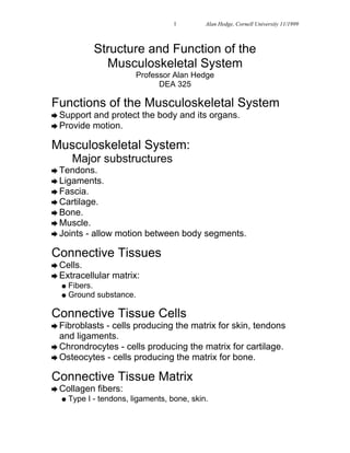
  • 1. 1 © Alan Hedge, Cornell University 11/1999 Structure and Function of the Musculoskeletal System Professor Alan Hedge DEA 325 Functions of the Musculoskeletal System è Support and protect the body and its organs. è Provide motion. Musculoskeletal System: Major substructures è Tendons. è Ligaments. è Fascia. è Cartilage. è Bone. è Muscle. è Joints - allow motion between body segments. Connective Tissues è Cells. è Extracellular matrix: l Fibers. l Ground substance. Connective Tissue Cells è Fibroblasts- cells producing the matrix for skin, tendons and ligaments. è Chrondrocytes - cells producing the matrix for cartilage. è Osteocytes - cells producing the matrix for bone. Connective Tissue Matrix è Collagen fibers: l Type I - tendons, ligaments, bone, skin.
  • 2. 2 © Alan Hedge, Cornell University 11/1999 l Type II - cartilage. l Type III - blood vessel walls. è Mechanicalproperties depend on fiber types and fiber arrangements. Tendons Ligaments Fascia Mechanical Properties of Fibers è Stress - force on a fiber e.g. weight. è Strain - % stretching of fibers. è Elastic limit - point at which the elasticity of the fiber is lost. è Failure - point at which the fiber breaks. Collagen fibers è Strengthof collagen is 50% strength of bone. è Under tension, collagen fibers first elongate slightly and then become increasing stiff until failure. Elastic fibers è Weak and brittle fibers. è At low loads they strain greatly and can increase ~200% in length before failure. Tendon structure è Tendon è Tendon sheath: l Synovium - lubricant producing tissue. l Synovial fluid - lubricant fluid. Tendons and Ligaments è Atcertain points, ligaments surround parts of the tendon sheath to act as: l pulleys.
  • 3. 3 © Alan Hedge, Cornell University 11/1999 l tendon guides. è Ligament attachments allow tendons to work around corners, as in the fingers and toes. Cartilage: 3 types è Hyaline cartilage - present in the growth plates at the end of bones and on the articular surfaces of joints. Also present in the respiratory tract (e.g. trachea). è Fibrocartilage - present in intervertebral discs, which consist of islands of hyaline cartilage in collagen fibers. è Elastic cartilage - present in ears, nose, epiglottis etc. Consists mainly of elastic fibers. Bone è 99% of bodies calcium is in bone. è 80% of bone tissue by weight is minerals. Bone groups è Axial (appendicular) bones - flat bones, such as the skull, pelvis, ribs, vertebrae è Long bones - bones of the extremities. Round bones comprising: l diaphysis - shaft l epiphyses - 2 expanded ends Types of Bone è Cortical (compact) bone - tissue with high proportion of bone. 5-30% porosity. è Cancellous (spongy) bone - less dense bone tissue found at the epiphyses of the long bones and in axial bones, such as the skull. 30-90% porosity. Cortical (Compact) Bone Cancellous (Spongy) Bone
  • 4. 4 © Alan Hedge, Cornell University 11/1999 Anisotropy è All bone is anisotropic (i.e. its mechanical property changes when loads are applied in different directions). Anisotropic behavior of cortical bone. Bone: Mechanical properties è Most important properties are strength and stiffness of bone. è Fractures occur when the bone is loaded to failure. è Compression fractures are commonest in cancellous bone (e.g. fractured skull) è Bending and torsional fractures are commonest in cortical bone (e.g. broken tibia). Speed of loading to fracture. Bone Structure and Growth è Ossification - processes of bone formation. è Osteoblasts - cells that form the bone matrix. They transform into: l Osteocytes - cells isolated inside the mineralized bone matrix. è Osteoclasts - cells important to bone remodeling. Skeletal muscle è Skeletal muscle is ~50% body weight. è Skeletal muscle is striated (striped), and excludes cardiac and smooth muscle. è Several hundred muscles in the body. è Skeletal muscle is under voluntary control. è Each muscle is a separate organ. è Each muscle is attached to bone by tendons that cross one or more joints. è Generally, skeletal muscles generate moments about joints.
  • 5. 5 © Alan Hedge, Cornell University 11/1999 Properties of Skeletal Muscle è Isometric (static) contraction - muscles contract to change muscle tone without changing length. This increases postural stability. è Static contraction is prone to rapid fatigue. è Isotonic (dynamic) contraction - muscles contract to change muscle length. This produces movement at joints. è Dynamic contraction increases blood flow through muscles which slows fatigue. Muscle Organization at Joints è Agonist muscle - the muscle directly engaged in contraction (e.g. in flexing the elbow the biceps brachii is the agonist). è Antagonist - the opposing muscle in relaxation (e.g. in flexing the elbow the triceps is the antagonist). Structure of Skeletal Muscle è Skeletal muscle is covered by a fascia called the epimysium. è The epimysium penetrates and subdivides the muscle into muscle fiber bundles called the fascicles (fasciculi). è Each fascicle is covered by connective tissue called the perimysium. è Each individual muscle fiber is covered by connective tissue called the endomysium. è Connective tissue provides pathways for nerves and blood vessels + contributes to the mechanical properties of the muscle. Skeletal Muscle Innervation è Nerves (neurons) connect from the brain and spinal cord to muscles. è Efferent nerves carry motor instructions for contraction to the muscles. è Afferent nerves carry sensory information to the brain. l Proprioceptive feedback - muscle tone. l Kinesthetic feedback - muscle + joint status.
  • 6. 6 © Alan Hedge, Cornell University 11/1999 è Mixed nerves carry a mix of both sensory and motor neurons (e.g. median nerve). Neuromotor Junction Motor Units è Motor unit - group of muscle fibers innervated by branches of the same efferent neuron. è Functional unit of the muscle. è Motor units are small in muscles requiring precise control (e.g. eye muscles), and large in coarse acting muscles (e.g. gastrocnemius muscle). è Motor units work in an “all-or-none” way. è Progressive contraction occurs because of recruitment of motor units. Muscle Junctions è Myotendinal junction - area where tendon fibers insert into muscle connective tissue and also into bone matrix (Sharpey’s fibers). è Neuromuscular junction - motor endplate where nerve fibers connect with myofibrils. Muscle fibers è Long, cylindrical cells containing several nuclei. è Most of the fiber volume is occupied by the contractile elements - myofibrils. è Myofibrils show a banding pattern (transverse striation) of thick myofilaments (myosin) and thin ones (actin). è The contractile unit of the myofibril is called the sarcomere. Sarcomere Sarcomere and Contraction Joints èA joint is the union of 2 or more bones. 3 types of joint are found in the body.
  • 7. 7 © Alan Hedge, Cornell University 11/1999 è Synovial joint (diarthrodial joint) - no tissue between the articular surfaces. Most of the body joints are this type. è Fibrous joint - fibrous tissue bridge between bones (e.g. skull). è Cartilagenous joint - cartilage bridge between bones (e.g. intervertebral disc in spine). Structure of a Synovial Joint è Joint capsule - fibrous capsule around the joint. è Synovial membrane - membrane lining the joint capsule and surrounding the synovial cavity. This tissue secretes the lubricating synovial fluid. è Synovial cavity (joint cavity) - cavity containing synovial fluid + bone ends è Articular cartilage (hyaline cartilage) - cartilage covering articular surfaces of the bone ends. Joint degeneration è Cartilage does not have a capillary blood supply. è Cartilage has a poor ability to repair and regenerate itself. è With age, overuse and/or disease the articular cartilage degenerates and bone ends make more direct contact, causing pain. Intervertebral Disc Structure è Nucleus pulposus - incompressible watery gel contained within an elastic sac. è Annulus fibrosis - fibrocartilage lamellae arranged in layers around the nucleus. Fiber orientation varies from layer to layer. è End plates - hyaline cartilage end plates protect each end of the disc. è Motion segment - 2 vertebrae either side + intervertebral disc. Fibrocartilage: Intervertebral Discs