2. Blood Film or Peripheral Blood
Smear
o Is a thin layer of blood
o Smeared on a microscope slide
o And then stained in such a way to allow the
various blood cells to be examined
microscopically
Blood films are usually examined to
investigate hematological problems
3. Prepare a Blood Film
1. Place a blood drop on one end of a dry slide.
2. Then using a spreader slide to disperse the blood
over the slide’s length.
3. Air dry the film about 5 minutes.
4. Add 8 drops of leishmann stain onto the slide and
leave 2 minutes.
5. At the end of 2 minutes, add 16 drops of distilled
water onto the slide and leave for another 7
minutes.
6. Then pour off mixture and wash the film with fresh
tap water.
7. Dry it and see under microscope
4.
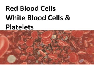
5. Red Blood cells
• Also called erythrocytes.
• Has Biconcave Disk shape.
– Provides 20-30% greater surface area than a
sphere
– Allows the erythrocytes to deform readily
• No Nucleus, Organelles, Centrioles or
Division.
• Count - Male 5.2-5.8 million/mm3
Female 4.3-5.2 million/mm3
• Erythropoiesis is occur in bone marrow.
6. • Life span of an erythrocyte is 120 days.
• Old erythrocytes become rigid and fragile and
their Hb begin to degenerate.
• Red cell destroy is occur in spleen.
In a blood film red cells are stained in pink due
to high contain of Hb( A basic Protein )
7. Multipotent uncommitted stem cells
Committed stem cells
Early erythroblast
Pronomoblast
Early normoblast
Late normoblast
Reticulocyte
Erythrocyte
BONE MARROW
BLOOD STREAM
10. Granules are two types…….
1. Specific Granules
• Binds to neutral, acidic or basic
component of the dye
2. Azurophilic granules
• These are with lysosomes and stain
purple blue.
11. All granulocytes…………..
– Non dividing terminal cells
– Have less cellular organs
– Have less protein synthesis
– Have less mitochondria
– Contain glycogen
Count 6000 – 11000 cells/mm3
12. Neutrophils
• 40% to 70% in WBC
• 12-15 µm in diameter
• 2 – 5 lobes in nucleus
• In females inactive X chromosome appear as a
drumstick appendage on one lobe
• Cytoplasm contains granules, granules are
small and numerous and purple
• Function – phagocytosis of bacteria
• Half life time 6-7 days in blood
1-4 days in connective tissue
• Increases in bacterial infection
13. Eosinophil
• 1% to 6% in WBC
• Bilobed nucleus
• Contain Histamine
• Increase in number
– Helminthic infections
– Filariasis
– Allergic conditions
• Show phagocytic function.
14. Basophils
• Less than 1% in WBC
• Irregular lobe
• Specific basic granules
– Granules cover nucleus so difficult to see nucleus
• Contain Histamine and Heparin
• Increase in number in allergic conditions
15.
16. Lymphocytes
• 20% to 45% in WBC
• Spherical shape with indentation
• Cytoplasm continued to peripheral area
• Few azurophilic granules
• Life span- few days to several years
• Only cell type that return to blood stream
after diapedesis
• Two types T – directly attack cell
B – produce antibody
• Smallest cell type
17. Monocytes
• 2% to 8% in WBC
• Dark bean shaped nucleus
• Less condensed chromatin than lymphocytes
• Basophilic cytoplasm with azurophilic granules
• Bluish color cytoplasm
• Precursor of macrophages
• Show high phagocytic activity
• Larges cell type
18.
19. PLATELETS
Synthesis
• Produced in bone marrow – Megakaryocytes
• Regulated by – Thrombopoietin
• each megakaryocyte formed around 4000
platelets
• From differentiation of
stem cell to platelets takes
around 10 days
20. • Smallest blood cells
• Colorless, spherical appear as dark pink in stained
sections
• No nucleus cannot reproduce
• Covered with a glycoprotein surface coat
• In healthy individuals 1/3 remain in the spleen
• Life span 7 – 10 days
Secretions
Fibrin Stabilizing Factor
Platelet Derived Growth Factor
Von Willebrand Factor
Serotonin