Mammalian MSC from Selected Species: Features and Applications Christiane Ude...
Cassidy Wang Poster Final
1. Physically Confined Microenvironments
Impair Mitotic Progression
Cassidy Wang, Xiaohu Wan, Konstantinos Konstantopoulos
Department of Chemical and Biomolecular Engineering, Johns Hopkins University, Baltimore, MD, USA
0
10
20
30
40
50
60
30 60 90 120 150 180 210 240 270 300 330 360 390 420 450 480 510 540
Frequency (%)
Duration (minutes)
HT1080 Mitosis Duration Distribution
Unconfined
10 μm channel
6 μm channel
3 μm channel
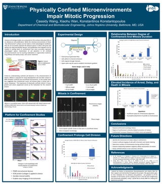
Introduction
Conclusions
Elevated Incidence of Arrest, Delay, and
Death in Mitosis
Mitosis in Confinement
Future Directions
Acknowledgments
I extend my thanks to Konstantinos Konstantopoulos and Xiaohu Wan for
their support and mentorship throughout this project. My appreciation goes
to Chris Yankaskas for his patience and guidance. I am grateful to all the
members of the Kostas Lab for making my time in the lab both enlightening
and immensely enjoyable. Lastly, I would like to thank INBT for making this
experience possible.
References
Critical to understanding confined cell dynamics is the characterization of
mitosis. Mitosis is required for tissue development and has been shown to
differ between confined and unconfined environments. In vivo studies have
also suggested that intravascular tumor cell division plays a critical role in
cancer metastasis.1 The importance of physically confined mitosis in tissue
growth and cancer progression serves as the motivation for this research
project.
Platform for Confinement Studies
• PDMS microchannel devices
• ECM protein (collagen-I) applied to device
• Variable channel widths
• Enables easy imaging of microchannels
Confinement Prolongs Cell Division
Relationship Between Degree of
Confinement And Mitosis Duration
• Physical confinement prolongs mitosis
• Degree of confinement is directly related to duration of mitosis
• Confinement increases frequency of mitotic arrest and apoptosis
• Identify the mechanism of mitotic delay in confinement
• Conduct confocal imaging of cells with chromatin and tubulin labeling
• Determine number of chromosomes during confined mitosis
• Examine relationship between migratory ability and duration of mitosis
• Study confined mitosis in non-cancerous cell lines
• Forms bipolar spindle similar to that seen in unconfined mitosis
• More elongated morphology
1. Xi, W., Schmidt, C. K., Sanchez, S., Gracias, D. H., Carazo-Salas, R. E., Butler, R., ...
Schmidt, O. G. (2016). Molecular Insights into Division of Single Human Cancer Cells in
On-Chip Transparent Microtubes. ACS Nano, 10(6), 5835-5846.
2. Hung, W.-C., Chen, S.-H., Paul, C. D., Stroka, K. M., Lo, Y.-C., Yang, J. T., &
Konstantopoulos, K. (2013). Distinct signaling mechanisms regulate migration in
unconfined versus confined spaces. The Journal of Cell Biology, 202(5), 807–824.
Experimental Design
Classical cell biology studies are conducted on flat surfaces that pose little to
no obstacle to cell proliferation. While the chemical environments of these
“two-dimensional” surfaces can be made to mimic physiological conditions,
they do not accurately replicate the physical spaces in which cells grow and
spread. By using microfluidic devices, cell proliferation and migration can be
studied in varying levels of confinement that more accurately represent
physiological spaces. Examination of cell dynamics in 3D confined
microenvironments has far-reaching implications in regenerative medicine,
drug efficacy and toxicity assays, and cancer treatment.
Figure 1. Cancer
cells in confinement.
Xi et al. (2016)1
0
10
20
30
40
50
60
70
30 60 90 120 150 180 210 240 270 300 330 360 390 420
Frequency (%)
Duration (minutes)
MDA-MB-231 Mitosis Duration Distribution
Unconfined
3 μm channel
Mitosis in unconfined space. HeLa cells stained with SiR-Tubulin (green) and
mCherry H2B (red). (Daniel Gerlich and Claudia Blaukopf, Institute of Molecular
Biotechnology, Vienna)
• Fluid pressure generates flow
• Cells adhere to channel entrances
• Cells migrate through channels
• Upper wells may be used to establish chemotactic gradient
Changes in cell morphology with
varying degrees of confinement.
Hung et al. (2013)2
Cells
Pressure-driven
flow
• 3, 6, 10 μm width
• 1 mm length
• 10 μm height
• 3 μm width
• 200 μm length
• 10 μm height
Fluorescence images
(tubulin)
Phase images
65
158
0
50
100
150
200
250
300
Unconfined 3 μm channel
Duration (minutes)
Average Duration of MDA-MB-231 Mitosis Under Physical Confinement
N=74 N=25
6 μm channel
47
77
130
348
0
100
200
300
400
500
600
Unconfined 10 μm channel 6 μm channel 3 μm channel
Duration (minutes)
Average Duration of HT1080 Mitosis Under Physical Confinement
N=53 N=62 N=37 N=11
Hypothesis
• Insufficient space for chromosome alignment
• Incomplete attachment of spindle to chromosomes
0
11
35
97
0
20
40
60
80
100
Unconfined 10 μm channel 6 μm channel 3 μm channel
Percentage of Mitotic Events (%)
Relative Frequencies of Mitotic Delays/Deaths
Arrest/delay
threshold:
3 hours
N=53 N=66 N=46 N=30
Migration
Device designs used in study