1. Macrophage Cytokine Secretion is Modulated by Black Tea
Cassandra Houser and Christopher Thompson
Loyola University Maryland, Department of Biology
ABSTRACT
MATERIALS AND METHODS
RESULTS
RESULTS
CONCLUSIONS
Macrophages are key modulators of both the innate and adaptive branches of the immune system and emit potent,
pleiotropic signaling molecules: cytokines that regulate inflammation.1 Many studies aim at understanding and modulating these
responses as a way to combat disease. Previous studies have shown that black tea and its bioactive molecule, theaflavin, have the
ability to inhibit transcription factors involved in the synthesis of pro-inflammatory cytokines.3 However, many of these findings are
not physiologically relevant because they do not consider the effects of the digestive system upon black tea or its polyphenol
constituents, which are often consumed orally.
Our study focused upon digested black tea and found that a simulated human digest of black tea induces secretion of tumor
necrosis factor-α and interleukin-6, both pro-inflammatory cytokines, in RAW 264.7 macrophages. RAW 264.7 macrophages were
treated with 225µL of simulated digest of black tea, simulated digest fluid alone, or water for 30 min, 2 hr, 4 hr, 8hr, 12 hr, or 24 hr
before the supernatant was collected. The supernatant was used to perform ELISAs to determine the concentration of TNF-α, IL-6,
and IL-10 secreted by macrophages under all treatments. Our findings indicate that digested black tea induces pro-inflammatory
responses in macrophages via secretion of TNF-α and IL-6 and does not induce secretion of IL-10.
Camellia sinensis
(Black Tea)
Simulated
Digestion (SD)
Black Tea
SD
BTSD SD H2O
Capture Ab binds to plate
Cytokine binding
Avadin-HRP binding
Substrate addition
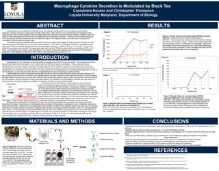
Figure 1. Methods. Prepared a black tea
simulated digest (BTSD) and treated RAW
264.7 macrophages with 225µL of BTSD,
simulated digest (SD), or DI H2O. The cells
were then incubated at 37°C. Supernatant
from samples was collected after prescribed
incubation periods: 30 min, 2hr, 4hr, 8hr,
12hr, and 24hr. The supernatants were
diluted and used to perform ELISAs to test
for the concentration of TNF- α, IL-6, and
IL-10.
0
2000
4000
6000
8000
10000
12000
14000
TNF-αconcentration(pg/mL)
Treatment time
30min 2hr 4hr * 8hr * 12hr * 24hr *
Figure 2.
BTSD
H2O
SD
TNF-α Secretion
0
50
100
150
200
250
300
350
400
450
500
IL-6concentration(pg/mL)
Figure 3.
30min 2hr 4hr 8hr 12hr 24hr
Treatment time
BTSD
IL-6 Secretion
Figure 2. Results: Black tea simulated digest (BTSD) modulates
RAW 264.7 cells secretion of tumor necrosis factor-α
RAW 264.7 macrophages were treated and the supernatant collected
according to the protocol in Figure 1. BTSD and simulated digest (SD)
induced secretion of TNF-α in RAW 264.7 macrophages. RAW 264.7
cells treated with BTSD secreted more TNF-α than those treated with
SD at all time points. Secretion of TNF-α increased over time in BTSD
treated cells and peaked after 8 hours of exposure.
Figure 3. Results: Black tea simulated digest (BTSD)
modulates RAW 264.7 cells secretion of interleukin-6
RAW 264.7 macrophages were treated and supernatant collected
according to the protocol in Figure 1. BTSD induced secretion of
IL-6 in RAW 264.7 macrophages after 4 hours of exposure, but
SD did not. Similar to Figure 2, secretion of IL-6 increased over
time in BTSD treated cells and peaked after 8 hours of exposure.
-Digested black tea induces an inflammatory response in macrophages via secretion of TNF-α and IL-6, especially after 8 hrs of
exposure
-Digested black tea does not induce secretion of IL-10, an anti-inflammatory cytokine
-Simulated digest fluid alone induces an inflammatory response in macrophages, but to a far less extent than black tea simulated
digest.
-Black tea’s pro-inflammatory properties could be beneficial for wound healing and fighting off infections and bacteria
Future Directions
-Dilute the supernatants further to quantitatively measure TNF-α after 4hrs of treatment with black tea digest
-Perform ELISAs to test the concentration of other pro-inflammatory and anti-inflammatory cytokines
-Evaluate modulations in cytokine secretion in the presence of black tea digest and a pathogen.
-Introduce bacteria and evaluate the microbicidal activity of macrophages in the presence of black tea digest
Figure 4. Results: Black tea simulated digest (BTSD) has no effect
upon RAW 264.7 cells secretion of interleukin-10
BTSD, SD, and DI H2O did not induce secretion of IL-10 in RAW 264.7
cells over all prescribed incubation times. The bottom of our IL-10 standard
was 125 pg/mL.
*Note: The top of our TNF-α standard was 4000 pg/mL, therefore, our data after 4 hrs of
treatment are estimations.
0
25
50
75
100
125
150
175
200
IL-10concentration(pg/mL)
30 min 2hr 4hr 8hr 12hr 24hr
Treatment time
IL-10 SecretionFigure 4.
ELISA
Supernatant
from BTSD,
SD, and H2O
INTRODUCTION
Macrophages are key modulators of both the innate and adaptive branches of the immune system. They express a phenotypic
plasticity that allows them to specialize in their resident area and efficiently respond to pathogens.1 These responses include
phagocytosis and secretion of potent, pleiotropic signaling molecules called cytokines.2 Cytokines are essential to the function of
macrophages and modulate cellular inflammation processes. The type and quantity of cytokines produced depends upon the stimuli
introduced into a macrophage’s microenvironment.2 As many studies aim at understanding and modulating these responses as a way to
combat disease, our study focused on the influence of digested black tea upon macrophage cytokine secretion.
Camillia sinensis is used to make green tea and black tea and is one of the most-consumed plants worldwide. Research has
been growing on this plant due to its bioactive tea polyphenols that have been indicated to have potent effects upon the immune system.
Although they are produced from the same plant, black tea has a distinctively different catechin profile from green tea. Black tea derives
from fermentation, where its catechins undergo enzymatic oxidation into theaflavins and thearubigens. Although they only account for
2-6% of the total dry weight of black tea; theaflavins have shown to exhibit numerous health benefits.3
Studies have found that theaflavins have an affinity for the lipid bilayer
surface via hydrogen bonding, potentially preventing the binding of bioactive
molecules and disrupting the cell membrane.4 In particular, theaflavin-3,’3-digallate
(TF-3) has been found to inhibit nuclear factor-kB (NF-kB) in endotoxin-induced
macrophages, decreasing gene expression of pro-inflammatory cytokines.3 5 NF-kB
plays a critical role in our immune system’s inflammatory processes and can be activated
by a variety of stimuli, such as pro-inflammatory cytokines, endotoxins, and reactive
oxygen species (ROS).5 Proper regulation of NF-kB is key to controlling inflammatory
processes that can cause detrimental diseases if left unmonitored, such as cancer
and diabetes.5 These findings indicate that black tea, specifically theaflavins, help mediate inflammatory cellular processes.
Although black tea polyphenol constituents have been found to have anti-inflammatory properties, not many studies have
focused on the effects of black tea upon macrophages without endotoxin, inflammatory, or pathogenic stimuli. Additionally, black tea is
most consumed orally in brewed form, and studies have not considered the effects of the digestive system upon black tea. The objective
of this study is to better identify the influence of ingested black tea upon macrophages cytokine secretion in vitro. The studies described
herein add to our understanding of the biological activities of black tea and its relation to macrophage function. We treated RAW 264.7
cells with black tea simulated digest, simulated digest, or DI water over prescribed incubation periods before collecting the supernatant.
The supernatant was used to perform ELISAs to test the concentration of TNF-α, IL-6, and IL-10. We hypothesize that black tea
simulated digest will proliferate RAW 264.7 cells and increase secretion of these cytokines.
REFERENCES
Oxidative stimuli
ROS
IkB kinase
Pro-inflammatory
cytokines
Theaflavin-3,’3-digallate
1 Sasmono, R Tedjo, and David A. Hume. 2004. The Biology of Macrophages, p 71-94. In Kaufmann S, Medzhitov R, Gordon S (ed), The Innate Immune
Response to Infection. ASM Press, Washington, DC.
2 Duque, Guillermo, and Albert Descoteaux. “Macrophages: involvement in immunity and infectious diseases”. Frontiers in Immunology. 5 (2014): 1-12.
Web. 5 February 2016.
3 Gutierrez-Orozco, Fabiola. et al. “Green and black tea inhibit cytokine-induced Il-8 production and secretion in AGS gastric cancer cells via inhibition of
NF-kB activity”. Planta Med. 76 (2010): 1659-1665. Web. 10 February 2016.
4 Sirk, Timothy W. “Molecular Binding of Black Tea Theaflavins to Biological Membranes: Relationship to Bioactivities”. Journal of Agricultural and Food
Chemistry. 59 (2011): 3780-3787. Web. 10 February 2016.
5 Pan, Min-Hsiung, et al. “Suppression of lipopolysaccharide-induced nuclear factor-kB activity by theaflavin-3,3’-digallate from black tea and other
polyphenols through down-regulation of IkB Kinase Activity in Macrophages”. Biochemical Pharmacology. 59 (2000): 357-367. Web. 5 February 2016.