Call Girls Service Pune Vaishnavi 9907093804 Short 1500 Night 6000 Best call ...
Lymph Node Structure and Function
1. Lymph node (nodus lymphaticus)
• They act as filters of the lymph: lymph reaching the node through the afferent
lymphatic vessels is filtered for pathogens and cancer cells. (one of the organs
where the adaptive immune cells can meet with antigens the first time)
• This is the place where the antigens that entered the lymphatic system will be
recognized by the adaptive immune cells followed by cell proliferation and
differentiation.
• Tremendous clinical significance: Infectious agents and cancer cells may spread via
the lymphatic system.
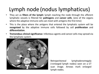
Retroperitoneal lymphadenomegaly
(=enlarged lymph nodes) seen on a CT
scan image. Arrows mark enlarged
lymph nodes.
2. Structure of lymph nodes 1.
• Have outer fibrous capsule from which trabeculae radiate towards the inner part
of the organ.
• Layers from outermost to innermost: cortex, paracortex and the medulla.
• Afferent lymphatic vessels enter through the convex surface; the efferent
lymphatic vessels and blood vessels (artery and venule) are located at the hilum.
• Reticular connective tissue forms the frameworks of the lymph nodes.
• Sites where immune cells enter:
– From the bloodstream: high endothelial venules (HEV)
– From the lymphatic system: afferent lymphatic vessels
• Cellular zones:[9.]
– Cortex: B cells organized into follicles, cells that recognized an antigen
proliferate and form germinal centers
– Paracortex: T cells and dendritic cells diffusely
– Medulla: mainly antibody-producing plasma cells
4. Structure of lymph nodes 3.
Dendritic cell
Chemokine attracting T cells
and denditic cells
B cell-specific chemokine
Afferent
lymphatic vessel
B cell zone
T cell zone
Naive
B cell
Naive T cell
HEV
Naive T cell
Naive B cell
Artery
T cell zone
(paracortex)
B cell zone
(follicle)
Immunofluorescence microscopy
(see later)
The cellular organization is
controlled by chemokines. (see
later in lectures)
5. Lymphoid follicle (folliculus lymphaticus)
1. Primary follicle:
Naive B cells that haven’t yet
met with an antigen
2. Secondary follicle (germinal
center):[9.]
Dark zone: centroblasts
(proliferating B cells)
Light zone: centrocytes (B
cells undergoing antigen-
dependent maturation, see
later)
Mantle zone: transient B cells
(=passing through)
Main cellular components:
B cells, macrophages,
follicular helper T cells,
follicular dendritic cells
(FDC)
Capsule Afferent vessel
Follicle (B cells)
Secondary follicle, germinal center
Primary follicle
Paracortical zone (T cells)
Medulla
Mantle
zone
Light
zone
Dark
zone
6. High endothelial venules (HEV)
• Lymphocytes use HEVs to enter
lymphoid organs. (through L-selectin,
see later)
• Found in all secondary lymphoid
organs (e.g. lymph nodes, tonsils,
Peyer’s patches), EXCEPT THE
SPLEEN[10.]
T cells binding to HEV
(frozen section assay)
T cells binding to the luminal surface of a HEV
(electron microscopy image)
HEVs
L-selectin ligand on
endothelial cells (IHC)
HEVs in a lymph node
HEVT cells
7. Filtration of lymph by nodes 1.
1. Infection on the periphery
2. The same antigen may enter the
lymphatic vessels in different
forms:
• Native bound antigen (e.g.
living bacteria)
• Native soluble form (e.g.
proteins derived from dead
bacteria)
• Processed form: dendritic
cells phagocytose the
antigen and present it as a
peptide to helper T cells
(see later)
3. Lymphocytes enter lymph nodes
either through afferent lymph
vessels or HEVs and meet with
the antigens (see the lectures for
more details)
Site of infection
(peripheral tissue)
Lymph node
without antigen
Lymph node
with antigen
Naive
T cell
Activated
T cellHEV
HEV
Blood vessels
Efferent lymphatic
vessel
Efferent lymphatic
vessel
Afferent
lymphatic vessel
Lymphatic
vessel
Blood
vessel
Thoracic duct
Vena cava
Microbes
8. DRAINING
LYMPH NODE
INFECTION
Cervical lymph
nodes
Intercostal
lymphatic vessels
Axillary lymph
nodes
Cisterna
chyli
Paraaortic
lymph nodes
Lymphatic
vessels from
the gut
Inguinal
lymph nodes
Dendritic cell
with the antigen
Free
antigen
Lymphatic
vessel
Connective tissue
ANTIGEN CAPTURE
AND TRANSPORT
ANTIGEN
PRESENTATION AND
T CELL RESPONSE
Lymph node
Dendritic cell
![Structure of lymph nodes 1.
• Have outer fibrous capsule from which trabeculae radiate towards the inner part
of the organ.
• Layers from outermost to innermost: cortex, paracortex and the medulla.
• Afferent lymphatic vessels enter through the convex surface; the efferent
lymphatic vessels and blood vessels (artery and venule) are located at the hilum.
• Reticular connective tissue forms the frameworks of the lymph nodes.
• Sites where immune cells enter:
– From the bloodstream: high endothelial venules (HEV)
– From the lymphatic system: afferent lymphatic vessels
• Cellular zones:[9.]
– Cortex: B cells organized into follicles, cells that recognized an antigen
proliferate and form germinal centers
– Paracortex: T cells and dendritic cells diffusely
– Medulla: mainly antibody-producing plasma cells](data:image/gif;base64,R0lGODlhAQABAIAAAAAAAP///yH5BAEAAAAALAAAAAABAAEAAAIBRAA7)