1. THE QUARTERLY JOURNAL OF THE GEMOLOGICAL INSTITUTE OF AMERICA
SPRING2007PAGES1–94VOLUME43NO.1
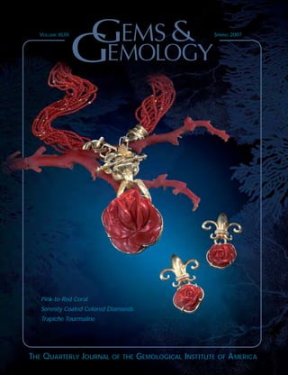
VOLUME XLIII SPRING 2007
GEMS&GEMOLOGY
Pink-to-Red Coral
Serenity Coated Colored Diamonds
Trapiche Tourmaline
2. ®
Carat Points
pg. 18
Volume 43, No. 1Spring 2007
1
3
16
36
4
47
56
83
81
86
94
REGULAR FEATURES _____________________
Lab Notes
Blue diamonds showing multiple phosphorescence colors • Diamond with
“etched dislocation loops” • HPHT-annealed yellow-orange diamond
• Translucent greenish yellow diamonds • Unusual natural-color black diamond
• Yellow synthetic diamond, possibly grown at higher temperatures • Heat-
treated blue sapphire with unusual dendrites • YAG with a dislocation spiral
Gem News International
Tucson report • “Emerald” green fluorite from India • Cat’s-eye leifite from
Canada • Play-of-color opal from Brazil • Prehnite from Tanzania • Colorado
rhodochrosite • Serpentinite from Argentina • Variscite from Western Australia
• Portable Raman spectrometer • Amethyst from the DRC • Color-zoned andra-
dite from Iran • Emerald with unusual growth features • Grossular and clino-
zoisite from California • Pegmatite gems from Madagascar • Pezzottaite from
Myanmar • Cat’s-eye prehnite • Cat’s-eye topaz from Sri Lanka • Tourmaline
from northeastern Mozambique • Turquoise from Sonora, Mexico • Lead
glass–filled rubies with hollow backs • October 2006 Myanmar Gem Emporium
2007 Gems & Gemology Challenge
Book Reviews
Gemological Abstracts
The Last Page: Take the G&G Challenge Today
EDITORIAL _____________
The Dr. Edward J. Gübelin Most Valuable Article Award
Letters
FEATURE ARTICLES _____________
Pink-to-Red Coral: A Guide to Determining Origin of Color
Christopher P. Smith, Shane F. McClure, Sally Eaton-Magaña, and David M. Kondo
This research report explores the identifying characteristics of natural
vs. dyed pink-to-red coral.
Serenity Coated Colored Diamonds: Detection and Durability
Andy H. Shen, Wuyi Wang, Matthew S. Hall, Steven Novak, Shane F. McClure,
James E. Shigley, and Thomas M. Moses
A new multi-layer coating technique produces a variety of evenly distributed,
natural-appearing colors on diamonds, but with limited durability.
Trapiche Tourmaline from Zambia
Thomas Hainschwang, Franck Notari, and Björn Anckar
This unusual green tourmaline displays a striking growth pattern in slices
cut from the crystals.
pg. 60
pg. 51
pg. 5
4. MOST VALUABLE ARTICLE AWARD GEMS & GEMOLOGY SPRING 2007 1
FIRST PLACE
THE IMPACT OF INTERNAL WHITISH AND REFLECTIVE GRAINING
ON THE CLARITY GRADING OF D-TO-Z COLOR DIAMONDS
AT THE GIA LABORATORY
John M. King, Thomas M. Moses, and Wuyi Wang
John M. King is technical director of the GIA Laboratory in New York
and the editor of Gems & Gemology in Review: Colored Diamonds.
Mr. King, who is also a noted artist, received his Master of Fine Arts
degree from Hunter College, City University of New York. Thomas M.
Moses is senior vice president, GIA Laboratory and Research, New York.
Wuyi Wang is manager, Research Projects, GIA Laboratory, New York.
Dr. Wang holds a Ph.D. in geology from the University of Tsukuba in
Japan, and has considerable research experience in diamond geochemistry
and diamond treatment.
he first-place article was “The Impact of
Internal Whitish and Reflective Graining on
the Clarity Grading of D-to-Z Color Diamonds at
the GIA Laboratory” (Winter 2006), which reviewed
the elusive nature of this type of graining in dia-
monds and the methodology that GIA graders use to
determine its impact on diamond clarity grades. Receiving
second place was “Identification and Durability of Lead
Glass–Filled Rubies” (Spring 2006), a study of this new corundum
treatment and techniques to identify it. Third place was awarded to “‘Paraíba’-type Copper-bearing Tourmaline from
Brazil, Nigeria, and Mozambique: Chemical Fingerprinting by LA-ICP-MS” (Spring 2006), which described how this
analytical technique can separate—on the basis of chemical composition—bright blue-to-green copper-bearing tourma-
line from the localities in which it has been found.
The authors of these three articles will share cash prizes of $2,000, $1,000, and $500, respectively. Following are brief
biographies of the winning authors.
Wuyi Wang
John M. King Thomas M. Moses
T
Congratulations
to Margrethe Gram-Jensen
of Overijse, Belgium,
whose ballot was drawn from the
many entries to win a three-year
subscription to GEMS & GEMOLOGY,
and copies of the two
GEMS & GEMOLOGY IN REVIEW
volumes, COLORED DIAMONDS and
SYNTHETIC DIAMONDS.
is pleased to
announce the winners of the
DR. EDWARD J. GÜBELIN
MOST VALUABLE ARTICLE AWARD
for 2006, as voted by the journal’s readers.
We extend our sincerest thanks to all the
subscribers who participated
in the voting.
5. SECOND PLACE
IDENTIFICATION AND DURABILITY OF LEAD GLASS–FILLED RUBIES
Shane F. McClure, Christopher P. Smith, Wuyi Wang, and Matthew Hall
Shane F. McClure is director of Identification Services at the GIA
Laboratory in Carlsbad, California. A popular lecturer, Mr. McClure is also
well known for his articles on gem identification and is coeditor of G&G’s
Lab Notes section. Christopher P. Smith is vice president and chief
gemologist at American Gemological Laboratories, New York City; at the
time the article was written, he was director of Identification Services at
the GIA Laboratory in New York. Mr. Smith has written numerous arti-
cles for G&G and other publications, and is a member of the G&G
Editorial Review Board. Wuyi Wang was profiled in the first-place entry.
Matthew Hall is manager of Identification Services at the GIA Laboratory
in New York. Mr. Hall has a bachelor’s degree in geology from Franklin
and Marshall College and a master’s in geology and geochemistry from
the University of Maryland.
THIRD PLACE
“PARAÍBA”-TYPE COPPER-BEARING TOURMALINE FROM BRAZIL,
NIGERIA, AND MOZAMBIQUE: CHEMICAL FINGERPRINTING BY LA-ICP-MS
Ahmadjan Abduriyim, Hiroshi Kitawaki, Masashi Furuya, and Dietmar Schwarz
Ahmadjan Abduriyim is manager of the Research Laboratory at the
Gemmological Association of All Japan (GAAJ) in Tokyo. Dr. Abduriyim
has a B.Sc. in geochemistry, petrology, and mineralogy from Beijing
University, and M.Sc. and Ph.D. degrees in mineralogy from Kyoto
University. Hiroshi Kitawaki is director of the Research Laboratory at
GAAJ. Mr. Kitawaki has a B.Sc. degree in geology from Niigata
University, Tokyo, and more than 15 years of experience in diamond and
colored stone research and education. Masashi Furuya is director of the
Japan Germany Gemmological Laboratory in Kofu, Japan. Mr. Furuya has
multiple degrees in German from Reitaku University, Kashiwa, Japan,
and over 30 years of experience in the jewelry industry. Dietmar Schwarz
is research manager at the Gübelin Gem Lab in Lucerne, Switzerland.
Dr. Schwarz is a former scientific lecturer at the German Academic
Exchange Service and professor of mineralogy and gemology at Ouro
Preto Federal University, Minas Gerais, Brazil.
Masashi Furuya
2 MOST VALUABLE ARTICLE AWARD GEMS & GEMOLOGY SPRING 2007
Ahmadjan Abduriyim Hiroshi Kitawaki
Matthew Hall
Dietmar Schwarz
Shane F. McClure Christopher P. Smith
6. LETTERS GEMS & GEMOLOGY SPRING 2007 3
MORE ON SYNTHETIC CORUNDUM
“GEM ROUGH”
The report on synthetic corundum “gem rough” in
Tanzania (Winter 2006 Gem News International, p. 282)
is both interesting and helpful, and Mr. Farooq Hashmi is
kind to alert the industry and provide these samples for
analysis. However, I would add that although laser abla-
tion–inductively coupled plasma–mass spectrometry (LA-
ICP-MS) is wonderful when it is accessible, simple pocket
instruments such as rare-earth magnets can instantly pro-
vide useful information for those dealing with gems in
remote locations.
By applying the magnetic separation approach that I
presented at last summer’s Gemological Research
Conference in San Diego (see the Fall 2006 Gems &
Gemology, p. 124) to distinguish between spessartine
rough and orange sapphire rough, gem dealers might
avert this potentially costly misrepresentation. Mn-rich
spessartine generally can be characterized by a relatively
strong magnetic response, using the direct or pendulum
method. Though not diagnostic, the lack of such a
response (as with sapphire) could quickly flag this materi-
al as something other than spessartine.
Your readers may find the magnetic separation table in
the G&G Data Depository (http://lgdl.gia.edu/pdfs/
gumpesberger-table.pdf) helpful; there is also a related
article in the Winter 2006 issue of Canadian Gem-
mologist (S. Gumpesberger, “Magnetic separation of gem-
stones,” Vol. 27, No. 4, pp. 120–124).
Sylvia Gumpesberger
Canadian Gemmological Association
Toronto, Ontario
FALL 2006 ISSUE REQUIRED READING
Unaccustomed as I am to writing letters of this kind—
especially to G&G—scientific integrity compels me to
acknowledge and commend your Fall 2006 issue.
As one unable to attend, I found your coverage of the
4th International Gemological Symposium extremely
interesting.
On the other hand, your 2006 GIA Gemological
Research Conference was simply outstanding. The infor-
mation imparted both by the speakers and through the
poster presentations should be required reading for all
who would call themselves gemologists.
Keep up the good work.
Dr. W. Wm. Hanneman
Poulsbo, Washington
IN MEMORIAM:
DAVID HARGETT (1953–2007)
Gemologist and author David Hargett passed
away recently in New York, the city he called
his home for most of his adult life. He was 53.
Dave worked for the GIA Laboratory in New
York for 16 years as a colleague of current GIA
Laboratory senior vice president Tom Moses
and their gemological mentor, G. Robert
Crowningshield. Dave began his career with
GIA in January 1977 after obtaining his Graduate
Gemologist (G.G.) diploma in Santa Monica, California,
followed later by a bachelor’s degree from New York
University.
At the East Coast GIA Laboratory, Dave served for
several years as manager of the Gem Identification
Department, where he was instrumental in developing
many of the techniques still used today to recognize
synthetic and treated colored stones, as well as cultured
pearls and enhanced diamonds. The author or co-author
of many Gems & Gemology Lab Notes on unusual gem
materials, in 1989 he also co-authored the
award-winning Koivula et al. article on “The
Characterization and Identification of Filled
Diamonds.” Dave was very fond of gemologi-
cally related travel, and he particularly
enjoyed exploring the gem mining areas of
Mexico and Central America. In its Summer
1990 issue, G&G published Dave’s definitive
paper on the jadeite deposits of Guatemala.
Dave left GIA in 1992 and started his own
consulting business, which allowed him to focus on
areas of gemology he had come to enjoy.
Those who were fortunate enough to know Dave
Hargett as a friend remember him as a kind and generous
person with an amazing sense of humor. Those who
worked with him remember him as an excellent gemolo-
gist with a keen and curious mind. Through his dedicat-
ed gemological research and an overall strong work ethic,
he gained the respect and admiration of gemologists all
over the world. He will be missed by many, both within
and outside the gemological community.
Letters
8. GENERAL BACKGROUND
Formation/Biology. Because it shows aspects of the
mineral, animal, and plant kingdoms, coral was not
properly classified until the 17th century (Liverino,
1989). A branch of coral is the skeletal remains of a
colony of tiny animals called coral polyps (figure 4).
The term coral can refer to both the marine animal
and the material produced from its skeletal
remains. Polyps are simple organisms with a
mouth surrounded by tentacles and gastrovascular
function. A polyp colony consists of three different
parts: the sclerax, the coenosarc, and the polyps
themselves. The sclerax is the hard skeleton left
behind by the coral polyps, and it is this material
that can be worked as a gem material. The
coenosarc is the tissue that binds the polyps to the
skeleton. In addition to organic macromolecules
such as proteins, polysaccharides, and lipids, the
coral skeleton also contains biogenic (i.e., biologi-
cally generated) carbonates (CaCO3). The two
major polymorphs are calcite and aragonite (see,
e.g., Rolandi et al., 2005; Bocchio et al., 2006). The
polyps precipitate the major components of most
coral branches from sea water (Liverino, 1989):
calcium carbonate (82–87%) and magnesium
carbonate (~7%).
A new coral colony is originally created through
sexual reproduction, but it continues to grow
through gemmation, whereby a polyp separates off a
portion of itself to create a new polyp. Coral requires
a sturdy foundation such as rocks, other coral, or
debris (e.g., a shipwreck), and it has a growth rate of a
ORIGIN OF COLOR IN PINK-TO-RED CORAL GEMS & GEMOLOGY SPRING 2007 5
Figure 1. For thousands
of years, coral like the
branch shown here has
been fashioned for use
as carvings and in jew-
elry. Although the har-
vesting of gem-quality
pink-to-red coral has
decreased steadily in
recent years because of
environmental and
other problems, leading
to a proliferation of
dyed material, fine
pieces such as these
beads (13–17 mm) and
cabochons (24 mm)
continue to enter the
marketplace. Courtesy
of R. H. & Co.,
Glendale, California;
photo by Harold &
Erica Van Pelt.
11. epoxy, reformed into blocks, dyed, and then used to
make jewelry (Weldon, 2003). It does not show a sur-
face pattern or a ribbed structure.
A careful R.I. reading, S.G. determination, and
infrared spectroscopy—combined with observation
of specific growth-structures—may provide clues to
the particular species of the coral. Such tests may
also help determine if the coral species is predomi-
nantly composed of calcite or aragonite (Kaczo-
rowska et al., 2003; Pederson, 2004; Rolandi et al.,
2005); however, these topics are outside the scope of
this article. Because so little has been published in
the recent gemological literature on the identifica-
tion of dye in coral, especially given the analytical
techniques that are now available, we conducted
numerous tests—standard gemological exams and
more sophisticated spectroscopic analyses—on
known treated and untreated samples to determine
which methods were most effective.
MATERIALS AND METHODS
One of the authors (CPS) acquired seven strands of
coral beads covering four species of the
Coralliidae order—Corallium elatius, Corallium
rubrum, Corallium secundum, and Melithaea
ochracaea—as well as samples of unknown
species (figure 5), all of which were represented as
natural color. They ranged from pinkish orange to
pink and red. In addition, 11 strands of coral—all
represented as dyed—were obtained (see, e.g., fig-
ure 6). Most of these belonged to the Corallium
rubrum species, with the dyed colors ranging from
pink-orange and various shades of light pink to
red. Included in this group were two strands of
Melithaea ochracaea that had been treated with a
colored polymer. We also analyzed 10 known nat-
ural-color and dyed coral specimens from the GIA
reference collection and 24 dyed samples from the
collection of one of the authors (SFM). In all, well
over 1,000 pieces of natural-color and dyed coral
were included in this study.
All of the samples were examined using a stan-
dard GIA Gemolite binocular microscope with
8 ORIGIN OF COLOR IN PINK-TO-RED CORAL GEMS & GEMOLOGY SPRING 2007
Figure 5. Natural-color coral can span a wide range
of hues, including these examples (2.5–11.8 mm) of
the most valuable shades of pink to red. These
strands of coral belong to the following species: (1)
Melithaea ochracaea, (2) Corallium elatius, (3)
Corallium rubrum, (4) Corallium elatius, (5)
Corallium species, (6) Corallium secundum, and
(7) Corallium rubrum. Photo by Jessica Arditi.
Figure 6. Coral (here,
3.5–18.0 mm) can be dyed
almost any color, but it is
seen most commonly in the
gem trade as pink to red.
Photo by Jessica Arditi.
12. fiber-optic illumination. Acetone and a cotton
swab, such as a Q-tip, were used to test inconspicu-
ous areas of the coral on a random sample of
approximately 50 pieces of each group (natural-color
and treated) for the presence of dye.
A Renishaw System 1000 Raman micro-spec-
trometer with an argon-ion laser (514 nm excita-
tion) was used to analyze 40 natural-color and 40
dyed samples (including approximately 10 with a
colored polymer), covering the complete color
range. For the Raman analysis, we used a variety of
magnifying lenses, from 5× to 50×. The integration
times varied from 3 to 20 seconds per grating
sequence. The spectra were taken over two different
ranges. The first extended from 2000 to 100 cm−1
and covered the standard range used to identify the
key Raman bands of a material. The second extend-
ed from 517 to 1000 nm, with the intent of measur-
ing Raman bands outside of the previous range as
well as any photoluminescence bands that might be
present (refer to Raman Spectroscopy below).
Reflectance spectra of 10 natural-color and 14
dyed coral samples that varied from pink to red were
acquired for the 200–850 nm range with a Perkin
Elmer Lambda 950 ultraviolet/visible/near-infrared
(UV-Vis-NIR) spectrometer utilizing an integrating
sphere, with a 1.0 nm scan interval and 141 nm per
minute scan speed. Although reflectance spectra are
typically shown as % reflectance (%R), the authors
have elected to portray the spectra in absorbance, as
the negative log of %R divided by 100 (i.e., −log[%R
100
]),
since most spectra in gemological publications are
shown using absorbance. To confirm the validity of
this approach, we performed absorption spec-
troscopy on thin slices made from seven of the
samples (four pink-to-red natural-color and three
dyed) using the same spectrometer and measuring
conditions. The two types of spectra, reflectance
(converted to absorbance) and absorbance, were vir-
tually identical.
RESULTS AND DISCUSSION
Microscopic Examination and General Obser-
vations. The structures of the most common orna-
mental corals—Corallium elatius, Corallium
rubrum, and Corallium secundum—typically con-
sist of two patterns. The first is a ribbed or striated
pattern that extends roughly parallel to the length
of the coral branch (figure 7). The other is a concen-
tric, scalloped structure (figure 8). Natural features
ORIGIN OF COLOR IN PINK-TO-RED CORAL GEMS & GEMOLOGY SPRING 2007 9
Figure 7. The parallel striations that are obvious in
“rough” coral are also clearly evident after the mate-
rial has been fashioned, so they readily separate
coral from its simulants. Note, however, that this
structure is visible in both natural-color and dyed
coral. Photomicrograph by C. P. Smith; fiber-optic
illumination, magnified 18×.
Figure 8. A concentric
scalloped pattern is also
characteristic of natural
coral and will readily
distinguish it from its
most common imitators
shell and plastic.
Photomicrographs by
C. P. Smith; fiber-optic
illumination, magnified
15× (left, dyed coral)
and 25× (right, natural-
color coral).
13. dotting the coral surface, which may be described as
pits and pock marks, also are common and charac-
teristic of many Corallium species. The unusual
patterns (figure 9) and the open and porous structure
(figure 10) of the red coral species Melithaea ochra-
caea are particularly distinctive. Such features were
observed in all the coral beads examined in this
study, both natural-color and dyed, depending on
their species.
Coral occurs naturally in a broad range of attrac-
tive pinkish orange to pink and red colors (again,
see figure 5). Similarly, coral may be dyed to virtual-
ly any color; however, only results for coral that has
been dyed pale pink to deep red and pink-orange are
reported in this study (again, see figure 6). In some
instances, the colors achieved by dyeing appear very
similar to those seen in natural-color coral.
Commonly, though, the dyed coral is very different
in appearance from its natural-color counterpart
(see, e.g., figure 11). Therefore, visual observation of
color may provide an important clue to the pres-
ence of dye. In addition, the surface pits and cavities
common to coral readily act as receptacles for dye
concentrations (figure 12), as do fractures or separa-
tions in the concentric growth pattern. In such
instances—where visual observation is insuffi-
cient—the use of acetone and a cotton swab may be
effective to identify the presence of dye (figure 13).
This reaction was observed in many of the dyed
samples we tested; however, it was very faint in
several samples and not evident at all in a few. A
piece of coral may also be immersed in acetone to
see if any dye leaves the coral structure and turns
the acetone pink (although we did not conduct this
test on our samples). However, both of these tech-
niques are somewhat destructive, as a positive test
removes some of the color imparted by the dye. For
this reason—and because, as our testing with a cot-
ton swab showed, the results are not always conclu-
sive—we also investigated two nondestructive ana-
lytical techniques.
10 ORIGIN OF COLOR IN PINK-TO-RED CORAL GEMS & GEMOLOGY SPRING 2007
Figure 9. The red coral species Melithaea ochracaea
(sometimes referred to as “red king coral”) is notable
for its distinctive patterning. This piece measures 2.8
cm at the base. Courtesy of Holly Smith; photo by
Elizabeth Schrader.
Figure 10. The characteristic “open” structure of Melithaea ochracaea is evident whether the coral is fashioned
with the channels open (left, magnified 15×) or if it has been impregnated with a polymer (right, magnified
25×). Today it is common to treat this species of coral with a polymer to reduce its rough texture and allow it
to take a better polish. The polymer has a yellow-orange color, as may be seen in the open pores of the treated
sample. Photomicrographs by C. P. Smith.
14. Raman and Photoluminescence Spectroscopy. The
Raman spectra of these four species of coral typical-
ly exhibit a combination of bands associated with
both the coral matrix (CaCO3) and the compounds
responsible for the color (figure 14). A distinct band
positioned at approximately 1087 cm−1
, with subor-
dinate peaks at approximately 714 and 283 cm−1
, is
indicative of the calcium carbonate phase forming
the skeleton of the coral. All of the samples we test-
ed with Raman spectroscopy, both natural-color
and dyed, showed this feature.
Natural color in pink-to-red coral may be readily
identified by the presence of certain organic pigments
incorporated into the coral skeleton. A pair of
distinct Raman peaks positioned at approximately
1520 and 1123 cm−1
, with subordinate peaks at
approximately 1297 and 1020 cm−1
, identify carotene
ORIGIN OF COLOR IN PINK-TO-RED CORAL GEMS & GEMOLOGY SPRING 2007 11
Figure 11. Often the dyes used to treat coral will
impart a vivid red hue, as in this 14 mm bead, that is
not found in nature. Photo by Jessica Arditi.
Figure 12. Coral can be a brittle material, as well as
somewhat porous. Breaks or cavities at the surface
commonly contain concentrated remnants of the dye
that was used to color it. The very dark red masses
obvious near the drill hole in this sample readily iden-
tified this bead as dyed, even without magnification.
Photomicrograph by C. P. Smith; magnified 13×.
Figure 13. Acetone has traditionally been used to
confirm the presence of dye in coral. Commonly this
test involves dipping a cotton swab in acetone and
then rubbing the cotton on an inconspicuous area of
the sample to see if any of the color rubs off.
Occasionally, the item is immersed in acetone to
see if the liquid will discolor slightly. In both cases,
this test may be destructive to the color of the sam-
ple. Photo by Jessica Arditi.
Figure 14. The Raman spectrum of coral identifies
the biogenic calcium carbonate phase of the skeleton
(CaCO3), with peaks positioned at approximately
1087, 714, and 283 cm−1
. The Raman spectrum of
natural-color coral typically reveals additional peaks
related to organic pigments, such as those positioned
here at 1520, 1123, and 1020 cm−1
.
15. (Urmos et al., 1991; Kaczorowska et al., 2003;
Rolandi et al., 2005). In our experience, the intensity
of these carotene peaks directly correlates to the satu-
ration of the pink-to-red color, as was the case with
the natural-color samples we tested (figure 15).
Using the full spectral range of the 514 nm Ar-
ion laser (517–1000 nm; figure 16), we observed a
series of peaks positioned at approximately 543
(1020 cm−1
), 546 (1123 cm−1
), 551 (1297 cm−1
), 558
(1520 cm−1
), 578, 581, 587, 591, 595, 601, 607, 609,
618, 637, 653, 670, 685, and 702 nm, as well as other
subordinate peaks, all of which are due to carotene
pigments. In contrast, the dyed pink-to-red coral
samples did not exhibit the Raman spectrum associ-
ated with carotene. Instead, they typically showed
an increase in underlying photoluminescence in the
standard Raman spectral range of 2000–100 cm−1
, in
addition to a series of very weak peaks positioned at
approximately 1608, 1492, 1482, 1455, 1378, 1353,
1327, 1290, 1032, 906, 817, and 701 cm−1
(figure 17;
details of many of these peaks cannot be observed at
the scale shown). With the extended spectral range
(517–1000 nm), we observed that the dominant pho-
toluminescence of the dye was centered between
approximately 630 and 665 nm, the asymmetry of
this photoluminescence structure indicating that it
likely consists of multiple bands (figure 18). In the
Melithaea ochracaea samples that had been treated
with a colored polymer, we noted a compound spec-
trum of carotene peaks and a dominant broad lumi-
nescence band.
Since the dye or colored polymer may be incor-
12 ORIGIN OF COLOR IN PINK-TO-RED CORAL GEMS & GEMOLOGY SPRING 2007
Figure 15. Carotene is the organic pigment responsible
for the pink-to-red shades of natural-color coral. In the
Raman spectra of these three natural-color corals, the
intensity of the carotene peaks (with two dominant
peaks at approximately 1520 and 1123 cm−1
and
minor peaks at approximately 1297 and 1020 cm−1
)
correlates directly with the saturation of color. The
bands marked in blue, with the main peak positioned
at ~1087 cm−1
, identify the calcium carbonate phase
(CaCO3) of the coral.
Figure 16. By scanning the full Raman spectral range
capable with a 514 nm excitation Ar-ion laser
(517–1000 nm), carotene pigments are seen to be
responsible for a range of peaks in the same samples
as illustrated in figure 15.
Figure 17. The Raman spectra of dyed pink-to-red
coral are identified by the increasing photolumines-
cence extending beyond 2000 cm−1, as well as the
absence of peaks associated with carotene. Often a
series of very weak peaks attributed to dye may also
be evident, such as the structure seen here between
1700 and 1300 cm−1 in the green spectrum. The red
spectrum illustrates a common occurrence, where the
luminescence of the dye is so strong that no other
structure or Raman peaks are evident.
16. porated unevenly, our samples also confirmed the
wisdom of analyzing several areas on the piece in
question. In some areas with a partial natural col-
oration, we noted a combination of spectral fea-
tures: the presence of carotene-related peaks indi-
cating natural color, together with the significantly
increased broad luminescence indicating dyed color.
In such instances, the intensity of the carotene-
related peaks was not consistent with the saturation
of color in the area analyzed.
Reflectance Spectroscopy. The UV-Vis-NIR spectra
of the pink-to-red coral samples were dominated by
a series of broad absorption bands. In the natural-
color samples, the primary absorption responsible
for the color consisted of a multiple-band structure
composed of at least three independent bands locat-
ed at approximately 465, 498, and 525 nm (figure 19).
At the tail of this absorption on the high-wavelength
side, most of the natural-color samples showed
another broad absorption feature positioned at ~665
nm. Also seen in most of the samples were absorp-
tions positioned at approximately 370, 392, 415, and
445 nm, as well as other broad bands deeper in the
UV region at approximately 280 and 315 nm.
In the dyed samples, the spectra were dominated
by an absorption feature that saturated the detector
in the spectral range between approximately 400 and
550 nm (figure 20). The 465, 498, and 525 nm bands
noted in the natural-color corals were not evident.
We also did not record the weak broad bands at
approximately 315, 370, 392, 415, and 445 nm that
are present in natural-color coral. However, the
ORIGIN OF COLOR IN PINK-TO-RED CORAL GEMS & GEMOLOGY SPRING 2007 13
Figure 19. As these three representative spectra indi-
cate, natural-color pink-to-red coral has a series of
broad absorption bands in the UV-Vis-NIR region. The
predominant absorption consists of a multiple-band
structure with individual positions located at ~465,
498, and 525 nm. The same general combination of
absorptions was recorded in all the natural-color coral
tested during this study.
Figure 18. Photoluminescence in the 517–1000 nm
spectral range clearly illustrates the differences
between the series of very strong carotene peaks in the
area of ~540–700 nm in natural-color coral and the
dominant photoluminescence band structure centered
at ~630–665 nm in dyed red coral.
Figure 20. The absorption spectra of these three
samples of dyed coral have a predominant absorp-
tion in the 400–550 nm range, similar to that seen
in the natural-color coral shown in figure 19.
However, several of the associated, subordinate
broad absorption bands present in natural-color
coral are not seen. In addition, there is a slightly
modified absorption trend in the deep UV region.
17. broad feature at 665 nm was weakly present in a few
of the dyed specimens. In addition, we also noted
three weak bands in the UV region of the spectrum
positioned at approximately 237, 285, and 325 nm.
IDENTIFICATION
Once it has been established that an item is coral,
several tests may be conducted to determine if its
pink-to-red color is natural or dyed. The visual
appearance of the coral may provide an indication of
the origin of color, but such observations should not
be considered conclusive. Additionally, although
microscopic examination alone can prove the pres-
ence of dye in color-treated coral (through concen-
trations of color in surface pits, cavities, or frac-
tures), the lack of such features is insufficient to
prove that the color of a specimen is natural.
Historically, gemologists have used acetone to con-
firm the presence of dye, but as our experiments
showed, not all dyed coral will respond to acetone.
In those cases where acetone is inconclusive or the
use of this potentially destructive test is not advis-
able, the coloring agent may be conclusively and
nondestructively identified with Raman spec-
troscopy. By establishing the presence of carotene in
the Raman spectrum—with its two dominant peaks
located at approximately 1520 and 1123 cm−1
—and
correlating it to the intensity of color, it is possible
to confirm the natural color of pink-to-red coral. In
contrast, dyed pink-to-red coral is characterized by a
dominant underlying photoluminescence band cen-
tered between approximately 630 and 665 nm, usu-
ally without specific carotene-related peaks (again,
see figure 18). Because dye may be unevenly dis-
tributed, it is important to analyze several areas on
the sample. In areas that contain a partial natural
coloration, the spectra will show a combination of
carotene-related peaks (that will not correspond to
the intensity of color, as would be the case with
untreated pink-to-red coral) and significantly
increased broad photoluminescence centered at
approximately 630–665 nm.
The study revealed some potentially interesting
trends in the UV-Vis-NIR spectra of the natural-
color and dyed samples. However, more testing is
needed to establish the consistency of these findings
and their usefulness in making this distinction.
SUMMARY AND CONCLUSION
Coral has been used in jewelry and objets d’art for
thousands of years, and it continues to be very popu-
lar in many markets today (figure 21). Attractive
14 ORIGIN OF COLOR IN PINK-TO-RED CORAL GEMS & GEMOLOGY SPRING 2007
Figure 21. Pink-to-red
coral, with its natural
variations in color, can be
carved to create unique
pieces of jewelry such as
this image of a woman’s
face (56 mm high).
Courtesy of Castelnuovo
d’Aiassa Designs, Mount
Hamilton, California;
photo by Harold &
Erica Van Pelt.
18. shades of pink to red are typically considered the
most valuable. Unfortunately, the global supply of
gem-quality coral is diminishing, as overharvesting
and other factors have had a detrimental effect on
existing coral beds. This has led to an increased use
of dyes to expand the availability of the most sought-
after colors. In some cases, microscopy or testing
with acetone is sufficient to identify the presence of
a dye. However, the use of acetone to remove color
may be somewhat destructive, and the results are
not always conclusive. UV-Vis-NIR reflectance spec-
troscopy may provide clues to the natural or dyed
condition of a piece of coral, but further work is nec-
essary to confirm the applicability of this testing pro-
cedure. However, Raman analysis is a nondestruc-
tive method that can conclusively determine the
natural origin of such colors, as well as the presence
of dye. Carotene, the natural coloring agent for pink-
to-red coral, may be readily identified by its signa-
ture Raman spectrum.
ORIGIN OF COLOR IN PINK-TO-RED CORAL GEMS & GEMOLOGY SPRING 2007 15
REFERENCES
Bauer M. (1969) Precious Stones. Charles E. Tuttle & Co.,
Rutland, VT.
Bierman F. (2005) Coral reefs in peril, report says. New York Times,
March 13, http://query.nytimes.com/gst/fullpage.html?sec=
travel&res=9801E4D61E3DF930A25750C0A9639C8B63.
Bocchio R., Bracco S., Brajkovic A., Comotti A., Rolandi V. (2006)
Gem corals: X-ray diffraction, solid state NMR, elemental
analysis. Australian Gemmologist, Vol. 22, pp. 524–532.
Caley E.R., Richards J.C. (1956) Theophrastus on Stones. Ohio
State University Press, Columbus, Ohio.
Chadwick D.H. (1999) Coral in peril. National Geographic, Vol.
195, No. 1, pp. 30–37.
Doney S.C. (2006) The dangers of ocean acidification. Scientific
American, Vol. 294, No. 3, pp. 58–65.
Fountain H. (2004) When coral turns white. New York Times,
June 15, http://www.nytimes.com/2004/06/15/science/
15obse.html?ex=1402632000&en=a14415ae73ca3ef4&ei=
5007&partner=USERLAND#.
Henn U. (2006) Corals in the gem and jewellery trade.
Gemmologie: Zeitschrift der Deutshen Gemmologischen
Gesellschaft, Vol. 55, No. 3/4, pp. 77–104 (in German with
English abstract).
Gemological Institute of America (2005) Gem Identification Lab
Manual, 5th ed. Gemological Institute of America, Carlsbad,
CA.
Kaczorowska B., Hacura A., Kupka T., Wrzalik R., Talik E.,
Pasterny G., Matuszewska A. (2003) Spectroscopic characteri-
zation of natural corals. Analytical and Bioanalytical
Chemistry, Vol. 377, No. 6, pp. 1032–1037.
Laurs B. (2000) Gem News: Coral exploration resumes in Hawaii.
Gems & Gemology, Vol. 36, No. 3, pp. 263–264.
Liverino B. (1989) Red Coral. Transl. by J. H. Johnson, Analisi-
Trend s.c.r.l., Bologna, Italy.
Merlin J.C. (1985) Resonance Raman spectroscopy of carotenoids
and carotenoid-containing systems. Pure and Applied
Chemistry, Vol. 57, pp. 758–792.
Merlin J.C., Delé M.L. (1983) Étude par spectroscopie Raman de
résonance de la pigmentation des sequelettes calcaires de cer-
tains coreaux. Bulletin de la Société Zoologique de France,
Vol. 108, pp. 289–301.
O’Donoghue M. (2006) Gems: Their Sources, Descriptions and
Identification, 6th ed. Butterworth-Heinemann, Oxford, U.K.
Pederson M.C. (2004) Gem and Ornamental Materials of Organic
Origin. Elsevier Butterworth-Heinemann, Oxford, U.K.
Pizzolato V. (2005) Coral and Torre del Greco. http://www.gimav.it/
glassinstyle/giugno/coral.pdf.
Prost M.A. (2001) In the red. Colored Stone, Vol. 14, No. 4, pp.
32–33.
Rolandi V., Brajkovic A., Adamo I., Bocchio R., Landonio M.
(2005) Gem corals: Classification and spectroscopic features.
Australian Gemmologist, Vol. 22, No. 7, pp. 285–297.
Tsounis G. (2005) Demography, reproductive biology and trophic
ecology of red coral (Corallium rubrum L.) at the Costa Brava
(NW Mediterranean): Ecological data as a tool for manage-
ment. Alfred Wegener Institute for Polar and Marine Research,
Bremerhaven, Germany, http://web.awi.de/Publications/
Tso2005d.pdf.
Urmos J., Sharma S.K., Mackenzie F.T. (1991) Characterization of
some biogenic carbonates with Raman spectroscopy.
American Mineralogist, Vol. 76, No. 3–4, pp. 641–646.
Walton J. (1959) Coral: Classification of species, method of for-
mation and characteristics. Gemmologist, Vol. 28, No. 335,
pp. 105–116.
Weldon R. (2003) The sea’s vanishing gift. Professional Jeweler,
Vol. 16, No. 8, pp. 35–36.
ABOUT THE AUTHORS
Mr. Smith (chsmith@aglgemlab.com) is vice president and
chief gemologist at American Gemological Laboratories
(AGL), New York City; at the time the article was originally
written, he was director of Identification Services at the GIA
Laboratory in New York. Mr. McClure is director of
Identification Services at the GIA Laboratory in Carlsbad,
California. Dr. Eaton-Magaña is technical editor of Gems &
Gemology, and Mr. Kondo is a research technician at the
GIA Laboratory, New York.
ACKNOWLEDGMENTS
The authors thank Peter Rohm of Rohm GesmbH & Co. KG,
Linz, Germany, for supplying both natural-color and dyed
samples of known coral species. They also thank the follow-
ing for helpful discussions on the origin of color in coral: Dr.
Carolyn Van der Bogert, former research scientist at the GIA
Laboratory, New York; Wendi Mayerson, former senior staff
gemologist in the Identification Department, GIA Laboratory,
New York; and Shane Elen, analytical equipment supervisor at
GIA Research, Carlsbad.
20. microscope. She used a combination of reflected
and diffused transmitted light to examine the facets
for iridescent colors, scratches, a spotty or blotchy
appearance to the color, or areas where the color
had been removed (e.g., along facet junctions).
Coated diamonds continued to be reported periodi-
cally in the Lab Notes column of Gems & Gemo-
logy over the next several decades (see, e.g., Crown-
ingshield, 1965; Liddicoat, 1966; Fryer, 1983;
Hargett, 1989; Sheby, 2003).
The coating materials used in the past varied in
adhesion, durability, and extent of coated area on
the diamond’s surface. Although we do not know
for certain, we believe that a variety of coating
materials were used. These older color-treatment
processes may seem unsophisticated or outdated
in comparison to permanent techniques such as
irradiation and high-pressure, high-temperature
(HPHT) annealing, which are most prevalent
today. However, recent technological advances
could have significant implications for the applica-
tion of coatings onto diamonds with respect to
their color, thickness, and durability. This “mod-
ernization” of coating materials and application
technologies was evident in diamonds recently
identified as having a thin coating of calcium fluo-
ride (CaF2) doped with gold to produce a pink color
(Evans et al., 2005; Wang et al., 2006b).
The number of coated diamonds being submit-
ted to the GIA Laboratory has increased dramatical-
ly in recent months. Most of these diamonds have
CaF2 coatings and appear pink and strongly saturat-
ed, but others are much lighter in tone (e.g., figure
2). These lighter coated diamonds have proved to be
a challenge for even our most experienced gemolo-
gists, as the interference colors associated with the
coating are very subdued and areas (such as facet
junctions) where the film might have been polished
off are not readily discernable. Therefore, the pres-
ence of the CaF2 coating can be missed if careful
examination protocols are not followed.
SERENITY COATED COLORED DIAMONDS GEMS & GEMOLOGY SPRING 2007 17
Figure 1. A variety of col-
ors can be produced on
polished diamonds by a
new coating technique
from Serenity Techno-
logies, Inc. These color-
treated diamonds are
now being sold commer-
cially in the jewelry mar-
ket. The stones shown
here range from 0.01 to
0.70 ct. Composite photo
by Jessica Arditi and Jian
Xin (Jae) Liao.
Figure 2. The light pink coloration of these diamonds
(each about 1 ct) is created by a thin coating of calci-
um fluoride doped with gold. This gives a convincing
resemblance to the color of some natural pink
diamonds and creates a challenge in gem identifica-
tion. Photo by C. D. Mengason.
21. Consequently, when we learned that a new
source was introducing large quantities of coated
diamonds with various colors into the marketplace,
we undertook the following investigation into the
characterization and identification of this treated
material. We also studied the durability of these
coated diamonds to standard jewelry manufactur-
ing, repair, and cleaning procedures, as well as to
household products they might come into contact
with during consumer wear.
MATERIALS AND METHODS
Materials. A total of 102 coated diamonds were
obtained from dealers who had their diamonds treat-
ed by Serenity Technologies (see, e.g., figure 1). The
hues of these diamonds included blue (16 samples),
green (16), orange (16), pink (20), purple-pink (18), and
yellow (16). The green and yellow diamonds were all
round brilliant cuts with weights of 0.01–0.03 ct. No
larger coated diamonds with these colors were avail-
able in the market during this study. The other colors
ranged from 0.03 to 0.70 ct and were faceted into var-
ious styles. According to the treater, there is no limi-
tation on the size of diamond that can be coated by
this process (J. Neogi, pers. comm., 2007). In addition,
Serenity Technologies agreed to treat six colorless to
near-colorless natural diamonds that GIA supplied
(0.34–0.40 ct; see figure 3 and table 1), which allowed
us to examine their gemological and spectroscopic
properties before and after treatment.
Gemological Examination. All samples were
observed with a gemological microscope and various
lighting conditions. Electrical conductivity of two
blue-coated diamonds was tested using the method
previously described by King et al. (1998). Reactions
to UV radiation for all samples were checked in a
darkened room with conventional four-watt long-
wave (366 nm) and short-wave (254 nm) Ultraviolet
Products lamps, and—for the six GIA before/after
stones—with the DTC DiamondView deep-ultravio-
let (<230 nm) imaging system (Welbourn et al., 1996).
The colors of these treated diamonds were described
in accordance with the GIA colored diamond grading
system (King et al., 1994) to facilitate discussion of
color changes during these experiments. Note that
GIA does not issue grading reports on coated dia-
monds, so these color designations should be inter-
preted as equivalent colors only.
Spectroscopic Analysis. Absorption spectra were
recorded for all six of the before/after samples and sev-
eral randomly selected market samples (eight blue
and 21 orange, purple-pink, or pink coated diamonds;
the yellow and green samples were too small to ana-
lyze). They were recorded in the mid-infrared
(6000–400 cm−1
, 1 cm−1
resolution) and near-infrared
(up to 11,000 cm−1
, 4 cm−1
resolution) ranges at room
temperature with a Thermo-Nicolet Nexus 670
Fourier-transform infrared (FTIR) spectrometer
equipped with KBr and quartz beam splitters. A dif-
fuse reflectance apparatus was used to focus the inci-
dent beam on the sample, and a total of 512 scans per
spectrum were collected to improve the signal-to-
noise ratio. Absorption spectra in the ultraviolet/visi-
ble/near-infrared (UV-Vis-NIR) range were recorded
on two samples of each color except green with a
Thermo-Spectronic Unicam UV500 spectrophotome-
ter over 250–850 nm (sampling interval of 0.1 nm).
The samples were mounted in a cryogenic cell and
cooled to liquid nitrogen temperature (–196°C).
Raman and photoluminescence (PL) spectra were
recorded on four (gray-violet, orange, purplish pink,
and yellow) of the six diamonds treated for this
study—both before and after treatment—using a
Renishaw inVia Raman confocal microspectrometer
with an Ar-ion laser operating at two excitation
wavelengths, 488.0 and 514.5 nm. Raman spectra
were collected at room temperature. Up to five scans
were accumulated to achieve a better signal-to-noise
ratio. For the PL analyses, the samples were cooled
by direct immersion in liquid nitrogen. The lasers
were carefully focused on the coated surfaces.
18 SERENITY COATED COLORED DIAMONDS GEMS & GEMOLOGY SPRING 2007
Figure 3. These six diamonds (0.34–0.40 ct) were col-
orless or near-colorless before being subjected to a
new coating process to create their gray-violet, yellow,
purplish pink, and pink-to-orange color appearances.
Additional information is provided in table 1.
Composite photo by Jian Xin (Jae) Liao.
22. Chemical Analysis. We used several techniques to
determine the chemical composition of the coating
materials. Three coated diamonds (orange, pink, and
blue; 0.30, 0.30, and 0.59 ct, respectively) were
imaged and chemically analyzed using a high-reso-
lution analytical scanning electron microscope
(LEO 1550 VP FESEM) at the California Institute of
Technology in Pasadena. This instrument was
equipped with an Oxford INCA Energy 300 X-ray
energy-dispersive spectrometer (EDS) system. The
analysis was performed using an accelerating volt-
age of 20 kV and electron beam current of 10 nA; no
additional coating (e.g., carbon) was applied.
Chemical analysis was also performed with a
Thermo-Noran Spectrace QuanX energy-dispersive
X-ray fluorescence (EDXRF) spectrometer on a few
samples of each color from the market group as well
as on the six GIA samples after treatment. A colli-
mator of 3.5 mm diameter was used. Beam current
was automatically controlled to maintain a 50%
dead time in data collection. The live time for data
accumulation was 100 seconds. All spectra were
collected under vacuum at ~0.01 Pa.
Analysis of the pink, purple-pink, yellow (no.
79539; table 1), orange, and blue surface coatings
was carried out by secondary ion mass spectroscopy
(SIMS) sputter depth profiling by the Evans
Analytical Group, East Windsor, New Jersey
(Wilson et al., 1989). This technique measures ele-
mental signals while a small cavity (80 × 80 μm) is
slowly etched into the surface of the sample.
Polished diamond facets provide ideal flat surfaces
for this analysis so that profiles with high depth res-
olution can be acquired. Therefore, we could dis-
cern the chemical composition of any chemically
distinct layer with a thickness of several nanome-
ters within the coatings. This analysis was carried
out using a Phi 6600 quadrupole-based SIMS instru-
ment. Depth profiles were acquired at one location
on each diamond using an oxygen ion beam focused
to a diameter of approximately 10 μm. This beam
was rastered over an area of 50–80 μm2
with a sput-
tering rate of 0.2 nm/second depth. This low sput-
tering rate was necessary to adequately sample the
very thin coatings on these diamonds. Mass spectra
were acquired for each sample to detect the chemi-
cal elements present in the films, and then the most
abundant signals were selected to be followed in the
depth profiles. The depth scales were calibrated
against a SiO2 film of known thickness. Note that
the green stones acquired from the market were too
small to be tested by this method.
RESULTS
Gemological Features. These coated diamonds
were remarkable for the large variety of natural-
looking colors (again, see figure 1). All of the com-
mercial samples except the yellow diamonds were
strongly saturated, corresponding to the GIA
grades of Fancy Intense to Fancy Vivid. Most of
the yellow samples showed some degree of brown
modifier, and only a few of the blue diamonds had
a modifying hue. A yellow modifier was seen in
many of the green and orange diamonds. The
before/after experiments confirmed the produc-
tion of yellow, orange, orangy pink, purplish pink,
and pink colors by this new coating method
(again, see figure 3; table 1 lists the corresponding
color grades). It is interesting to note that a Fancy
SERENITY COATED COLORED DIAMONDS GEMS & GEMOLOGY SPRING 2007 19
TABLE 1. Characteristics of six diamonds before and after coating.a
Fluorescence to Fluorescence to
Color short-wave UV long-wave UV
Before After Before After Before After
79538 0.36 VS1 I Fancy gray-violet Inert Inert Weak blue Weak blue
79539 0.37 VS2 I Fancy Light yellow Weak yellow Weak yellow Strong blue Strong blue
79540 0.38 VS1 J Fancy Intense Inert Inert Moderate blue Moderate blue,
orangy pink chalky
79541 0.34 VVS1 E Fancy Vivid orange Inert Inert Inert Inert
79542 0.40 VVS2 E Fancy Intense pink Inert Inert Weak blue Very weak blue
79543 0.40 VS1 F Fancy Deep Inert Inert Moderate blue Weak blue
purplish pink
a
Note that although these six samples were of relatively high color and clarity, the industry typically sends lower-quality diamonds for this coating treatment.
Sample Weight
no. (ct)
Clarity
23. gray-violet color was produced on sample 79538,
instead of the pure blue color we observed in many
samples obtained from the market. Microscopic
examination did not reveal any damage to the dia-
monds from this treatment process.
The color of all the coated samples appeared
evenly distributed when they were examined face-
up with magnification and diffused light. However,
when their pavilion facets were viewed in diffused
reflected light, the coating was visible as an interfer-
ence-related colored film on the surface of all sam-
ples. This feature was particularly evident in the
green-coated diamonds, which displayed an intense
purple interference color (figure 4). Often, the color
appeared bronzy. Randomly distributed colorless
spots, dark stains, and scratch lines were also
observed on some coated facets (figure 5), but many
of the facets did not show such damage. For all the
samples examined in this study, the coating was
seen only on the pavilion facets; it was not observed
on the table or crown facets.
Unlike natural-color pink diamonds, no pink
graining was evident in the pink-coated diamonds
when they were examined with magnification and
darkfield illumination. Common internal features
in natural-color diamonds (e.g., the high clarity
observed in type IIb blue diamonds and the patchy
distribution of color in natural yellow-orange dia-
monds) were generally missing in their coated coun-
terparts. Instead, inclusions of varying sizes were
typically seen, indicating the relatively low clarity
of some of the stones selected for this treatment.
The coating had little effect on the UV-activated
fluorescence in the six before/after samples (see
table 1). After treatment, these six samples exhibit-
ed the same color of fluorescence to both long- and
short-wave UV radiation, but the intensity to long-
wave UV was weaker in two of them (nos. 79542
and 79543). In addition, chalky fluorescence devel-
oped in sample 79540 after the treatment.
Likewise, there seemed to be no correlation
between UV-activated fluorescence and the coated
color for the diamonds obtained from the market.
Blue fluorescence of varying intensity was the
most common (~80%) reaction to long-wave UV
among the 102 diamonds, followed by weak yel-
low fluorescence (~20%). Weak-to-moderate yel-
low fluorescence to short-wave UV radiation was
very common (~80%), while some samples
appeared inert (~15%).
The DiamondView instrument did not show any
obvious fluorescence variations on either the table
or the pavilion after the coating treatment for any
colors except orange. Before treatment, sample
79541 showed blue fluorescence in the Diamond-
View; after the orange coating was deposited, we
observed a moderately strong orange fluorescence
from the pavilion.
20 SERENITY COATED COLORED DIAMONDS GEMS & GEMOLOGY SPRING 2007
Figure 4. An interference-related colored film—here, a
purple film on a green-coated diamond—was
observed when the diamonds were viewed with dif-
fused reflected light. Photomicrograph by W. Wang;
magnified 105×.
Figure 5. Colorless spots, dark stains, and scratches
occurred randomly on some coated facets. Photo-
micrograph by W. Wang; magnified 112×.
24. Spectroscopic Features. Infrared absorption spectra
for the 21 randomly selected orange, purple-pink,
and pink treated diamonds indicated that they were
all type IaAB with very high concentrations of
nitrogen. The spectral features of many samples
(>50%) were similar to those of cape yellow dia-
monds. Eight blue samples analyzed in this study
showed moderately high to very high concentra-
tions (well over 250 ppm; type IaAB) of nitrogen.
Two of these stones had higher concentrations of
B-aggregate and were rich in hydrogen, as evi-
denced by the 3107 cm−1
peak in figure 6. No
boron-related absorptions, such as those observed
in natural or synthetic type IIb diamonds, were
detected in any of the blue diamonds analyzed.
Identical mid- and near-infrared absorption spectra
were recorded in the six diamonds before and after
treatment, and all showed cape series features. We
did not observe any absorption features in these
regions of the spectrum that could be attributed to
the coatings.
Consistent absorption features in the UV-Vis-
NIR region were recorded from samples of the same
color categories; typically, they differed from those
associated with their natural-color counterparts (fig-
ure 7). Absorption by the N3 system (zero phonon
line at 415 nm) was detected in all of these samples
with varying intensity, indicating the natural origin
of these treated diamonds. The pink-coated dia-
monds showed an absorption band centered at ~525
nm that was so broad that a significant amount of
blue light was absorbed (full width at half maxi-
mum [FWHM] ~89 nm). A similar broad absorption
band occurred in the purplish pink diamonds.
However, the center of this band shifted to ~540
nm, and it was slightly narrower (FWHM ~82 nm).
As a result, absorption of blue light was less intense
SERENITY COATED COLORED DIAMONDS GEMS & GEMOLOGY SPRING 2007 21
Figure 7. The visible spectra of the Serenity coated diamonds (left) exhibited absorption features that were dis-
tinctly different from the spectra of their natural-color counterparts (right). Spectra are offset vertically for clarity.
Figure 6. This absorption spectrum of a blue-coated
diamond in the mid-infrared region shows that it
is type IaAB with a moderately high to very high
concentration of nitrogen (>250 ppm) and high
hydrogen. No boron-related absorption was
detected in any of the treated blue diamonds.
25. than in the pink-coated diamonds. In the blue
samples, a broad band extended from 500 to ~700
nm, with the maximum at ~576 nm—absorbing a
wide range of visible light and creating a transmis-
sion window in the blue region. In the orange-coat-
ed diamonds, we observed a decrease in absorption
from the UV region to ~560 nm, as well as a weak
feature at ~530 nm. The yellow-coated diamonds
displayed a smooth decrease in general absorption
from the UV region to ~550 nm. Although the other
coated diamonds showed absorption features in the
region of 520–580 nm, no such features were
observed in the yellow-coated diamonds.
No Raman or photoluminescence signals
attributable to the coatings were detected. The spec-
tra before and after treatment were nearly identical.
Electron Imaging and Chemical Composition. The
back-scattered electron images generated by the scan-
ning electron microscope confirmed the presence of a
thin surface film on the pavilion facets of all three
diamonds tested (e.g., figure 8). The thickness of the
coating could not be determined with this technique,
but these images showed it to be <100 nm. Some vis-
ible damage to the coating, or lack of deposition, was
seen along facet junctions of each of the three dia-
monds. The irregular embayments near the facet
junctions are not known to occur on the surface of a
polished diamond, and they are a strong indication
that a coating has been applied. EDS chemical analy-
sis detected Fe, Si, O, and C in the orange-coated
diamond, and Si, Au, O, and C in the pink- and blue-
coated diamonds. Since these films are very thin,
the concentrations of these chemical elements could
not be determined reliably. Only carbon was detected
on the crown facets.
Using EDXRF analysis, we detected Fe and Si in
an orange-coated diamond, and Au and Si in the
22 SERENITY COATED COLORED DIAMONDS GEMS & GEMOLOGY SPRING 2007
Figure 10. The SIMS depth profile of a blue coating
shows similar chemical components as in the pink
film, but with the added presence of Ti. (The boron
signal is likely due to contamination.) However, the
signal intensity of Ag is much higher than that of Au,
and parallel trends for Al and Ti are observed. The
blue coating also exhibits fewer variations in the
apparent number of film layers. The elemental inten-
sities are shown on a logarithmic scale.
Figure 9. This SIMS depth profile reveals a silicon
dioxide (SiO2) coating with oscillating metal-bearing
layers (Au>Ag) on one of the pink samples. The ele-
mental intensities are shown on a logarithmic scale.
Figure 8. Backscattered electron imaging taken with
the SEM reveals a thin film on the pavilion of this dia-
mond. Some damage to the coating is visible along
this facet junction. Image by Chi Ma.
26. blue, pink, and purple-pink diamonds, similar to the
observations of other researchers (Kitawaki, 2007).
Signals of Fe and Au were usually very clear.
Although Si has been shown by other techniques to
be a major element of the coating, with this type of
analysis it appeared as a weak “shoulder” beside a
strong diffraction peak.
The depth profiles as measured by SIMS
showed that these films, except the orange, consist
of silicon and oxygen (likely as SiO2) with various
impurity elements added as coloring agents (fig-
ures 9–12). The interface between the coating and
the diamond is marked by an increase in the inten-
sity of the carbon signal, which then levels off
within the diamond.
In the pink-coated diamond, Au had a higher
intensity than Ag in the SiO2 film (figure 9).
Oscillations in the Au and Ag intensities with
depth indicate that the film is composed of three
SiO2 layers (about 30 nm total thickness) that con-
tain varying amounts of Au and Ag. A very thin
layer of Al metal appears to be present between the
diamond and the SiO2 layers.
The blue-coated diamond had a SiO2 layer only
14 nm thick that was doped with both Au and Ag
(figure 10); Al is evident as well. This film also con-
tained significant Ti. A very weak boron signal was
also seen in the blue coating; this likely is due to
contamination. Its intensity decreased gradually
with depth before reaching the diamond, and then
it decreased sharply. In addition, there was no
increase in boron intensity as the sputtering ion
beam reached the diamond surface, whereas the
intensity of carbon increased gradually.
The SiO2 film of the yellow-coated diamond was
found to be Ag-doped, with virtually no Au detected
(figure 11). It is interesting to note that a high con-
centration of carbon was observed in the outermost
layer of the coating. After attaining a maximum in
the near-surface region, the intensity of the carbon
signal decreased with increasing sputtering depth
before reaching the diamond—a profile that was not
observed for other elements including Si. An ~9 nm
layer of Al and Ti was identified between the SiO2
coating and the diamond. The total thickness of the
coating on the yellow diamond was about 52 nm.
In contrast to the other colored samples, the orange-
coated diamond had a surface coating of nominally
undoped iron oxide approximately 39 nm thick (figure
12). Beneath the iron oxide film was a SiO2 film about
19 nm thick, with an underlying Al layer of ~4 nm.
DURABILITY TESTING
When a new gem treatment method is introduced,
inevitably the question arises “Is it durable?” To test
this, we set up a limited series of experiments that
SERENITY COATED COLORED DIAMONDS GEMS & GEMOLOGY SPRING 2007 23
Figure 11. In the yellow coating, the concentration of
Ag is very high, while Au is virtually undetectable.
Unlike the other colors analyzed, the outermost layer
(˜6 nm) of the yellow coating appears to contain
carbon. Two isotopes of silver (107
Ag and 109
Ag) were
identified for this sample. The elemental intensities
are shown on a logarithmic scale.
Figure 12. The orange coating was the only color ana-
lyzed that had a surface layer of iron oxide (likely as
Fe2O3). Beneath the iron oxide layer is a SiO2 film and
an Al adhesion layer. The orange coating contains
virtually no Au or Ag. The elemental intensities are
shown on a logarithmic scale.
27. focused mainly on the conditions such treated dia-
monds might encounter in real-life situations. The
tests included controlled heating; standard jewelry
setting, repair, and cleaning procedures; exposure to
common household cleaners; and scratching.
Methods. Controlled Heating. Controlled heating
experiments were performed using a Lindberg/Blue M
Moldatherm box furnace. One sample of each color
(pink, purple-pink, yellow, green, blue, and orange)
was heated at 300–800°C for 30 minutes in air, with a
100°C increment between each step. In each experi-
ment, the target temperatures were reached and then
the samples were placed in the oven.
Jewelry Setting, Repair, and Cleaning. Three coated
samples (pink, blue, and orange) were exposed to a
series of procedures designed to assess this treat-
ment’s durability to standard jewelry manufactur-
ing, repair, and cleaning practices. The samples had
to be larger than 0.30 ct to fit in a standard solitaire
setting that would allow adequate viewing of the
pavilion. After each test, the stones were examined
carefully with a standard gemological microscope
and photographed as appropriate. Only one of the
stones (the pink) was used for the rhodium plating
test, and seven other coated diamonds were exposed
to sulfuric acid.
1. Setting: The diamonds were mounted in standard
14K white gold rings, the prongs filed, and then one
pavilion facet of each mounted diamond was pol-
ished using a wheel charged with a Tripoli or Rouge
compound for about 5 seconds (as would be the case
if the jeweler inadvertently touched the stone while
polishing a prong).
2. Retipping of Prongs: The procedures used were simi-
lar to those previously performed on lead glass–filled
rubies (McClure et al., 2006):
A. The rings were soaked in a borax solution and
gently heated. A small bead of low-temperature
solder (“easy flow,” melting temperature 621°C
[1150°F]) was dipped in Battern’s flux (a mixture
of borax and ammonium chloride) and placed
on top of a prong. Using a bench jeweler’s torch,
the solder was heated until it flowed around the
broken prong.
B. Following the same procedure, a high-tempera-
ture solder (“hard flow,” melting temperature
788°C [1450°F]) was tested on a different prong,
where the adjacent coating seemed undamaged.
C. A small drop of Battern’s flux was placed on the
coating and the stone was heated to the melting
temperature of the low-temperature solder.
3. Rhodium Plating: This simple electrode plating
process for white-metal jewelry involves immers-
ing the set object in a sodium hydroxide bath, as
well as in a sulfuric acid bath containing dissolved
rhodium. In both baths, the jewelry serves as an
electrode with current passing through it. The ring
set with the pink-coated diamond was plated and
cleaned with ultrasonic and steam cleaners.
4. Steam Cleaning: A standard steam cleaner (40–50
psi) was used on the three mounted diamonds, held
~2.5 cm from the nozzle for 10–30 seconds.
5. Immersion in a Pickling Solution (Sodium
Bisulfate): The three rings were immersed for one
minute in a warm pickling solution after the low-
and high-temperature retipping processes, and then
steam cleaned for a several seconds.
6. Exposure to Sulfuric Acid: Seven other coated dia-
monds were boiled in sulfuric acid for 30 minutes, a
method commonly used to clean master stones. We
included one sample of each color (two were pink).
Except for one orangy pink sample (0.50 ct), all of
these samples were small (0.01–0.07 ct).
Exposure to Common Household Cleaners. The
durability of 15 coated diamonds (pink, yellow,
green, blue, and orange) was tested by exposure to
kitchen detergent (water-diluted Palmolive), rubbing
alcohol (isopropyl), acetone (reagent grade), and
bleach (undiluted Clorox, which is 5.25% sodium
hypochlorite). The diamonds were immersed in each
substance and removed for observation after one
minute, 5 minutes, and every 10 minutes up to one
hour, then every hour for 6 hours. Another observa-
tion was made after 24 hours of immersion, and the
last was made after 48 hours. All samples were then
reimmersed and transferred to an ultrasonic cleaner
for 60 minutes. Note that the limited time span of
the testing may not correlate with the long-term
durability of the coatings.
Scratch Tests. We tested seven coated diamonds of
pink, blue, and orange colors using the following
materials: a household sandpaper (Mohs hardness ~
9), a synthetic corundum boule (Mohs hardness = 9),
an abrasive powdered cleanser (Mohs hardness ~ 7),
and a stainless steel needle (Mohs hardness ~ 5.5).
The coated surface was gently rubbed against these
materials. The powdered cleanser was made into a
slurry and spread on a cloth, and the diamonds were
gently rubbed between two layers of the cloth for 10
minutes.
24 SERENITY COATED COLORED DIAMONDS GEMS & GEMOLOGY SPRING 2007
28. Results. The results of the durability testing are list-
ed in table 2 and summarized below.
Controlled Heating. All colors remained virtually
the same up to 500°C (figure 13). Only the blue-
coated stone turned pink after heating at 600°C.
However, severe damage to the coatings (as well as
the diamonds) was observed after heating at
700–800°C: Almost all samples became cloudy,
and on all of them microscopic examination
SERENITY COATED COLORED DIAMONDS GEMS & GEMOLOGY SPRING 2007 25
TABLE 2. Summary of durability tests on different colors of coated diamonds.
Testing method Blue Green Orange Pink Purple-Pink Yellow
Jewelry setting and repair
Controlled heating No damage No damage No damage No damage No damage No damage
in air up to 500°C
(figure 13)
Controlled heating Color became pink Cloudy appearance Cloudy appear- Cloudy appear- Cloudy appear- Cloudy appear-
in air from 600°C to at 600°C; cloudy ap- at 700°C, coating ance, coating ance, coating ance, coating ance, coating
800°C (figure 13) pearance at 800°C loss at 800°C loss at 800°C loss at 800°C loss at 800°C loss at 800°C
Setting—filing No damage N/Ta
No damage No damage N/T N/T
Setting—polishing No damage N/T No damage No damage N/T N/T
(Tripoli or Rouge on
metals)
Setting—polishing Damaged N/T Damaged Damaged N/T N/T
(Tripoli or Rouge on
a pavilion facet;
figure 15)
Retipping—low- Color changed N/T Burned appear- Reacted to N/T N/T
temperature (easy (figure 16)/reacted ance (figure 17) flux (figure 18)
flow) solder to flux
Retipping—high- Color changed N/T Changed color/ Reacted to N/T N/T
temperature (hard (figure 16)/reacted burned appear- flux (figure 18)
flow) solder to flux ance (figure 19)
Exposure to Reacted N/T N/T Reacted N/T N/T
Battern's flux (figure18)
Rhodium plating N/T N/T N/T Dissolved (fig- N/T N/T
ures 20 and 21)
Jewelry cleaning
Steam cleaning No damage N/T No damage No damage N/T N/T
(10–30 seconds)
Pickling solution No damage N/T No damage No damage N/T N/T
(one minute)
Sulfuric acid bath Damaged Damaged Damaged Damaged Damaged Damaged
(boiling; 30 minutes) (figure 22)
Ultrasonic cleaning No damage No damage No damage No damage No damage No damage
Exposure to household chemicals
Kitchen detergent No damage No damage No damage No damage N/T No damage
(diluted Palmolive)
Isopropyl alcohol No damage No damage No damage No damage N/T No damage
Acetone No damage No damage No damage No damage N/T No damage
Bleach (undiluted Color changed Color changed No damage No damage N/T Color changed
Clorox—two days) (figure 23) (figure 23)
Scratch tests
Sandpaper (3M Scratched N/T Scratched Scratched N/T N/T
Wetordry Tri-M-ite
silicon carbide paper)
Synthetic corundum Scratched N/T Scratched Scratched N/T N/T
boule
Powdered cleanser Scratched N/T Scratched Scratched N/T N/T
slurry (Waxie brand) (figure 24)
Stainless steel Not scratched N/T Not scratched Not scratched N/T N/T
needle point
a N/T = not tested.
29. revealed that most of the coating had peeled off,
with only small remnants remaining (figure 14). It
should be mentioned that even untreated diamonds
would display surface damage if heated at this tem-
perature in air.
Epelboym et al. (2006) studied the impact of
annealing similar coated diamonds in a vacuum,
which reduced the effects of oxidation. They report-
ed that while some stones began to change color at
temperatures as low as 300°C, most changed color
at 900–1000°C.
Jewelry Setting, Repair, and Cleaning. No damage
was noted from the filing or polishing of the prongs,
but the coatings on all three diamonds were removed
where the pavilion facet was polished with a wheel
charged with either Tripoli or Rouge (figure 15).
The most dramatic changes during retipping
were seen in the blue-coated diamond. This Fancy
gray-blue sample turned Fancy purplish gray after
the low-temperature solder procedure (figure 16).
This is not surprising, given the results of the heat-
ing tests (again, see figure 13). The blue-coated dia-
mond turned pink at 600°C, and the easy-flow sol-
der has a nominal melting temperature of 621°C.
The orange coating showed a burned appearance on
a relatively large area adjacent to the retipped prong
(figure 17). The pink-coated diamond did not show
any obvious color alteration after retipping with
either type of solder. However, the pink and blue
26 SERENITY COATED COLORED DIAMONDS GEMS & GEMOLOGY SPRING 2007
Figure 15. Note the difference in reflection on this
pavilion facet of the orange sample after polishing
(with Tripoli), which removed the coating where it
touched the stone. Photomicrograph by A. H. Shen;
magnified 40×.
Figure 14. After heating at 800°C for 30 minutes,
most of the green coating peeled off this diamond and
only a small portion remained. Photomicrograph by
W. Wang; magnified 70×.
Figure 13. Heating of the coated diamonds in air
demonstrated that the coatings are stable up to
500°C, as shown here, but substantial change could
occur between 600°C and 800°C. The blue coating
turned pink after heating at 600°C for 30 minutes.
Composite photo by Jessica Arditi.
Not
heated
300˚C
400˚C
500˚C
600˚C
700˚C
800˚C
30. coatings reacted with the Battern’s flux, as shown
by mottled features that did not exist before testing
(figure 18).
After the higher-temperature retipping process,
the blue-coated diamond appeared more purple, but
retained its equivalent grade of Fancy purplish gray
(again, see figure 16). The original Fancy Vivid
orange sample changed to a Fancy Intense yellowish
orange and had a burned appearance (figure 19).
After retipping and rhodium-plating, the pink-
coated diamond remained Fancy Intense pink, but
there was a slight change of color and obvious damage
to the surface (figure 20), as the film showed signs of
dissolution (figure 21). This is not surprising because
the coating can be completely dissolved in strong sul-
furic acid, as described below.
No damage was seen in any of the stones with
steam cleaning, ultrasonic cleaning, or immersion
in a pickling solution. However, boiling in sulfuric
acid entirely removed the coatings from six of the
seven samples, restoring their original light yellow
or light brown coloration (e.g., figure 22). However,
a very light pink hue was preserved in the purplish
pink coated diamond, and examination with the
microscope revealed some coating remnants. In
comparison, examination of such coated dia-
monds at intervals shorter than 30 minutes by Kita-
waki (2007) found no effect after three minutes of
boiling with aqua regia (25% nitric acid and 75%
hydrochloric acid) and no macroscopic evidence of
damage after 10 minutes of boiling.
Exposure to Common Household Cleaners. There
was no observable damage to the pink and orange
coatings with exposure to the various chemicals.
The blue, green, and yellow samples were stable in
SERENITY COATED COLORED DIAMONDS GEMS & GEMOLOGY SPRING 2007 27
Figure 16. Retipping of a
prong with a low-temper-
ature solder caused a pro-
nounced change in the
appearance of this 0.36 ct
coated diamond, from
Fancy gray-blue (left) to
Fancy purplish gray (cen-
ter). The subsequent use
of high-temperature sol-
der caused the color to
become slightly more
purple (right). Photos by
Jian Xin (Jae) Liao (left)
and Robison McMurtry
(center and right).
Figure 17. The film on the orange-coated diamond was
damaged (“burned”) over a relatively large area during
the retipping process with a low-temperature solder.
Photomicrograph by A. H. Shen; magnified 20×.
Figure 18. Exposure to Battern’s flux, which was used
during the retipping process, caused a mottled appear-
ance on this area of the pavilion in the pink-coated
stone. Photomicrograph by A. H. Shen; magnified 40×.
31. the detergent, alcohol, and acetone, but the colors of
all three had faded after two days of immersion in
the bleach (e.g., figure 23). Although prolonged expo-
sure to bleach for this amount of time is unlikely in
normal household use, regular exposure over
months or years could produce the same result.
Scratch Tests. The stainless steel needle did not
scratch the coatings, but the sandpaper, the tip of
the synthetic corundum boule, and the powdered
cleanser slurry did cause damage. For example, after
the stones were rubbed with a powdered cleanser
slurry, their coatings showed a mottled pattern (fig-
ure 24). According to the Materials Safety Data
Sheets (MSDS) database, the powdered cleanser we
used contained a bleach agent, calcium carbonate,
and crystalline silica.
DISCUSSION
Nature of the Treatment. Diamond treatments to
alter color are now routine. Well-known methods
include irradiation (with and without subsequent
heat treatment) and HPHT annealing. The treated
colors produced by these methods generally are
caused by defects similar to those present in natural
diamonds. Consequently, most such treated colors
are considered permanent with respect to the condi-
28 SERENITY COATED COLORED DIAMONDS GEMS & GEMOLOGY SPRING 2007
Figure 21. After the rhodium plating process, this
pavilion facet on the pink-coated diamond in figure 20
showed damage that was not evident prior to the test.
The pattern on the area adjacent to the girdle suggests
dissolution of the coating. Photomicrograph by A. H.
Shen; magnified 40×.
Figure 22. When this pink-coated diamond (0.50 ct)
was boiled in sulfuric acid for 30 minutes, the
coating was completely removed and the sample
regained its original light yellow coloration. Photos
by Jian Xin (Jae) Liao.
Figure 19. This orange-coated diamond (0.30 ct) was
Fancy Vivid orange before the retipping test, but
changed to Fancy Intense yellowish orange during
exposure to the high-temperature solder. The sample
also developed a burned appearance. Photos by Jian
Xin (Jae) Liao (left) and Robison McMurtry (right).
Figure 20. These images show that the jewelry repair
tests had some effect on this 0.40 ct pink-coated dia-
mond’s apparent color. While the stone was graded
Fancy Intense pink both before and after the tests, fol-
lowing rhodium plating approximately 10% of the sur-
face showed evidence of damage in the form of uneven
color distribution (see, e.g., circled areas). Photos by
Jian Xin (Jae) Liao (left) and Robison McMurtry (right).
32. tions that a diamond would be subjected to during
jewelry manufacture, repair, cleaning, and wear. In
contrast, the treated diamonds in this study all
received their perceived color from the presence of a
thin coating, as confirmed by microscopic examina-
tion as well as SEM and SIMS analyses. Compared
to some other coatings seen in the laboratory, the
Serenity Technologies coating appeared to be very
even, and the numerous colors produced were quite
natural. Strong saturation was seen in all the hues
except yellow.
The consistently high concentrations of Si and O
in all the coatings demonstrate that the film has
SiO2 as a major component, while other metals or
metal oxides appear to have been intentionally
introduced as dopants or additional layers. Au
and/or Ag likely are the major dopants in the pur-
ple-pink, blue, and yellow diamonds, while a high
content of Fe was detected in an orange diamond.
These thin coatings have strong selective absorp-
tions in the visible light region, which induce the
various colors that are observed. These observations
are consistent with a recently published patent
application (Neogi and Neogi, 2006).
According to the SIMS analyses, the coatings are
very thin (<60 nm). Interference of light reflected
from the coating surface and from the interface with
the diamond creates the colors observed with dif-
fused reflected light (again, see figure 4). However,
the presence of the thin coatings cannot be detected
using standard techniques for recording Raman spec-
tra or infrared absorption spectra. The sampling
depth of the Raman microspectrometer used in this
study is ~3 μm, which is about two orders of magni-
tude thicker than the coatings. As a result, the
Raman spectra were dominated by the underlying
diamond, and no scattering signal could be detected
from the coatings. Although many details, such as
the presence of a silica layer and gold impurities, cor-
respond to those reported by Epelboym et al. (2006),
this study could not confirm their observation of a
Raman peak associated with silicon carbide (SiC).
The SIMS depth profiles showed gradual, rather
than abrupt, changes in the elemental concentra-
tions at the transition between the coating and the
diamond. However, this should be interpreted as an
artifact of the SIMS sputtering method and resolu-
tion. In reality, there is likely a distinct interface
between the coating and the diamond, with no sig-
nificant interdiffusion of the elements.
Metal layers in these coatings are commonly
used to help the subsequent nonmetal layers
adhere to the underlying substrate material
(Mattox, 1998). Most commonly, Cr, Ti, or Pt have
been used as adhesion promoters; however, thin
films of these metals may be more opaque than Al
(Ohring, 1992). Very thin layers of Al and/or Ti
(several nanometers) detected immediately above
the diamond surface on all the samples we tested
by SIMS were probably applied to help the coatings
SERENITY COATED COLORED DIAMONDS GEMS & GEMOLOGY SPRING 2007 29
Figure 23. The coatings were removed from these dia-
monds (0.01–0.02 ct) by immersion in undiluted
Clorox bleach for 48 hours. Top—a Fancy Deep green-
yellow diamond (left) received a color grade of N on
the D-Z color scale (right). Bottom—a Fancy Light yel-
low diamond (left) was graded V color (right). Photos
by Robison McMurtry.
Figure 24. A mottled pattern was evident on this
pavilion facet of a pink-coated diamond after being
rubbed with a slurry of a powdered household
cleanser for 10 minutes. Photomicrographs by A. H.
Shen; magnified 40×.
Before After