
Recommended
More Related Content
What's hot
What's hot (20)
Similar to LOWER LIMB EXAMINATION.pptx
Similar to LOWER LIMB EXAMINATION.pptx (20)
Recently uploaded
Recently uploaded (20)
LOWER LIMB EXAMINATION.pptx
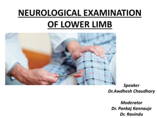
- 1. NEUROLOGICAL EXAMINATION OF LOWER LIMB Speaker Dr.Awdhesh Chaudhary Moderator Dr. Pankaj Kannauje Dr. Ravindu
- 3. ANATOMY OF LOWER LIMB • Lumbar Plexus: formed by the anterior rami (divisions) of L1, L2, L3 and L4. It also receives contributions from thoracic spinal nerve 12. • Cords then combine together to form the six major peripheral nerves: 1. Iliohypogastric Nerve (L1, T12) • Motor: Internal oblique and transversus abdominis. • Sensory: Posterolateral gluteal skin in the pubic region. -Moore’s Clinically Oriented Anatomy
- 4. 2. Ilioinguinal Nerve [L1] • Motor: Internal oblique and transversus abdominis. • Sensory: Skin on the upper middle thigh, over the root of the penis and anterior scrotum, the skin over mons pubis and labia majora. 3. Genitofemoral Nerve [L1, L2] • Motor: Cremasteric muscle. • Sensory: – The genital branch innervates the skin of the anterior scrotum or the skin over mons pubis and labia majora. – The femoral branch innervates the skin on the upper anterior thigh. -Moore’s Clinically Oriented Anatomy
- 5. 4. Lateral Cutaneous Nerve of the Thigh[L2,L3]: • Sensory: Anterior and lateral thigh down to the level of the knee.(no motor supply). 5. Obturator Nerve [L2, L3,L4]: • Motor: obturator externus, pectineus, adductor longus, adductor brevis, adductor magnus, gracilis. • Sensory: Skin over the medial thigh. 6. Femoral Nerve [L2,L3,L4] • Motor: Illiacus, pectineus, sartorius, all the muscles of quadriceps femoris. • Sensory: skin on the anterior thigh and the medial leg as a saphenous nerve. -Moore’s Clinically Oriented Anatomy
- 9. Sacral plexus: – formed by the anterior rami (divisions) of the S1, S2, S3 and S4. – Also receives contributions from L4 and L5. • Spinal roots and trunk divide into several cords. • Cords then combine together to form the five major peripheral nerves: 1. Superior gluteal nerve [L4, L5, S1] • Motor: gluteus minimus, gluteus medius and tensor fascia lata • Sensory: None -Moore’s Clinically Oriented Anatomy
- 10. 2. Inferior gluteal nerve[ L5, S1, S2] • Motor: Innervates gluteus maximus. • Sensory: None. 3. Sciatic Nerve [L4, L5, S1, S2, S3] • When the sciatic nerve reaches the apex of the popliteal fossa, it terminates bifurcating into – Tibial – Common fibular nerves.
- 11. • Motor: Tibial Portion – Innervates: – all of the muscles in the posterior compartment of the thigh, including the hamstring portion of adductor magnus, except the short head of the biceps femoris. • When it becomes the tibial nerve, it innervates: – All the muscles in the posterior compartment of the leg (superficial and deep) – all muscles in the sole of the foot as medial plantar nerve and lateral plantar nerve. -Moore’s Clinically Oriented Anatomy
- 12. • Motor: • Common Fibular Portion – Innervates the short head of the biceps femoris. – Then via its terminal branches, provide innervation to: Superficial fibular nerve: Innervates the muscles of the lateral compartment of the leg. Deep fibular nerve: Innervates the muscles of the anterior compartment of the leg and extensor digitorum brevis. -Moore’s Clinically Oriented Anatomy
- 13. Sensory Functions of Sciatic nerve: • Tibial Portion: – Innervates the skin of the postero-lateral and medial surfaces of the foot as well as the sole of the foot. • Common Fibular Portion: – Innervates the skin on the antero-lateral surface of the leg and the dorsal aspect of the foot. -Moore’s Clinically Oriented Anatomy
- 14. 4. Posterior Femoral Cutaneous Nerve: [S1, S2, S3} • Motor: None • Sensory: Skin on the posterior surface of the thigh and leg and skin of the perineum. 5. Pudendal Nerve: [S2, S3, S4] • Motor: – Skeletal muscles in the perineum, the external urethral sphincter, the external anal sphincter, levator ani. • Sensory: Penis and clitoris and most of the skin of the perineum. -Moore’s Clinically Oriented Anatomy
- 16. Action Muscle Nerve ,nerve root FLEXION Iliopsoas Lumbar plexus, femoral, L1,L2,L3 ADDUCTION Adductor longus /magnus/brevis Obturator , L2,L3,L4 ABDUCTION Gluteus medius & minimus Superior gluteal , L4,L5,S1 EXTENSION Guteus maximus Inferior gluteal, L5,S1,S2 EXTENSION Quadriceps femoris Femoral L2,L3,L4 FLEXION Hamstrings Sciatic L4,5,S1,S2 DORSIFLEXION Tibialis anterior Deep peroneal ,L4,L5. PLANTARFLEXION Gastrocnemius &soleus Tibial, L5,S1,S2 EVERSION Peroneus longus and brevis Superficial peroneal, L5,S1 INVERSION Tibialis posterior Tibial , L5,S1 EXTENSION OF BIG TOE Extensor hallucis longus Deep peroneal, L5,S1 EXTENSION OF OTHERS Extensor digitorum longus and brevis Deep peroneal,L5,S1 FLEXION Flexor digitorum longus & flexor hallucis longus Tibial L5,S1
- 17. MUSCLES OF LOWER LIMB(anterior &posterior)
- 19. INTRODUCTION TO THE PATIENT • Wash hands • Introduce yourself • Confirm patient details – Name/Age(DOB)/Sex • Explain the examination • Gain consent • Expose patient appropriately • Ask if the patient currently has any pain/discomfort
- 20. EXAMINATION OF MOTOR SYSTEM • BULK • TONE • POWER • LOOK FOR ANY ABNORMAL MOVEMENTS • EXAMINATION OF COORDINATION AND GAIT ARE CLOSELY RELATED TO MOTOR SYSTEM • Before doing neurological examination, it is essential to differentiate between neurological and non-neurological disease. DeJong’s Neurological Examination 7e
- 21. Motor Unit: Lowest echelon of motor activity. Alpha motor neuron in spinal cord or brainstem ,its axon, and all muscle fibres it innervates. DeJong’s Neurological Examination 7e
- 22. BULK • INSPECTION • PALPATION • MEASUREMENTS INSPECTION : Look down along axis symmetrically • General appearance – any limb deformity or posturing? • Scars • Wasting of muscles • Involuntary movements – dystonia/chorea/myoclonus • Fasciculation – lower motor neuron lesions • Tremor – Parkinson’s DeJong’s Neurological Examination 7e
- 23. PALPATION: Assess muscle bulk , contour and consistency. • Normal muscle: Semi-elastic and regains its shape at once when compressed. • Hypertrophy: Muscles are firm • Pseudohypertropy: Hard muscle, may feel doughy or rubbery. • Atrophy: Soft and pulpy in consistency . DeJong’s Neurological Examination 7e
- 24. MEASUREMENTS OF BULK : • Upper Limb: At 10 cm above and below olecranon process. • Lower Limb: At 18 cm above patella and 10 cm below tibial tuberosity. • Difference of 1 cm between limbs is normal. • Muscle atrophy (amyotrophy) causes decrease in muscle volume, usually accompanied by change in shape or contour. DeJong’s Neurological Examination 7e
- 25. • Disease affecting, anterior horn cell, nerve roots, peripheral nerve, muscle cause ATROPHY. • Neuromuscular junction disorders do not cause muscle atrophy. • Atrophy may be due to: – DISUSE OR INACTIVITY – IMMOBILISATION – TENDENOTOMY – MUSCLE ISCHEMIA – MALNUTRITION – ENDOCRINE DISORDER: Hyperthyroidism , steroid – NORMAL AGING DeJong’s Neurological Examination 7e
- 26. • NEUROGENIC ATROPHY: – Diseases affecting any part of LMN. – Weakness and wasting are comparable. • MYOGENIC ATROPHY : – Due to muscle disease. – Weakness is disproportionally greater than wasting . • When a muscle appear wasted but not weak the cause is likely to be non-neurogenic e.g. disuse or atrophy related to arthritis. DeJong’s Neurological Examination 7e
- 27. • HYPERTROPHY: – excessive use of muscles, or pathological. – Not necessarily stronger than normal muscle • PSEUDOHYPERTROPHY: – enlargement is due to infiltration of muscle with fat and connective tissue – no increase in muscle fiber or number – e.g. DMD. DeJong’s Neurological Examination 7e
- 28. GENERALISED • Malignancy • TB • Thyrotoxicosis • Advanced myopathy/MND PROXIMAL • Muscular dystrophy, Inflammatory Myopathy • Myasthenia gravis • MND, neuralgic amyotrophy • Neuronopathy/ plexopathy, radiculopathy DISTAL • AHC: polio, MND, syringomyelia, cord tumors • Anterior root: cervical spondylosis/ tumors • Plexus lesions/ individual nerve lesions Wasting Bickerstaff’s Neurological Examination, 6e
- 29. TONE • Tone: Tension in relaxed muscle or resistance to passive movement. • Ask the patient to keep their legs fully relaxed and “floppy” throughout your assessment. METHODS 1. Leg roll – • Roll the patient’s shin and watch the foot – it should flop independently of the leg- Normal or reduced tone. • In hypertonic state –ankle and foot move all `all one piece’ DeJong’s Neurological Examination 7e
- 30. 2. Leg lift – • Place both hands under knee , briskly flex joint with an upward movement of hand– the heel should remain in contact with the couch. • When spastic, the leg raises all in one. 3.Passsive flexion and extension of Hip, knee, ankle. 4.Flex the hip and raise the lower limb until it forms more than right angle at knee and allow it to fall. 5.With the flat of finger, flap calf muscle on each side DeJong’s Neurological Examination 7e
- 32. HYPOTONIA: • muscles are lax, is found in LMN lesions, cerebellar disorders and spinal shock. • Common causes of hypotonia : 1.Lesion of motor side of reflex arc: anterior horn cell disease, neuropathies, peripheral nerve injury. 2.Lesion of sensory side of reflex arc: neuropathies, tabes dorsalis, herpes zoster 3.Combined motor and sensory lesion: syringomyelia, cord or root compression, gross destruction of cord. 4.Lesions of muscle: myopathies, SMA, myasthenia gravis, periodic paralysis. 5.Cerebellar lesions: ipsilateral hypotonia with prolong and pendular reflex rather than loss. DeJong’s Neurological Examination 7e
- 33. TONE CORD Syringomyelia, cord/ root compression, neurologic ‘shock’ SENSORY Neuropathy, tabes dorsalis, herpes zoster CEREBELLAR (ipsilateral hypotonia) MOTOR Myopathy, neuropathy, AHC disease HYPOTONIA
- 34. HYPERTONIA SPASTIC TYPE: • Tone and resistance are greater in one group muscle than antagonist, resistance is most at start of movement then it suddenly overcome, producing clasp knife effect. • Signs of pyramidal pathway lesion. • It is velocity dependent(i.e. more noticeable with fast movements). • More evident in flexors and pronator of upper limb and extensors and adductors in lower limb.
- 35. RIGIDITY: • Seen in extrapyramidal lesions. • It is not velocity dependent. • Equal resistance in both agonist and antagonist at any point i.e. plastic or lead pipe rigidity • Antagonist and agonist contract alternately producing cogwheel rigidity • COGWHEEL RIGIDITY: Present only on first movement, feel on wrist . • PARATONIA(gegenhalten): – seen in diffuse frontal lobe disease – involuntary contraction of muscle when patient ask to relax and allow movements – Patient appear to actively opposing any movement at joint
- 36. SPASTIC • One group of muscles • Clasp-knife/ supinator catch • UMN- flexors UL, extensors LL • Velocity dependent PLASTIC/ RIGID • Both groups of muscles • Lead pipe/ cog wheel • Extra- pyramidal involvement PARATONIA • Resistance increases in proportion to passive movement • Gegenhalten • Diffuse frontal lobe disease HYPERTONIA
- 37. CLONUS • Sudden stretching of hypertonic muscle produces reflex contraction. • Sign of upper motor neuron lesions. Ankle clonus: • Position the patient’s leg so that the knee and ankle are slightly flexed, supporting the leg with your hand under their knee, so they can relax. • Rapidly dorsiflex and partially evert the foot. • Keep the foot in this position. • Clonus is felt as rhythmical beats of dorsiflexion/plantarflexion. (>5 is abnormal) Patellar clonus: • by sharply moving patella downward
- 38. POWER • MRC scale for muscle power • 0= No muscle contraction is visible • 1= A Flicker or trace of contraction. • 2= Active movement with gravity eliminated • 3=Active movement against gravity • 4-=Active movement against gravity and slight resistance(muscle can be broken by using 1 finger) • 4= Active movement against gravity and moderate resistance(muscle can be broken by using 3 finger) • 4+= Active movement against gravity and strong resistance(muscle can be broken by whole hand firm push) • 5 Full and normal power against resistance DeJong’s Neurological Examination 7e
- 39. • MRC scale is nonlinear. • Patient who has 4/5 does not have mild or equivocal weakness but has serious involvement. • Examiner patient mismatch should be recognized • By varying length of lever and the shortening of muscle permitted, examiner may give or take mechanical advantage as necessary to compensate for strength mismatch. • Some clinician use grade 5-, 3-, 2-. • 5- =borderline or equivocal weakness • 3- =muscle can move against gravity but not through full range • 2- = muscle can move with gravity eliminated but not through full range • Mayo clinic grades: more linear, five point scale, normal is zero, mild weakness is -1, and total paralysis is -4.
- 40. HIP • FLEXION :(L1-L3): • Supine: patient is supine , attempt to flex his thigh against resistance, knee is flexed and and leg rest on examiner arm. • Sitting position: ask to flex hip, normal hip flexor can not be overcome by examiner`s arm and hand unless examiner stand close and uses his body weight to defeat hip flexors.
- 41. • TEST FOR MILD WEAKNESS – Supine patient attempts to maintain both lower limb flexed at hip and extended at knee, the leg is at about 45 degree angle off the bed, feet apart. – Drifting of leg rapidly towards bed compared to other leg suggests corticospinal tract invovement (leg drift).
- 42. HIP EXTENSOR: L5-S2 • Patient lying prone: – raise the flexed knee up from the table against resistance (knee flexion minimizes any contribution from hamastring). • Patient lying on side and extending the hip • Patient seated: – trying to press raised knee as back down as examiner holds it up. DeJong’s Neurological Examination 7e
- 43. •Marked weakness of hip extensors suggests muscular dystrophy. •And Gower’s sign is positive. DeJong’s Neurological Examination 7e
- 44. HIP ADDUCTION :L5-S1 • Can be tested in supine sitting or lying down • Attempt to adduct leg in supine position against resistance with extended knee. DeJong’s Neurological Examination 7e
- 45. HIP ABDUCTION:L4-S1 • The supine patient attempts to move the extended leg outward against resistance, contraction of gluteus medius and tensor faciae latae can be patlpated. • Ask patient to push your leg out . DeJong’s Neurological Examination 7e
- 46. INTERNAL/MEDIAL ROTATION AT HIP : • Carried out by hip abductors(glutei medius and minimus and tensor facie lata) with some contribution from adductors. • Patient is supine with knee extended and patient is asked to rotate the foot medially as if to touch big toe to the bed. • Patient lying prone with leg flexed at knee, attempt to carry foot laterally against resistance. • With unilateral cortico-spinal tract lesion internal rotators are weak e.g. acute stroke. DeJong’s Neurological Examination 7e
- 47. EXTERNAL OR LATERAL ROTATOR OF THIGH • Muscles: – gluteus maximus (primarly), – obturator externous(Obturator nerve L3,L4) – obturator internus (L5,S1,nerve to obturator internus). • Piriformis externally rotates extended thigh but abduct the flexed thigh. • It is tested by maneuvers similar to those for testing internal rotation, but patient rotate hip externally by attempting to carry the foot medially against resistance with knee flexed. DeJong’s Neurological Examination 7e
- 48. KNEE Flexion (L4-S2) –hamstring (biceps femoris, semimembranosus, semitendinosus), tested in prone, supine, sitting. • Bend your knee and stop me from straightening it. • Prone patient attempt to maintain flexion of leg while examiner attempts to extend it. • Weak knee flexor suggest pyramidal tract lesion. DeJong’s Neurological Examination 7e
- 49. Extension (L2-4): Quadriceps femoris(powerful muscle). • Sitting or supine position ,“kick out your leg” • Quadriceps can be seen contacting or palpated • Take mechanical advantage by holding leg near ankle. DeJong’s Neurological Examination 7e
- 50. BARKEEPER’S HOLD
- 51. Ankle Dorsiflexion (L4,L5) • “keep your legs flat on the bed, push your foot up towards your face, don’t let me push it down “ • Powerful muscle, subtle weakness can some time be detected by placing patient at maximum mechanical disadvantage . • Ask patient to “Walk on heel” Plantarflexion (L5/S1/2) – “push down like on a pedal” • Plantar flexor are among powerful muscle of body • Ask patient “to stand on tip toes”. Inversion (L5/S1) – “push your foot in against my hand” Eversion (L5/S1) – “push your foot out against my hand” DeJong’s Neurological Examination 7e
- 54. Big toe • Extension (L5) – “don’t let me push your big toe down” • Flexion: “don’t let me push your big toe up”
- 55. REFLEX AND REFLEX ARC • DTR • SUPERFICIAL REFLEX
- 56. REFLEXES EXAGGERATED/ BRISK • UMN • Increase in reflexogenic zone • Spread of reflex to nearby muscles • Crossed extension REDUCED • LMN • Initial Spinal ‘ shock’ • Extreme rigidity/ spasticity/ contracture CLONUS • Ill sustained • Well sustained NORMAL
- 57. REFLEX Deep tendon reflexes(DTR) • Ensure the patient’s lower limb is completely relaxed. • Hold at the end of the tendon hammer handle and allow gravity to aid a good swing. • If a reflex appears absent: Make sure the patient is fully relaxed and then perform a reinforcement manoeuvre. • Ask the patient to hook their hands together in a monkey grip and try to pull them apart, while you hit the tendon. -Bickerstaff’s Neurological Examination, 6e
- 58. GRADING OF REFLEX • By convention the deep tendon reflexes are graded as follows: • 0 = no response; always abnormal • 1+ = a slight but definitely present response; may or may not be normal • 2+ = a brisk response; normal • 3+ = a very brisk response; may or may not be normal • 4+ = a tap elicits a repeating reflex (clonus); always abnormal. -Bickerstaff’s Neurological Examination, 6e
- 59. KNEE JERK (L3,L4),FEMORAL NERVE • Hold knee with one palm and then stuck tendon lightly ,watch the movement of lower leg and quadriceps • Absence of patellar reflex is known as Westphal’s sign. • Inverted patellar reflex: Lesion of nerve supplying quadriceps, cord lesion at L3, L4. -Bickerstaff’s Neurological Examination, 6e
- 60. ANKLE JERK:(S1),MEDIAL POPLITEAL • Patient’s leg should be externally rotated and slightly flexed at knee, dorsiflex the foot with other hand. • For left leg, move to other side of the bed. • Observe for movement of foot and contraction of calf. • Also called as Achilles or triceps surae reflex. -Bickerstaff’s Neurological Examination, 6e
- 61. PLANTAR REFLEX(S1): • Position the patient such that knee is slightly flexed and thigh is externally rotated, outer aspect of foot resting on couch, explain that sole will be scratched and ask him to try to let foot remain loose. • Outer aspect of foot is firmly stroked with blunt point such as key or end of hammer. • Stimulator should move forward and then curved inward toward middle metatarso-phalangeal joint . • Observe the great toe. • Normal result = Flexion of the great toe and flexion of the other toes • Abnormal (Babinski sign) = Extension of the great toe and spread of the other toes – upper motor neuron lesion -Bickerstaff’s Neurological Examination, 6e
- 62. • True- Extension of big toe (MTP and IP), fanning of other toes • Minimal- contraction of hamstrings and tensor faciae latae • Crossed extensor- bilateral response (bilateral cerebral/ cord disease) BABINSKI REFLEX
- 63. BABINSKI REFLEX Fallacies: • Pes cavus/ deformity of great toe. • Normal extensor response in children up to 12 months of age. • Identify anxiety, painful stimulus causing a withdrawal. • Callosities of sole/ S1 neuropathy • Post ictal / deep anaesthesia/ deep sleep/ coma/ electroconvulsive therapy • Hypoglycemia/ alcohol intoxication/ narcosis -Bickerstaff’s Neurological Examination, 6e
- 66. Superficial reflex of lower extremities Cremastric reflex :(L1-L2): • elicted by stroking or lightly scratching or pinching the skin of upper inner aspect of thigh • response: contraction of cremastric muscle with elevation homolateral testicle Anal reflex :(S2-S5): • contraction of external anal sphincter in response to stroking or piercing the skin or mucous membrane in perianal region Bulbocavernous reflex: • related to anal reflex both causes contraction of external anal sphincter but in BCR ,stimulus is delivered to glans penis or clitoris • Response is best palpated with gloved finger in the rectum • Useful for assessing integrity of cauda equina , lower sacral root and conus medullaris. DeJong’s Neurological Examination 7e
- 67. SENSORY SYSTEM Modality of sensation to be tested : • 1.pain, light touch, temperature: extoceptive sensation • 2. position sense, passive movement, vibration and deep pain:proprioceptive • 3.stereognosis, graphaesthesia and two point discrimination. • 4. Visceral or interoceptive. -Bickerstaff’s Neurological Examination, 6e
- 70. Dermatome
- 72. Pin-prick sensation: • Assesses spinothalamic tracts. • Use sharp pin with rounded head, with the shaft of pin long enough to allow the examiner`s index finger and thumb to slide downward on impact. • Always start from impaired sensation to normal sensation. • If sensation is reduced peripherally, assess from a distal point and move proximally to identify ‘stocking’ sensory loss (peripheral neuropathy). • If necessary, keep going all the way up the leg and truck until normal sensation is felt. This may reveal a “sensory level”, which is suggestive of a spinal lesion -Bickerstaff’s Neurological Examination, 6e
- 73. Light touch sensation: • Assesses dorsal/posterior columns and spinothalamic tracts. • The patient’s eyes should be closed for this assessment. • Touch the patient’s sternum with the cotton wool wisp to confirm they can feel it. • Ask the patient to say “yes” when they are touched. • Using a wisp of cotton wool, gently touch the skin (don’t stroke) • Assess each of the dermatomes. • Compare left to right, by asking the patient if it feels the same on both sides
- 74. Vibration sensation • Assesses dorsal / posterior columns 1. Ask patient to close their eyes. 2. Tap a 128 Hz tuning fork. 3. Place onto patient’s sternum/clavicle and confirm patient can feel it buzzing. 4. Ask patient to tell you when they can feel it on their foot and to tell you when it stops buzzing. 5. Place onto the distal phalanx of the great toe. 6. If sensation is impaired, continue to assess more proximally – e.g. proximal phalanx.
- 75. Proprioception : • Dorsal / posterior columns • Hold the distal phalanx of the great toe by its sides • Demonstrate movement of the toe “upwards” and “downwards” to the patient (while they watch) • Then ask patient to close their eyes and tell you if you are moving the toe up or down. • If the patient is unable to correctly identify direction of movement, move to a more proximal joint. • (big toe > ankle > knee > hip)
- 76. Sterognosis • Ability of recognizing object purely from feel of its shape and size. • Test object must be familiar and easily identifiable. • It is defective if touch and position sense are defective. Graphaesthesia • Ability to recognize letter or number written on skin with a blunt point. • If peripheral sensation is lost it will also lost but if it is absent in intact peripheral nerve it suggest parietal cortical lesion . -Bickerstaff’s Neurological Examination, 6e
- 77. Two point discrimination: • Ability to detect that a stimulus consist of two blunt point when they are simultaneously applied. • Normal ability to distinguish the two points from one varies in different body parts, on finger it should be possible to differentiate less than 5 mm ,but on dorsum of foot separation of 5 cm may be required. • Depends on integrity of light touch: When light touch is normal, impaired two point discrimination suggests parietal lesion. -Bickerstaff’s Neurological Examination, 6e
- 78. • Co-ordination: requires both motor and sensory intact • Purpose of test : 1. cerebellar dysfunction, 2. proprioceptive defeciency • Heel to shin test –“put your heel on your knee, run it down your shin, lift it up and repeat” • An inability to perform this test may suggest loss of motor strength, proprioception or a cerebellar disorder. -Bickerstaff’s Neurological Examination, 6e
- 81. • Gait • • Abnormal gaits: • · Hemiplegia: the foot is plantar flexed and the leg is swung in a lateral arc • · Spastic paraparesis: scissors gait • · Parkinson's: starting hesitation, shuffling, freezing, festination • pro/retropulsion • · Cerebellar: drunken wide-based or reeling on a narrow base gait; staggers • towards side of cerebellar lesion • · Posterior column lesion: clumsy slapping down of the feet on a broad base • · Footdrop: high stepping gait • · Proximal myopathy: waddling gait • · Prefrontal lobe (apraxic): feet appear glued to floor when erect, but move • more easily when the patient is supine • · Hysterical: characterised by a bizarre, inconsistent gait
- 82. MUSCLE OF ANTERIOR AND POSTERIOR COMPARTEMENT OF LEG
- 83. • Romberg’s test • Ask the patient to stand with their feet together and eyes closed • Observe the patient (ideally for 1 minute) • Positive test – loss of balance (swaying without correction/falling over) – this suggests a sensory ataxia (proprioceptive deficit) • It’s important to stand close by the patient during this test to stop them falling over!
- 84. • Gait • 1. Ask patient to walk to the end of the • room & back – assess speed, symmetry • ,balance and arm swing • 2. Tandem (heel-to-toe) gait – ask to walk • in a straight line heel-to-toe – an • abnormal heel-to-toe test may suggest • weakness, impaired proprioception or a • cerebellar disorder • 3. stand on heel then on toes walking: • testing distal power. toes (L4/5) & heels • (S1)
- 85. Question :right side body movement is controlled by ? • 1.left cerebral hemisphere only • 2.left cerebral hemisphere and left cerebellar hemisphere • 3.right cerebral and left cerebellar hemisphere • 4.left cerebral and right cerebellar hemisphere
Editor's Notes
- Modified Ashworth grading of spasticity 0 no increase 1 catch/slight increase 1+ catch+ increase 2 more marked increase in tone 3 considerable increase 4 part rigid in flexion
- Pseudo-Babinski-choreoathetosis Inversion of plantar reflex- paralysis of toe flexors