
Recommended
Recommended
More Related Content
What's hot
What's hot (20)
Similar to IntraCranial Aneurysm by Dr. Avinash
Similar to IntraCranial Aneurysm by Dr. Avinash (20)
More from AvinashDahatre
Recently uploaded
Recently uploaded (20)
IntraCranial Aneurysm by Dr. Avinash
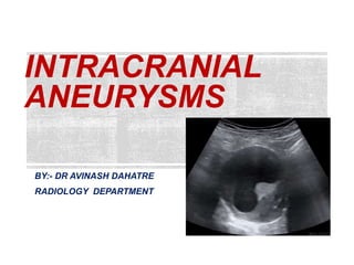
- 1. INTRACRANIAL ANEURYSMS BY:- DR AVINASH DAHATRE RADIOLOGY DEPARTMENT
- 5. SACCULAR ANEURYSM Rounded berry like Arise from arterial bifurcation Dilatation of vascular lumen due to weakness in vessel wall. Absent or reduced internal elastic membrane. T. adventitia may be infilterated by Lymphocyte and phagocyte.
- 7. CAUSES:- 1. Developmental/ degenerative aneurysm 2. Traumatic aneurysm 3. Mycotic aneurysm 4. Oncotic aneurysm 5. Flow related aneurysm
- 8. DEVELOPMENTAL/ DEGENERATIVE ANEURYSM In past few yrs intracranial aneurysm thought to be congenital origin occuring due to focal defect in T. media Most common intracranial aneurysm occurs as hemodynamically induced vascular origin. Abnormal hemodynamic shear stress on wall of larger cerebral arteries particularly at bifurcation point. Change in flow direction occurring during systole and diastole cause damage to T. intima.
- 9. Less common causes are:- TRAUMA INFECTION TUMOR DRUG ABUSE HIGH FLOW STATES
- 10. INCIDENCE:- Incidence is unknown. In patient undergoing coronary angio incidental Intracranial aneurysm were found in 5.6 % in cases. 1% of patient undergoing four vessel cerebral angio for indications other than SAH. Familial incidence have been reported.
- 11. Increase in incidence of ICA seen in Anamolous vessel Arterial coarctation Fibromuscular dysplasia MARFANs syndrome Ehlers danlos synd. High flow states (Fistula, vascular malformations)
- 12. 40-60yrs. In children they are post traumatic or mycotic. Aneurysm in children are larger than adult.(>17mm) LOCATION:- At bifurcation of larger arteries. Most common MCA bifurcation. 90% of aneurysm arise in anterior circulation. 30-35% occurs in A.com and P.com 20% in MCA
- 13. GRADE I GRADE II GRADE III GRADE IV GRADE V Asymptomatic patient or have mild headache Moderate to severe headache with 3rd nerve palsy Confusion and drowsy Hemiparesis Comatose or moribound.
- 14. B. T1WI MRI Aneurysm appears as iso to hyperdense mass with loss of signal intensity. C. DSA image Aneurysm in internal carotid artery near ophthalmic division A.NCCT axial section of brain showing well delineated mass located in suprasellar cistern.
- 15. Fig. Coronal section post contrast T1WI of brain showing partially thrombosed vessel with intensly enhanced lumen and outer wall enhancement.
- 16. Fig. non-contrast CT image showing slightly hyperdense, well- defined round extra-axial masses may demonstrate a peripheral calcified rim
- 17. TRAUMATIC ANEURYSM PENETRATING TRAUMA Due to high velocity missile head wound. Meningeal vessels are common site. Post traumatic aneurysm may be over looked on Ct because of the lesion is often obscured by hemorrhage. NON PENETRATING TRAUMA Due to skull trauma or skull fracture Frontolateral impact produces shearing force between inferior margins of falx cerebri and distal anterior cerebral artery and juxta falcine hematoma is seen.
- 18. PENETRATING TRAUMA ANEURYSM Fig. A 40-year-old pedestrian man was involved in a motor vehicle accident, his initial Glasgow Coma Scale (GCS) was 3. On day one, cerebral angiography revealed a right internal carotid artery paraophthalmic aneurysm measuring 4mm x 4 mm.
- 19. Fig. Axial NCCT showing hyperdense subdural hemorrhage along left side of anterior falx cerebri NON PENETRATING TRAUMA
- 20. MYCOTIC ANEURYSM mycotic aneurysm is a dilation of an artery due to damage of the vessel wall by an infection. “mycotic” referring to fungal is a misnomer as various organisms including predominantly bacterial can cause the aneurysm. most common organisms are:- Staphylococcus aureus Salmonella spp. There is disruption of muscular layer and adventitia of vessel wall causing aneurysmal dilatation. Thoracic aorta is most common site and intra cranial is less common.
- 21. MYCOTIC ABDOMINAL AORTIC ANEURYSM Contrast CT Abdomen Axial and Coronal section showing Mycotic aneurysm.
- 22. ONCOTIC ANEURYSM Neoplastic aneurysm result from direct vascular invasion by tumor or implant. Forms pseudoaneurysm. Eg. Squamous cell carcinoma and severe epistaxis.
- 23. Fig.3D MRA showing pseudoaneurysms of Internal carotid artery.
- 24. FLOW RELATED ANEURYSM Occur along proximal and distal feeding vessels. Proximal lesion- arises in circle of willis related to increased hemodynamic stress. Distal flow related aneurysm located within AVM nidus.
- 25. Fig. Digital substraction angiogram showing abnormal blood vessels (nidus)
- 26. 1. SLE 2. TAKAYASU ARTERITIS 3. FIBROMUSCULAR DYSPLASIA 4. DRUG ABUSE –COCAINE -HEROINE -Methamphetamine
- 27. Fig. T2WI Subcortical and periventricular hyperintense Fig. FLAIR axial image showing periventricular hyperintensities. Autoimmune disease causing small vessel dementia a/k/a Binswanger disease.
- 28. granulomatous large vessel vasculitis that predominantly affects the aorta and its major branches. USG showing smooth, homogeneous and moderately echogenic circumferential thickening of the arterial wall. this finding is termed as the 'macaroni sign' and is highly specific for Takayasu arteritis.
- 29. Contrast CT image showing aortic wall thickness of takayasu arteritis.
- 30. characterized by an idiopathic, non-inflammatory, and non-atherosclerotic angiopathy of small and medium- sized arteries. Most commonly seen in renal arteries then ICA and vertebral arteries. Fig. CECT axial image showing alternating stenoses and dilatations, causing a string of beads Appearence
- 31. FUSIFORM ANEURYSM Occurs due to damage to tunica media layer. Vertebrobasilar system is commonly affected. Intraluminal clots are more common. Produces brain stem infract and compresses adjacent brain. Causes cranial nerve palsies.
- 32. Fig. CT and MRI revealed bilateral giant fusiform aneurysms of the petrous portion of the internal carotid artery
- 33. Fig. DSA showing fusiform dilatation of petrous part of ICA
- 34. DISSECTING ANEURYSM Blood accumulates in the vessel wall through a tear in the Intima and internal elastic lamina. If intra luminal hematoma extend to sub adventitial plane sac like out pouching is formed called as false saccular aneurysm or dissecting aneurysm.
- 35. Fig. CT AXIAL section showing aneurysmal dilatation of the ascending aorta.
- 37. INTRACRANIAL ANEURYSMS SACCULAR Dilatation of vascular lumen due to weakness in vessel wall. Absent or reduced internal elastic membrane. FUSIFORM Occurs due to damage to tunica media layer. DISSECTING Blood accumulates in the vessel wall through a tear in the Intima and internal elastic lamina. SUMMARY POINTS:-
- 38. Osborn diagnostic neuroradiology.