2. Bacterial
Morphology
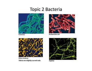
• Morphology:
– Spherical
=
Coccus
– Rod
shaped
=
Bacillus
– Comma
Shaped
=
Vibrio
– Spiral
=
Spirillium
–
Varied
shape
=
Pleiomorphic
• Generally
not
a
good
predictor
of
physiology,
ecology,
or
phylogeny
• Morphology
may
be
determined
by
selecBve
forces
– nutrient
uptake
efficiency
(surface-‐to-‐volume
raBo)
– spirals
allow
efficient
swimming
in
viscous
or
turbulent
fluids
(i.e.
near
surfaces)
– gliding
moBlity
(filaments)
• Bacteria
can
also
assume
mulBcellular
organizaBons
– hyphae
(branching
filaments
of
cells)
– mycelia
(tuNs
of
hyphae)
– trichomes
(smooth,
unbranched
chains
of
cells)
3. Cell
Sizes
• Prokaryotes
are
0.2
μm
to
>
700
μm
in
diameter
– most
rod-‐shaped
bacteria
between
0.5
μm-‐4.0
μm
wide
and
1-‐15
μm
long
– very
few
“large” prokaryotes
– ExcepBons:
• Thiomargarita namibiensis: up to 700 μm in
diameter"
• Epulopiscium fishelsoni: 200‒700 μm x 80
μm!
• EukaryoBc
cells
range
from
10
μm
to
>200
μm
• Minimum
size
simply
due
to
minimum
space
requirements
for
genome,
proteins,
ribosomes
– Diameters
<
0.15
μm
unlikely
– “Very
small”
cells
common
in
open
marine
environments
•
Advantages
to
being
small:
•
Higher
surface-‐to-‐volume
raBo
• greater
rate
of
nutrient/waste
exchange
per
unit
volume
• supports
higher
metabolic
rate
• supports
faster
growth
rate,
faster
evoluBon
4. What
Is
in
the
Cytoplasm
• inclusion
bodies
may
also
be
present
sulfur
globules:
sulfur
storage
for
energy
polyhydroxybutyrate
granules:
carbon
storage
gas
vesicles:
buoyancy
control
carboxysomes:
locaBon
of
carbon
fixaBon
reacBons
(RUBISCO)
magnetosomes:
organelle
associated
with
direcBon
finding
5. How
does
DNA
compress
within
the
nucleoid
of
bacteria?
• several
mechanisms
to
reduce
space
– use
of
caBons
(Mg2+,
K+,
Na+)
to
shield
negaBve
charges
on
sugar-‐phosphate
(PO4-‐)
backbone
– small,
posiBvely
charged
proteins
bind
to
the
chromosome
to
maintain
condensed
structure
– topoisomerases
modify
structure
of
DNA
to
enable
“supercoiling”
• No
membrane
surrounds
the
nucleoid
• No
histone
proteins
(like
those
found
in
Archaea
and
Eukaryotes)
6. Cytoskeleton
Proteins
• FtsZ
–
Forms
Z
ring,
is
used
to
divide.
–
If
you
didn't
have
this,
you
would
become
a
very
long
cell
with
no
mechanism
to
divide.
– Rips
apart
the
cell
wall
and
then
glues
it
back
together,
facilitates
cell
division.
– HOMOLOG
TO
TUBULIN.
• MreB
-‐
governs
the
shape
of
bacterial
cell.
– If
you
are
lacking
MreB
at
all,
you
will
be
cocci
shaped.
– If
you
do
have
MreB
then
you
will
polymerize
MreB
protein
that
acts
like
a
spring
that
will
support
the
shape
of
the
bacteria.
– HOMOLOG
TO
ACTIN.
• ParM
-‐
polymerize
(need
ATP)
to
push
the
plasmids
and
chromosomes
to
either
side
so
the
cell
can
divide.
– Alaches
to
ParR
7. Cell
Envelope
• All layers surrounding the cytoplasm of cells, which includes:"
– Cell membrane (plasma membrane):"
• Bilayer composed of a phospholipid bilayer (glycerol w/ fatty acids attatched with
ESTER linkages) with embedded proteins and hopinoids"
• Separates internal from external enviro (fluid mosaic model)"
• Capturing energy"
– electron transport chains create proton motive force (PMF)"
– can be used for respiration/photosynthesis "
– can be used to derive energy for motion (flagella)"
• Holding sensory systems (Chemotaxis)"
– embedded proteins can detect environment changes, alter gene expression in response"
– Cell wall"
• gives cells their shape. Without
it,
cell
can’t
resist
osmoBc
pressure
changes"
• protects from osmotic lysis/mechanical forces"
• a matrix of crosslinked strands of peptidoglycan subunits"
• Composed of Peptidoglycan subunits of NAG and NAM"
– Crosslinked by Petptide Crosslink or Peptidoglycine Interbridge "
– Outer membrane (if present)
8. How
do
items
cross
the
plasma
membrane?
• O2
and
CO2
are
small
and
can
diffuse
across
readily
• H2O
is
helped
across
by
aquaporin
protein
channels
(osmosis)
• Facilitated
diffusion
and
co-‐transport:
– protein
channel
moves
parBcles
WITH
a
concentraBon
gradient
– Co-‐transport
can
be
sym
(molecules
going
to
the
same
side)
or
anB
(molecules
going
to
opposite
sides)
– no
energy
• AcBve
transport
– protein
transporter
moves
parBcles
AGAINST
a
concentraBon
gradient
– requires
energy
– Includes
protein
secreBon
=
shipping
proteins
outside
the
cell
10. Breaking
the
Cell
Wall
• Lysozyme
cleaves
backbone
and
lysostaphin
cleaves
pepBdogylcine
interbridge
• β-‐lactam
anBbioBcs
– prevent
pepBdoglycan
crosslinking
• Ex
penicillin
– Inhibits FtsI transpeptidation
• AnBbioBc
Resistance
– Some bacteria can produce an enzyme to destroy the critical β-lactam ring
structure"
– second drug must be added to inhibit the enzyme"
11. Two
Types
of
Cell
Walls
• Gram
PosiBve
– thick
outer
layer
of
pepBdoglycan
– narrow
periplasmic
space
– negaBvely
charged
teichoic
acids
in
the
pepBdoglycan
• Gram
NegaBve
– very
thin
layer
of
pepBdoglycan
– periplasmic
space
of
varying
width
– outer
membrane
composed
of
lipopolysaccharide
(LPS)
• Composed
of
lipid
A
core
polysacharide
varying
O
chain
12. How
do
nutrients
get
through
the
cell
wall?
• Gram-‐posiBve
pepBdoglycan
layer
has
large
pores
throughout
its
matrix
• Gram-‐negaBve
cell
has
porin
and
TonB
proteins
in
its
outer
membrane
– transfer
molecules
into
the
periplasmic
space
– How
can
molecules
get
out
of
a
Gram-‐negaBve
cell’s
periplasmic
space?
• some
move
from
the
periplasm
to
outside
directly
(these
are
known
as
autotransporters
and
are
rare
• some
use
single-‐step
(never
entering
the
periplasm)
transport
systems
13. Cell
Movement
• Flagella
(Fillament-‐Hook-‐Basal
Body):
– MONOTRICHOUS
=
One
flagella
– AMPHITRICHOUS
=
Two
flagella
– LOPHOTRICHOUS
=
mulBple
but
polarized
– PERITRICHOUS
=
mulBple
from
all
ends
• Nonflagellar
MoBlity
– Gliding
moBlity
• smooth
sliding
over
a
surface,
not
well
understood
• e.g.
Myxobacteria,
Cyanobacteria
– Twitching
moBlity
• slow,
jerky
process
using
pili
that
extend,
alach
to,
and
pull
along
a
surface
14. Adherence Molecules
• allow
cells
to
sBck
to
surfaces
• pili
(s.
pilus),
fibers
of
pilin
protein,
possess
other
proteins
on
their
Bps
for
sBcking
– Ones
for
adherence
are
called
Fimbriae
• Some
microbes
will
use
an
extension
of
the
cell
envelope
Bpped
by
a
“holdfast”
of
polysaccharides
– Called
a
Stalk
– Provide
extra
surface
area
for
nutrient
absorpBon
15. Capsules
and
S-‐Layers
• Capsules:
– Thick
layer
of
polysaccharides
surrounding
some
cells
– provide
adhesion,
defense
against
host
immunity,
protecBon
against
desiccaBon
(biofilms)
• Surface
Arrays
– crystalline
array
of
interlocking
proteins
– can
protect
a
cell
against
predaBon
or
infecBon
with
bacteriophages
– found
in
both
Gram-‐posiBve
and
Gram-‐negaBve
cells
16. Bacterial
Taxonomy
• Are
named
by
Species
and
Genus
• ClassificaBon
depends
on
many
features:
– DNA
sequence
data
– size/shape
– Gram
type
– colony
morphology
– presence
of
structures
such
as
capsules/endospores
– physiologic/metabolic
traits
• Once
classified,
they
are
put
into
the
database
of
the
World
Federa?on
for
Culture
Collec?ons
– Become
a
“Type
strain”
is
a
referenced
specimen
deposited
in
a
culture
repository.
But
MOST
can
not
be
cultured!!!