
Recommended
Recommended
More Related Content
What's hot
What's hot (20)
Similar to Niosomes
Similar to Niosomes (20)
Recently uploaded
Recently uploaded (20)
Niosomes
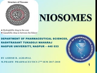
- 1. DEPARTMENT OF PHARMACEUTICAL SCIENCES, RASHTRASANT TUKADOJI MAHARAJ NAGPUR UNIVERSITY, NAGPUR – 440 033 BY ASHISH R. AGRAWAL M.PHARM PHARMACEUTICS 2ND SEM 2017-2018 1
- 2. CONTENTSIntroduction General characteristics of Niosomes Structure of niosomes Advantages & disadvantages of niosomes Niosomes vs. Liposomes Factors affecting the formation of Niosomes Types of Niosomes Materials used in the preparation of niosomes Methods of preparation Characterization In-vivo behavior of Niosomes Applications Marketed products Future prospects and Recent advances in niosomes Conclusion References 2
- 3. 3
- 4. INTRODUCTION • Development of new drug, improving safety and efficacy of existing drugs is difficult, expensive and time consuming. • At present, no available drug delivery system behaves ideally achieving all this goals. • Encapsulation of the drug in vesicular structures is one of the promising system such as liposomes, niosomes, transferosomes, ethosomes etc. • It delivers drug directly to the site of action, leading to reduction of drug toxicity with no adverse effects. • Vesicular drug delivery reduces the cost of therapy by improving bioavailability of medication and also solves the problems of drug insolubility, instability and rapid degradation. 4
- 5. • Niosomes are a novel drug delivery system, in which the medication is encapsulated in a vesicle, composed of a bilayer of non-ionic surface active agents and hence the name niosomes. • The niosomes are very small, and microscopic in size. • Their size lies in the nanometric scale. Definition • Niosomes are synthetic microscopic vesicles consisting of an aqueous core enclosed in a bi layer consisting of cholesterol and one or more nonionic surfactants or Niosomes are essentially non-ionic surfactant based multilamellar or unilamellar vesicles in which an aqueous solution of solute (s) is entirely enclosed by a membrane resulted from the organization of surfactant macromolecules as bilayers. • Niosomes made-up of self assembly of hydrated nonionic surfactant molecules (eg: tweens and spans) with or with out cholesterol and dicetyl phosphate. 5
- 6. • The assembly into closed bilayers is rarely spontaneous and usually involves some input of energy such as physical agitation or heat. • Niosomes also called as Nonionic surfactant vesicles or NSVS. Nios = non ionic surfactant somes = vesicles • NSVS results from the self assembly of hydrated surfactant monomers results in closed bilayer structures. • Most surface active agents when immersed in water yield micellar structures, how ever some surfactants can yield bilayer vesicles which are niosomes. 6
- 7. GENERAL CHARACTERISTICS OF NIOSOME • Biocompatible, biodegradable, non-toxic, non immunogenic and non-carcinogenic. • The ability of nonionic surfactant to form bilayer vesicles is dependent on the HLB value of the surfactant, the chemical structure of the components and the critical packing parameter. • Niosomes can be characterized by their size distribution studies • High resistance to hydrolytic degradation. • The properties of niosome depends both on composition of the bilayer & on method of their production. 7
- 8. Hydrophilic drug localized in aqueous region Polar heads facing hydrophilic region Hydrophobi c drugs localized in hydrophobic lamella Non-polar tails facing each other to form a hydrophobic region STRUCTURE OF NIOSOMES 8
- 10. ADVANTAGES OF NIOSOMES • Have long storage time compared to liposomes. • Access to raw materials is convenient. • Their surface formation and modification is very easy because of the functional groups on their hydrophilic heads. • Have high compatibility with biological systems. • Low toxicity because of their non-ionic nature. • They are biodegradable and non-immunogenic. • They can entrap lipophilic drugs into vesicular bilayer membranes and hydrophilic drugs in aqueous compartments. • Since the structure of the niosome offers place to accommodate hydrophilic, lipophilic as well as amphiphilic drug moieties, they can be used for a variety of drugs. • They can increase the oral bioavailability of drugs. • They can improve the therapeutic performance of the drug by protecting it from biological environment, resulting in better availability and controlled drug delivery. • Niosomes exhibits flexibility in their structural characteristics (composition, fluidity and size) and can be designed according to the desired situation. • Niosomes can act as a depot to release the drug slowly and offer a controlled release. • They are osmotically active and stable. • They increase the stability of the entrapped drug. • They can enhance the skin penetration of drugs 10
- 11. 11
- 12. DISADVANTAGES OF NIOSOME • Increase in the polyoxyethylene chain length enhances cytotoxicity (Hofland et al., 1992). • Preparation methods are time-consuming. • May require specialized equipment. • High Production cost. • Inefficient drug loading. • Physical and chemical instability. • Aggregation. • Fusion of vesicles. • Leakage of drug. • Aqueous suspension of niosome may exhibit fusion, aggregation leaching or hydrolysis of entrapped drug, thus limiting the shelf life of niosome dispersion. 12
- 13. LIPOSOMES VS. NIOSOMES Sr. No. Liposomes Niosomes 1. Vesicles made up of concentric bilayer of phospholipids. Vesicles made up of surfactants with or without incorporation of cholesterol. 2. Size ranges from 10-3000nm Size ranges from 10-100nm 3. Comparatively expensive Less expensive 4. Special storage condition are required No such special requirement 5. Phospholipids used are unstable Non-ionic surfactants are stable 6. Comparatively more toxic Less toxic 7 Made of neutral or charged double chain phospholipids. Made of uncharged single chain surfactant molecules 13
- 14. FACTORS AFFECTING THE FORMATION OF NIOSOMES • Several factors are involved in the formulation of niosomes. • These factors can depend on method of preparation, route of administration, and material consumption. 1. AMOUNT and TYPE OF SURFACTANTS 2. THERMODYNAMIC FEATURE 3. HYDROPHILIC–LIPOPHILIC BALANCE (HLB) 4. GEOMETRIC FEATURES OF AMPHIPHILIC MOLECULE 5. TEMPERATURE OF HYDRATION 6. ADDITIVE AGENTS 7. THE NATURE OF THE DRUG 8. METHOD OF PREPARATION 9. RESISTANCE TO OSMOTIC STRESS 14
- 15. 1. AMOUNT AND TYPE OF SURFACTANT • As the HLB value of surfactants like span 85 (HLB 1.8) to span 20 (HLB 8.6) increases, the mean size of niosomes also increases proportionally. • It is due to the fact that surface free energy decreases with increase in hydrophilicity of surfactant. • Alkyl chain is present in well ordered structure in gel state, while in the liquid state the structure of bilayer is more disordered. • Entrapment efficiency is also affected by phase transition temperature i.e. span 60 having higher TC, provide better entrapment efficiency. • Entrapment efficiency of the niosomes is affected by the HLB value for e.g. niosomes have high entrapment efficiency at HLB value 8.6 but HLB value 14 to 17 is not suitable for niosomes formulation 2. THERMODYNAMIC EFFECT • Thermodynamically stable niosomes are created only when a certain amount of surfactants and additives are mixed together. • The energy required for the formation of niosomes occurs through interaction energies (mechanical energy, chemical potential excess energy, and surface energy). 15
- 16. • The aggregation of nonionic surfactant monomers and the formation of niosomes is the result of two opposing forces: i. A high interfacial tension between lipophilic groups and water that causes these groups to associate, and ii. Repulsive forces between head groups (the steric and hydrophilic repulsion) (Uchegbu and Florence, 1995). • Niosomes in the range of 1–10 nm have been found to be more stable than those in the submicron size. • Thermodynamically, bigger niosomes have lower surface free energy and tend to segregate more than smaller ones in order to increase the excess free energy (Moazeni et al., 2010). 3. HYDROPHILIC–LIPOPHILIC BALANCE (HLB) • HLB is a dimensionless critical parameter in the selection of surfactant for the formation of niosomes. • This parameter affects the size and amount of drug loading. • Surfactants with a HLB number between 4 and 8 are compatible to vesicle formation. • Surfactants with HLB values higher than 6 are barely able to form vesicles; thus, for the formation of niosomes, cholesterol must be added (Yoshida et al., 1992). 16
- 17. • An important factor in determining the type of association formed in aqueous environments is geometric features. • The morphologies of the aggregate formed may be predicted by nominal geometric parameters of the surfactant molecule. 4. GEOMETRIC FEATURES OF AMPHIPHILIC MOLECULE (OR STRUCTURE OF SURFACTANT) • An important factor in determining the type of association formed in aqueous environments is geometric features. • The morphologies of the aggregate formed may be predicted by nominal geometric parameters of the surfactant molecule. • Concept of Critical Packing Parameter ( Israelachvili , 1985) • Prediction of vesicle forming ability is not a simply a matter of HLB CPP (Critical Packing Parameter )= 𝐯 𝐥 𝐜 𝐚 𝟎 where v - hydrophobic group volume, 𝒍 𝒄 - critical hydrophobic group length 𝒂 𝟎 - area of the hydrophilic head group 17
- 18. CPP between (0.5 ≤ CPP ≤ 1) likely to form bilayer vesicles. Spherical micelles (CPP ≤ 0.33) to cylindrical micelles (1/3 ≤ CPP ≤ 0.5), (indicating a large contribution from the hydrophilic head group area. Inverse micelles (CPP > 1) (indicating a large contribution from the hydrophobic group volume) , the latter presumably only in an oil phase, or precipitation would occur. 5. TEMPERATURE OF HYDRATION • Shape and size of niosome is also influenced by the hydration temperature. • Assembly of the niosomes vesicles is affected by the temperature change of niosomal system. • Temperature change can also induce the vesicle shape transformation 18
- 19. 6. ADDITIVE AGENTS • Stability of niosomes can be increased by the number of additives into niosomal formulation along with surfactant and drugs. • The membrane stability, morphology and permeability of vesicles are affected by numbers of additives e.g. addition of cholesterol in niosomal system increases hydrodynamic diameter of the niosomes and increases entrapment efficiency of drugs • Also increase the rigidity and decreases the drugs permeability through the membrane. • Other common additives are ionic compounds. These materials prevent vesicles sticking together and increase their stability by electrostatic repulsion. • Negatively charged ionic compounds, such as dicetyl phosphate (DCP) and positively charged molecules stearyl amine (STR) or stearyl pyridinium chloride are used in the preparation of niosomes. 19
- 20. 7. NATURE OF DRUG • The nature of the drug has an important role in drug entrapment in niosomes. • The drug entrapment is affected by multiple factors, such as interaction between drug and membrane of the noisome, chemical structure, molecular weight, and hydrophilicity and lipophilicity of the drug. • For example, a water-soluble drug, such as diclofenac sodium show the maximum loading in niosomes with Tween 60, and • Methotrexate as a hydrophobic drug has maximum entrapment in noisome with Span 60 (Chandraprakash et al., 1993; Raja et al., 1994; Stafford et al., 1988). 20
- 21. 21 EFFECT OFTHE NATURE OFTHE ENCAPSULATED DRUG ON THE PROPERTIES OFTHE NIOSOME DISPERSION
- 22. 8. METHOD OF PREPARATION • The average size of acyclovir niosomes prepared by hand-shaking process was larger (2.7µm) as compared to the average size of niosomes 1.5µm prepared by ether injection method which may be attributed to the passage of cholesterol and span-80 solution through an orifice into the drug solution. • Reverse phase evaporation can be used to produce smaller size vesicles. • Vesicles with smaller size and greater stability can be produced by microfluidization method. • Niosomes obtained by trans membrane pH gradient (inside acidic) drug uptake process showed greater entrapment efficiency and better retention of drug 9. RESISTANCE TO OSMOTIC STRESS • Diameter of niosomal vesicles was found to be decreased when niosomal suspension is kept in contact with hypertonic salt solution. • Firstly there is slow release with slight swelling of vesicles, in hypotonic solution, followed by faster release, which may be due to decrease in mechanical strength under osmotic stress. 22
- 23. TYPES OF NIOSOMES Based on the vesicle size, niosomes can be divided into three groups: i. Small Unilamellar Vesicles (SUV, Size=0.025-0.1 μm), ii. Large Unilamellar Vesicles (LUV, Size=>0.1-3 μm), iii. Multilamellar Vesicles (MLV, Size=> 5 -10 μm ), Other: 1. PRONIOSOMES Proniosomes are the niosomal formulation containing carrier and surfactant, which requires to be hydrated before being used. The hydration results in the formation of aqueous niosome dispersion. Proniosomes decreases the aggregation, leaking and fusion problem associated with niosomal formulation. 1. Carrier + Surfactants = Proniosomes 2. Proniosomes + 𝐻2O = Niosomes In case of Frusemide(Furosemide) delivery in the body, it has been found that proniosomal formulations have been found effective Examples of carriers include maltodextrin, sorbitol, mannitol, glucose monohydrate, lactose monohydrate and sucrose stearate. 23
- 24. 2. ASPASOMES Combination of acorbyl palmitate, cholesterol and highly charged lipid diacetyl phosphate leads to the formation of vesicles called aspasomes. Aspasomes are first hydrated with water/aqueous solution and then sonicated to obtain the niosomes. Aspasomes can be used to increase the transdermal permeation of drugs. Aspasomes have also been used to decrease disorder caused by reactive oxygen species as it has inherent antioxidant property (Bhaskaran and Lakshmi, 2009).. 3. VESICLES IN WATER AND OIL SYSTEM (V/W/O) It has been reported that the emulsification of an aqueous niosomes into an oil phase form vesicle in water in oil emulsion (v/w/o) (Yoshida et al., 1992; Hu et al., 2000). This can be prepared by addition of niosomes suspension formulated from mixture of sorbitol monostearate, cholesterol and solulan C24 (poly-24-oxyethylene cholesteryl ether) to oil phase at 60°C. This results in the formation of vesicle in water in oil (v/w/o) emulsion which by cooling to room temperature forms vesicle in water in oil gel (v/w/o gel) (Yoshioka et al., 1992). 24
- 25. The v/w/o gel thus obtained can entrap proteins/proteinous drugs and also protect it from enzymatic degradation after oral administration and controlled release. The immunogenic properties of v/w/o gel and w/o gel reported that both exhibit immuno adjuvant tendency. In this system aqueous niosomes (v/w) are emulsified into the oily phase. 4. BOLA-NIOSOMES • Bola-surfactant compounds have two hydrophilic heads that are connected by one or two long lipophilic spacers. • The early 1980s researchers found them in the membrane of Archaebacteria (Benvegnu et al., 2004; Meister and Blume, 2007). • Bola-surfactants have much lower surface activity but create strong assembling ability (Ikeda et al., 1989; Meguro et al., 1987). • Bola-niosomes showed a suitable tolerability both in in-vitro and in-vivo, • This group of vesicles are made of alpha omega-hexadecyl-bis-(1-aza- 18-crown-6) (Bola-surfactant)-Span 80-cholesterol (2:3:1 molar ratio) (Paolino et al., 2008). 25
- 26. 5. DISCOMES(large disc like niosomes) • Uchegbu et al., provided niosomes from hexadecyl diglycerol ether (C16), cholesterol, and dicetyl phosphate by mechanical shaking and sonication. • They reported that niosomes in the discome phase were large and gradually increased in size after sonication. • Discomes are thermoresponsive niosomes, which become leaky when temperature is raised above 37°C. • Hence, proposed as a potential as an ocular drug delivery system (Uchegbu et al., 1992; Uchegbu and Vyas, 1998). • Recently, Abdelkader et al. prepared niosome discomes for encapsulation of naltrexone hydrochloride (NTX), a promising agent in treatment of the diabetic complications affecting the cornea (diabetic Keratopathy). • In this study, niosomes were prepared by reverse phase evaporation method. The formed niosomes were capable of entrapping NTX up to 61.5% (w/w) (Abdelkader et al., 2011; McLaughlin et al., 2010). 26
- 27. 6. DEFORMABLE NIOSOMES OR ELASTIC NIOSOMES Elastic niosomes are prepared of nonionic surfactants, ethanol and water. They show superior to conventional niosomes due to their capability to increase penetration efficiency of a compound through intact skin by passing through pores in the stratum corneum, which are smaller than the vesicles. The flexibility of their structure allows them to pass through pores that are less than one-tenth of these vesicles (Cevc, 1996; Cevc et al., 1996). Manosroi et al. (2008) prepared elastic niosomes from Tween 61, Span 60, and ethanol for entrapment of diclofenac diethyl ammonium so that their deformability index values were 13.76 times higher than the conventional niosomes. 27
- 28. MATERIALS USED IN THE PREPARATION OF NIOSOMES Non ionic surfactants – Amphiphiles Sterols like cholesterol Charge inducers Non-ionic surfactant Hydrophilic head groups found in vesicle forming surfactants Glycerol head groups Ethylene oxide head groups Crown ether head groups Polyhydroxy head groups Sugar head groups + amino acids Sugar head groups (galactose, mannose, glucose, lactose) Glucosyl dialkyl ethers, Ester linked surfactants, polyoxyethylene alkyl ether and a series of spans and tweens 28
- 29. Hydrophobic moiety • One or two alkyl or perfluoroalkyl groups or in certain cases a single steroidal group. • Alkyl group chain length is usually from C12–C18 (one, two or three alkyl chains. • Perfluoroalkyl surfactants that form vesicles possess chain lengths as short as C10 • Additionally crown ether amphiphiles bearing a steroidal C14 alkyl or C16 alkyl hydrophobic unit have been shown to form vesicles. • The water soluble detergent polysorbate 20 also forms niosomes in the presence of cholesterol two portions of the molecule are linked by ether, ester & amide linkages. . 29
- 30. • The non-ionic surfactants orient themselves in bilayer lattices where the polar or hydrophobic heads align facing aqueous bulk (media) while the hydrophobic head or hydrocarbon segments align in such a way that the interaction with the aqueous media would be minimized. • To attain thermodynamic stability, every bilayer folds over itself as continuous membrane i.e. forms vesicles so that hydrocarbon/water interface remains no more exposed. • Mainly following types of non-ionic surfactants are used for the formation of niosomes: Alkyl Ethers: L’Oreal described some surfactants for the preparation of niosomes containing drugs/chemicals as 1) Surfactant-I (molecular weight (MW 473)) is C16 monoalkyl glycerol ether with average of three glycerol units. 2) Surfactant-II (MW 972) is diglycerol ether with average of the seven glycerol units. 3) Surfactant III (MW 393) is ester linked surfactant. Other than alkyl glycerol, alkyl glycosides and alkyl ethers bearing polyhydroxyl head groups are also used in formulation of niosomes. 30
- 31. Alkyl Esters: • Sorbitan esters are most preferred surfactant used for the preparation of niosomes amongst this category of surfactants. • Vesicles prepared by the polyoxyethylene sorbitan monolaurate are relatively soluble than other surfactant vesicles. • For example polyoxyethylene (polysorbate 60) has been utilized for encapsulation of diclofenac sodium. • A mixture of polyoxyethylene-10-stearyl ether : glyceryl laurate : cholesterol (27 : 15 : 57) has been used in transdermal delivery of cyclosporine-A. Alkyl Amides: Alkyl amide (e.g. galactosides and glucosides) have been utilized to produce niosomal vesicles. Fatty Acid and Amino Acid Compounds: Long chain fatty acids and amino acid moieties have also been used in some niosomes preparation. 31
- 32. STEROIDS – Cholesterol • Improves the fluidity and permeability of the bilayer. • Minimizes leaching out of water soluble drug. • Systems resulting in less leaky vesicles. • Improves stability in biological fluids – reduce interaction with plasma proteins • Incorporation of cholesterol affects properties of niosomes like membrane permeability, rigidity, encapsulation efficiency, ease of rehydration of freeze dried niosomes and their toxicity. • It prevents the vesicle aggregation by the inclusion of molecules that stabilize the system against the formation of aggregates by repulsive , steric or electrostatic forces. 32
- 33. CHARGE INDUCERS – Dicetyl Phosphate, Sod. Cholate, Stearylamine • Increase stability of niosomes by electrostatic repulsion • Prevents aggregation • Increases drug loading of water soluble drugs in MLV • The negatively charged molecules used are diacetyl phosphate (DCP) and phosphotidic acid. • Similarly, stearylamine (STR) and stearyl pyridinium chloride are the well known positively charged molecules used in niosomal preparations. • Only 2.5-5 mol % concentration of charged molecules is tolerable because high concentration can inhibit the niosome formation 33
- 34. Nonionic surfactants Examples Alkyl Ether 1. Alkyl glycerol ether 1. Polyoxyethylene glycol alkyl ether Hexadecyl diglycerol ether (C16G2) Brij 30, Brij 52, Brij 72, Brij 76, Brij 78 Crown ethers Bola Alkyl esters 1. Sorbitan fatty acid esters (Spans) 2. Polyoxyethylene sorbitan fatty acids(Tweens) Span 20, Span 40, Span 60, Span 80, Span 65, Span 85 Tween 20, Tween 40, Tween 60, Tween 80, Tween 65, Tween 85 Alkyl amides 1. Glycosides 2. Alkyl polyglucosides C-Glycoside derivative surfactant (BRM-BG) Octyl-decyl polyglucoside (OrCG110), decyl polyglucoside (OrNS10) Fatty alcohols or fatty acids 1. Fatty alcohols 2. Fatty acids Stearyl alcohol, cetyl alcohol, myristyl alcohol Stearic acid, palmitic acid, myristic acid Block polymer: 1) Pluronic Pluronic L64, Pluronic 105 Lipidic components Cholesterol 1-𝛼-Soya phosphatidyl choline Charged Molecules 1. Negative charged 1. Positive charged Diacetyl phosphate, phosphatidic acid, lipoamino acid, dihexadecyl phosphate Stearylamine, stearyl pyridinium chloride, cetyl pyridinium chloride 34
- 35. 35
- 36. METHOD OF PREPARATION Niosomes are prepared by different methods based on the sizes of the vesicles and their distribution, number of double layers, entrapment efficiency of the aqueous phase and permeability of vesicle membrane. Niosomes can be prepared by various methods[, including: 1) Preparation of Small Unilamellar Vesicles • Sonication • Micro fluidization method 2) Preparation of Multilamellar Vesicles • Hand shaking method (HSM) • trans membrane pH gradient 3) Preparation of Large Unilamellar Vesicles • Reverse phase evaporation method (REV) • Ether injection method (EIM) 4) Miscllaneous methods: • Multiple membrane extrusion method • Emulsion method • Lipid Injection method • the "Bubble" method • formation of niosomes from proniosomes (Proniosome technology (PT)) 36
- 37. 1) Preparation of Small Unilamellar Vesicles (a) Sonication: Drug in buffer(aqueous phase) + surfactant + cholesterol in 10 ml ↓ Above mixture is sonicated for 3 mints at 60°C using titanium probe ↓ niosomes. (b) Micro fluidization Two ultra high speed jets inside interaction chamber ( 100 mL/min) ↓ Impingement of thin layer of Liquid in micro channels ↓ High speed impingement and the energy involved leads to formation of uniform and small niosomes 37
- 38. 2) Preparation of Multilamellar Vesicles a) Hand shaking method (Thin film hydration technique) Surfactant + cholesterol + volatile organic solvent ↓ Remove organic solvent at Room temperature(20°C) by rotary evaporator ↓ Thin layer formed on the Wall of flask ↓ Film can be rehydrated with Drug and Aqueous phase ↓ Formation of multilamellar Niosomes. 38
- 39. b) Trans-membrane pH gradient (inside acidic) drug uptake process (remote loading): Surfactant + cholesterol in chloroform ↓ Solvent is evaporated under reduced pressure ↓ Thin film is deposited on the walls of RBF ↓ Hydrated with citric acid by vortex mixing ↓ 3 cycles of freezing and thawing then sonication ↓ Addition of aqueous drug solution and vortoxing ↓ pH raised to 7.0-7.2 by 1M disodium phosphate ↓ Heat mixture at 60°C for 10 min ↓ Multicellular vescicles 39
- 40. (iii) Preparation of Large Unilamellar Vesicles (a) Reverse phase evaporation technique (REV): Cholesterol + surfactant dissolved in ether + chloroform ↓ Sonicated at 5°C and again sonicated after adding PBS ↓ Drug in aqueous phase is added to above mixture ↓ Viscous niosomes suspension is diluted with PBS ↓ Organic phase is removed at 40°C at low pressure ↓ Heated on a water bath for 60°C for 10 mints to yield niosomes. 40
- 41. (b) Ether injection method Surfactant is dissolved in diethyl ether ↓ Then injected in warm water maintained at 60°C through a 14 gauze needle at 0.25 ml/min ↓ Ether is vaporized to form single layered niosomes. 4) Miscllaneous methods: (a) Multiple membrane extrusion method Cholesterol + surfactant + Diacetyl phosphate dissolved in chloroform ↓ Solvent Evaporation to form a thin film ↓ Aqueous drug solution is added to above mixture ↓ Pass the mixture through a series of 8 polycarbonate membranes ↓ Niosomes 41
- 42. (b) Emulsion method Organic sol of Surfactant+ Cholesterol+ Aqueous sol of Drug ↓ Prepare o/w Emulsion ↓ Evaporate the organic sol ↓ Niosomes dispersed in aqueous phase (c) Lipid injection method Lipid + Surfactant ↓ Melt ↓ Inject this melted mass in ↓ Higly agitated heated aqs phase of drug ↓ Drug gets dissolved in molten lipid ↓ Inject this mixture in heated aqs phase containing surfactant ↓ Niosomes (d) The “bubble” method RBF as bubbling unit with three necks in water bath ↓ Reflux, thermometer and nitrogen supply by three necks ↓ Cholesterol + surfactant dispersed in buffer pH 7.4 at 70ºC ↓ Above dispersion is homogenized for 15 sec and then bubbled with nitrogen gas at 70ºc ↓ To get niosomes. 42
- 43. (e) Formation of Niosomes from Proniosomes : 43
- 44. SIZE REDUCTION METHODS • Probe sonication 100 -140nm • Extrusion method in the range of 140nm • Sonication & filtration in the range of 200nm • Micro fluidizer <50nm • High pressure homogenization <100nm 44
- 45. Separation of Unentrapped Drug Dialysis Dialyzed in a dialysis tube against phosphate buffer or normal saline Gel Filtration The unentrapped drug is removed by gel filtration of niosomal dispersion through a Sephadex-G-50 column and elution with phosphate buffered saline Centrifugation The niosomal suspension is centrifuged and the supernatant is separated. The pellet is washed and re-suspended to obtain a niosomal suspension. 45
- 46. 46
- 48. a. Measurement of Angle of repose The angle of repose of dry niosomes powder was measured by a funnel method. b. Size, Shape and Morphology Freeze Fracture Electron Microscopy:- Visualize the vesicular structure of surfactant based vesicles. Photon Correlation spectroscopy :- Determine mean diameter of the vesicles. Electron Microscopy :- Morphological studies of vesicles. c. Entrapment efficiency After preparing niosomal dispersion, unentrapped drug is separated by dialysis and the drug remained entrapped in niosomes is determined by complete vesicle disruption using 50% n-propanol or 0.1% Triton X-100 and analysing the resultant solution by appropriate assay method for the drug. 48
- 49. d. Vesicle Suface Charge • Determined by measurement of electrophoretic mobility and expressed in expressed in terms of zeta potential. • More negative values is noticed with increasing the HLB values. • Neutral niosomes are more unstable against aggregation and fusion than charged niosomes. e. Bilayer Formation Assembly of non-ionic surfactants to form a bilayer vesicle is characterized by an X-cross formation under light polarization microscopy. f. Number of Lamellae This is determined by using nuclear magnetic resonance (NMR) spectroscopy, small angle X-ray scattering and electron microscopy. g. Membrane Rigidity Membrane rigidity can be measured by means of mobility of fluorescence probe as a function of temperature. 49
- 50. At various time intervals, the buffer is analyzed for the drug content by an appropriate assay method. The bag containing the vesicles is placed in 200 ml of buffer solution in a 250 ml beaker with constant shaking at 25°C or 37°C. The vesicle suspension is pipetted into a bag made up of the tubing and sealed. A dialysis sac is washed and soaked in distilled water. A method of in-vitro release rate study includes the use of dialysis tubing. 50 h. In- vitro release In vitro drug release can be done by • Dialysis tubing • Franz diffusion cell
- 51. i. Chemical characterization 1. Cholesterol concentration byCholesterol oxidase assay and HPLC 2. Osmolarity by Osmometer j. Biological characterization 1. Sterility by Aerobic or anaerobic cultures 2. Pyrogenicity by Limulus Amebocyte Lysate (LAL) test 3. Animal toxicity by Monitoring survival rates, histology and pathology. 51
- 52. STABILITY OF NIOSOMES The factors which affect the stability of niosomes are as following: • Type of surfactant; • Nature of encapsulated drug; • Storage temperature; • Detergents; • Use of membrane spanning lipids; • The interfacial polymerization of surfactant monomers in situ; • Inclusion of a charged molecule. Vesicles are stabilized based upon formation of 4 different forces: 1. Van der waals forces among surfactant molecules; 2. Repulsive forces emerging from the electrostatic interactions among charged Groups of surfactant molecules; 3. Entropic repulsive forces of the head groups of surfactants; 4. Short-acting repulsive forces. 52
- 53. IN-VIVO BEHAVIOR OF NIOSOMES • The size of niosomes is very impressive on tissue distribution or accumulation. • The large size niosomes stick to alveoli and will stop in the lungs, whereas the smaller size can easily pass through perforations in liver sinusoidal epithelium and thus have better access to spleen. • Due to the structural similarity niosomes behave in-vivo like liposomes. Interaction of cells and niosomes may be described by five mechanisms (Carter et al., 1989; Gregoriadis, 1985): 1. Intermembrane transfer: Intermembrane transfer to components that are between the two layers can be done without destroying the structure of niosomes. Interactions between niosomes and lipoproteins may be continued to demolish whole vesicle. 53
- 54. 2. Contact release: Contact release of niosomes occurs when vesicles contact with the cell increases permeability of the niosome membrane, this leads to release of high concentration of water soluble components in the area adjacent to the cell. In fact, it is an effective way to release the active ingredient without destroying the structure of all niosomes. 3. Endocytosis: The niosome enveloped by the cell and lysozyme digests and destroys the bilayer structure of niosomes. In this way, the entrapped material is released into the medium. 4. Fusion: In some cases, the property of the niosomal membrane allows the noisome to stick firmly to the cell; these fusion can cause niosomes to easily transfer their contents into the cytoplasm of the cell. 5. Adsorption: Adsorption can result from the interaction of physical forces between the cell surface and niosomes or the result of specific binding ligands on the surface of the niosomes and receptors of cell. 54
- 55. APPLICATIONS • Oral Delivery • Transdermal Delivery • Ocular Delivery • Pulmonary Delivery • Nasal Route • Cancer Therapy • Gene Delivery • Drug Targeting • Immunological Applications • Delivery of Peptide Drugs • Diagnostic Imaging • Leishmaniasis treatment • Carriers for haemoglobin • Cosmetics 55
- 56. 56 Application Examples Oral delivery 1. Span 40, Span 60, and cholesterol + Ganciclovir 2. Methotrexate 3. Cefdinir (BCS class 4 drug) Transdermal delivery 1. Gallidermin (Manosroi et al., 2010), 2. Curcumin (Gupta and Dixit, 2011), 3. Elagic acid (Junyaprasert et al., 2012), 4. Clomipramine (Mohawed et al., 2014), 5. Salidroside (Zhang et al., 2015). Occular delivery 1. Tacrolimus (Li et al., 2014) 2.Gatifloxacin (Zubairu et al., 2015) Pulmonary deliver 1. Proniosomes of the antiasthma steroid beclometasone dipropionate (Elhissi et al., 2013). Nasal delivery Span 60+cholesterol+ Sodium deoxycholate +melatonin (Priprem et al. 2012 ) Cancer treatment Span 40, cholesterol, and dicetyl phosphate (0.0475:0.0475:0.005) + Paclitaxel (Bayindir and Yuksel, 2010).
- 57. 57 Applications Examples Gene delivery Negatively charged niosomes were used for gene delivery of DNA (PUC18 supercoiled plasmid). Different amounts of Span, Tween, and cholesterol with or without dicetylphosphate were used by the film hydration method (Basiri et al., 2012). Drug Targeting A brain targeting niosomal formulation encapsulated with doxorubicin + consisted of Span, cholesterol, Solulan, and NPG, obtained via thin-layer evaporation. (Bragagni et al. ) Immunological applications Span 85/cholesterol for encapsulating of DNA encoding (Vyas et al., 2005). Delivery of Peptide drugs 1. Niosomal carrier for hemoglobin (Hb) was prepared from PEG 6000, Tween 80, and Span 80. (Liu et al., 2007) 2. Insulin entrapped in niosomes (Pardakhty et al., 2011). Diagnostic Imaging Niosomal carriers of Iobitridol prepared from PEG4000 stearate, D-alpha tocopheryl polyethylene glycol 1000 succinate, sorbitan monostearate, cholesterol, and dicetylphosphate (Muller et al., 2000) for diagnosing of tumour.
- 58. The applications of niosomes can be mainly classified into three categories 1) For Controlled Release of Drugs 2) To Improve the Stability and Physical Properties of the Drugs 3) For Targeting and Retention of Drug in Blood Circulation 1)To Prolong the Release Rate of Drugs 1.1 For Controlled Release • The release rate of drugs like withaferin and gliclazide from the niosomes was found slower as compared to other dosage forms 1.2 In Ophthalmic Drug Delivery • Experimental results of the water soluble antibiotic gentamicin sulphate showed a substantial change in the release rate. • Beside this, the percent entrapment efficiency of gentamicin sulphate was altered when administered as niosomes. • Also, as compared to normal drug solution, niosomes of drug show slow release • Niosomal formulation containing timolol maleate (0.25%) prepared by chitosan coating exhibited more effect on intra ocular tension with fewer side effects as compared to the marketed formulation. 58
- 59. 2) To Improve the Stability and Physical Properties of the Drugs 2.1 To Increase Oral Bioavailability • With the formulation of niosomes, the oral bioavailability of the acyclovir as well as griseofulvin was increased as compared to the drug alone. • Similarly, the absorptivity of poorly absorbed peptide and ergot alkaloid can be increased by the administration in the bile duct of rats when administered as micellar solution together with the POE-24-cholesteryl ester 2.2 For Improvement of Stability of Peptide Drugs • 8-arginin vasopressin, 9-glycinamide-ω • The in vitro release of insulin from niosomes formulated by span 40 and span 60 in simulated intestinal fluid was lower than the niosomes formulated by span 20 and span 80. • Niosomes prepared by the span 60 has high resistance against proteolytic enzyme and exhibit good stability in storage temperature and in presence of sodium deoxycholate. 59
- 60. 2.3 To Promote Transdermal Delivery of Drugs • Many drugs such as lidocaine, estradiol, cyclosporine etc. are used for topical and transdermal drug delivery system by formulating them as niosomes. • The niosomes of natural compound, ammonium glycyrrhizinate were formulated for effective anti-inflammatory activity using new non-ionic surfactant, bola surfactat-span 80-cholesterol (2 : 3 : 1 ratio). • Experimental study showed that the bola niosomes were able to promote the intracellular uptake of ammonium glycerrhizinic acid. 2.4 As a Tool for Improvement of Stability of Immunological Products • Important tool for immunological selectivity, low toxicity and more stability of the incorporated active moiety 2.5 To Improve Anti-inflammatory Activity • Niosomal formulation of diclofenac sodium prepared with 70% cholesterol showed greater anti-inflammatory effect as compared to the free drug. • Similarly, nimesulide and flurbiprofen showed greater activity than the free drug. 60
- 61. 3.For Targeting and Retention of Drug in Blood Circulation 3.1 (a) To reticulo-endothelial system (RES) • The cells of RES preferentially take up the vesicles. • The uptake of niosomes by the cells is also by circulating serum factors known as opsonins, which mark them for clearance. Such localized drug accumulation has, however, been exploited in treatment of animal tumors known to metastasize to the liver and spleen and in parasitic infestation of liver. (b) To organs other than RES • It has been suggested that carrier system can be directed to specific sites in the body by use of antibodies. • Immunoglobulins seem to bind quite readily to the lipid surface, thus offering a convenient means for targeting of drug carrier. Many cells possess the intrinsic ability to recognize and bind particular carbohydrate determinants and this can be exploited to direct carriers system to particular cells. 3.2 For Liver Targeting • Methotrexate was reported to be selectively taken up by liver cells after administration as a niosomal drug delivery system. 3.3 To Improve the Efficacy of Drugs in Cancer Therapy • Niosomes can also be used as a suitable delivery system for the administration of drugs like 5-FU. • Niosomes of doxorubicin prepared from C16 monoalkyl glycerol ether with or without cholesterol, exhibited an increased level of doxorubicin in tumor cells, serum and lungs, but not in liver and spleen • Niosomal preparation of methotrexate exhibited greater antitumor activity as compared to plain drug solution 61
- 62. 3.4 In Treatment of Localized Psoriasis • In the treatment of localized psoriasis, niosomes of methotrexate taking chitosan as polymer have shown promising results 3.5 In Leishmaniasis • The leishmaniasis parasite mainly infects liver and spleen cells. The commonly used drugs, antimonials, may damage the body organ like heart, liver, kidney etc. • The efficiency of sodium stibogluconate has been found to be enhanced by incorporation in niosomes. • The additive effect was observed for two doses given on successive days. • Moreover, the distribution of antimony in mice showed the higher level of antimony in liver after its intravenous (i.v.) administration via niosomes drug formulation. 3.6 In Diagnostic Imaging • It has been studied that niosomes can also act as a carrier radiopharmaceuticals and showed site specificity for spleen and liver for their imaging studies using 99mTc labelled DTPA (diethylene triamine pentaacetic acid) containing niosomes. • Conjugated niosomal formulations of gadobenate with (N-palmitoyl- glucosamine, NPG), PEG 4400 and both PEG and NPG can be used to increased tumor targeting of a paramagnetic agent 62
- 63. 3.7 Carrier for Haemoglobin • Niosomes play an important role as a carrier for haemoglobin. • The niosomal haemoglobin suspension was found to give superimposable curve on free haemoglobin curve Usefulness of Niosomes in Cosmetics. • Niosomes of N-acetyl glucosamine are prepared due to its potential in the delivery of hydrophilic and hydrophobic drugs in topical form and improved penetration into the skin. • Prepared formulations improved the extent of drug localized in the skin, as needed in hyperpigmentation disorders. • Elastic niosomes showed increased permeation through the skin which will be beneficial for topical anti-aging application. • Suitable for skin moisturising and tanning products • Niosomes were prepared as possible approach to improve the low skin penetration and bioavailability shown by conventional topical vehicle for minoxidil. • Niosomes with added solubilizers enhanced the permeation of ellagic acid (a potent antioxidant phytochemical substance which has limited use due to poor biopharmaceutical properties, low solubility and low permeability) into the skin with 55 increased efficacy of ellagic acid 63
- 64. 64
- 65. 65
- 66. 66
- 67. 67
- 68. NIOSOMES IN TARGETED DRUG DELIVERY 68
- 69. MARKETED PRODUCTS Lancôme has come out with a variety of anti-ageing products which are based on noisome formulations. L’Oreal is also conducting research on anti-ageing cosmetic products 69
- 70. 70
- 71. 71
- 72. FUTURE PROSPECTS • Niosomes represent a promising drug delivery system. • There is lot of scope to encapsulate toxic anti-cancer drugs, anti infective drugs, anti AIDS drugs, anti-inflammatory drugs, anti viral drugs, etc. in niosomes and to use them as promising drug carriers to achieve better bioavailability and targeting properties and for reducing the toxicity and side effects of the drugs. • The ionic drugs carriers are relatively toxic and unsuitable whereas niosomal carriers are safer. • In addition, handling and storage of niosomes require no special conditions. • Vesicular drug carriers like niosomes can be transported by microphages which are known to infiltrate tumour cells. • It may be possible to take advantage of these activated macrophage system in delivering the anti-tumour agents within vesicles more quantitatively to tumour sites. 72
- 73. RECENTADVANCES IN NIOSOMES Combination of PEG and glucose conjugates on the surface of niosomes significantly improved tumor targeting of an encapsulated paramagnetic agent assessed with MR imaging in a human carcinoma xenograft model. Phase I and phase II studies were conducted for Niosomal methotrexate gel in the treatment of localized psoriasis. These studies suggest that niosomal methotrexate gel is more efficacious than placebo and marketed methotrexate gel. A research article was published that Acyclovir entrapped niosomes were prepared by Hand shaking and Ether injection methods increases the oral bioavailability Lancome has come out with a variety of anti-ageing products which are based on niosome formulations 73
- 74. RECENT STUDIES ON NIOSOMES IN DRUG DELIVERY 74
- 75. DELIVERY OF DRUGS VIA NIOSOMES WITH THEIR REPORTED PRCLINICAL AND CLINICAL STUDIES 75
- 76. REFERENCES 1. Nonionic surfactant vesicular systems for effective drug delivery—an overview Gannu P.Kumarn, PogakuRajeshwarrao Department ofPharmaceutics,TallaPadmavathiCollegeofPharmacy,Warangal506002,India. 2. Niosomes: A Novel Drug Delivery System V. Pola Chandu1*, A.Arunachalam1, S.Jeganath2, K.Yamini1, K.Tharangini1, G.Chaitanya 1Department of Pharmaceutics, A.M.Reddy Memorial College of Pharmacy, Narasaraopet, Guntur, A.P, India. 2Department of Pharmaceutics, K.K. College of Pharmacy, Chennai, Tamilnadu, India. www.ijntps.org 3. NIOSOMES: A NOVEL APPROACH IN MODERN DRUG DELIVERY SYSTEMS Sepideh Khoee*, Morteza Yaghoobian** *University of Tehran, Tehran, Iran ,**Shahid Beheshti Medical University, Tehran, Iran . 4. Niosomes as Carrier in Dermal Drug Delivery Yahya Rahimpour and Hamed Hamishehkar http://dx.doi.org/10.5772/51729 5. Effect of formulation compositions on niosomal preparations Cheng Shu Chaw and Kwong Yioung Ah Kim Department of Pharmacy, Health and Well Being, University of Sunderland, Sunderland, UK DOI: 10.3109/10837450.2012.672988 76
- 77. 6.Niosomes as Drug Nanovectors: Multiscale pH-dependent Structural Response Carlotta Marianecci, Luisa Di Marzio, Elena Del Favero, L. Cantù, P. Brocca,Valeria Maria Rondelli, Federica Rinaldi, Luciana Dini, Antonio Serra, Paolo Decuzzi, Christian Celia, Donatella Paolino, Massimo Fresta, and Maria Carafa Langmuir, DOI: 10.1021/acs.langmuir.5b04111 http://pubs.acs.org 7. Niosome: A future of targeted drug delivery systems;Kazi Masud Karim, Asim Sattwa Mandal, Nikhil Biswas, Arijit Guha,Sugata Chatterjee, Mamata Behera,Ketousetuo Kuotsu,Department of Pharmaceutical Technology, Jadavpur University,Kolkata – 700 032, West Bengal, India http://www.japtr.org DOI:10.4103/0110-5558.76435 8. NIOSOMES AS APOTENTIAL CARRIER SYSTEM: A REVIEW Jindal Keshav Assistant Professor, Department of Pharmaceutics, Chandigarh University, Gharuan, district Mohali, Punjab, India. INTERNATIONAL JOURNAL OF PHARMACEUTICAL, CHEMICAL AND BIOLOGICAL SCIENCES www.ijpcbs.com 9. Niosomes as Nanoparticular Drug Carriers:Fundamentals and Recent Applications Didem Ag Seleci, Muharrem Seleci, Johanna-GabrielaWalter,Frank Stahl, and Thomas Scheper ,Institute for Technical Chemistry, Leibniz University Hannover, 30167 Hannover, Germany.http://dx.doi.org/10.1155/2016/7372306 77
- 78. 10. M. Bragagni, N. Mennini, S. Furlanetto, S. Orlandini, C.Ghelardini, and P.Mura, “Development and characterization of functionalized niosomes for brain targeting of dynorphin-B,” European Journal of Pharmaceutics and Biopharmaceutics, vol. 87, no. 1, pp. 73–79, 2014. 11. L. Tavano, R.Muzzalupo, L.Mauro, M. Pellegrino, S. And`o, and N. Picci, “Transferrin-conjugated Pluronic niosomes as a new drug delivery system for anticancer therapy,” Langmuir, vol. 29, no. 41, pp. 12638–12646, 2013. 12. M. Bragagni, N. Mennini, C. Ghelardini, and P. Mura, “Development and characterization of niosomal formulations of doxorubicin aimed at brain targeting,” Journal of Pharmacy and Pharmaceutical Sciences, vol. 15, no. 1, pp. 184–196, 2012. 13. C. Dufes, F. Gaillard, I. F. Uchegbu, A. G. Sch¨atzlein, J.-C. Olivier, and J.-M. Muller, “Glucose-targeted niosomes deliver vasoactive intestinal peptide (VIP) to the brain,” International Journal of Pharmaceutics, vol. 285, no. 1-2, pp. 77–85, 2004. 14. Akhtar N. Vesicular ocular drug delivery system: preclinical and clinical perspective of drugs delivered via niosomes. Int J Biopharm. 2013;4(1):38- 48. 78
- 79. THANK YOU! 79