1. Complications Following Various Esophageal Surgeries: Diagnostic Challenge and Pitfalls
S Selvarajan, R Madan, B Trotman-Dickenson, A R Hunsaker
Brigham and Women’s Hospital, Harvard Medical School, Boston MA, Email: sselvarjan@partners.org
Esophageal Surgeries: Indications
Feculent Vomiting following Nissen’s Fundoplication
Patient Presented with Classic
Triad of Symptoms: Diarrhea,
Weight Loss and Feculent
Vomiting
Fig 1A: Upper GI series reveals fistula
(yellow arrow) between fundus of
stomach and splenic flexure.
Fig 1B: CT shows gastrocolic and
gastrosplenic abscesses (red arrow).
A
Cutaneous Fistula Following Nissen Fundoplication
Patient with Fever and
Persistent Drainage at
Operative Site
Fig 2A: Upper GI series shows
gastrocutaneous fistula (red arrow)
with abscess (yellow arrow).
Fig 2B: Contrast CT confirms the
gastrocutaneous fistula (red arrow)
and reveals a perigastric collection
(yellow arrow).
• Treatment of Hiatal Hernia
Nissen fundoplication (+/- esophageal lengthening procedure like
Collis gastroplasty)
• Removal of Benign Esophageal Tumors
Esophageal myomectomy and enucleation of benign tumors like
leiomyomas
• Repair of Esophageal Perforation
Early (< 24 hours): Primary repair
Delayed (> 24 hours): Creation of controlled fistula over T tube,
esophageal exclusion and diversion, T tube
• Resection for Esophageal Cancer
Ivor Lewis procedure (Right Thoracotomy and laparotomy)
Transhiatal esophagectomy without thoracotomy
Minimally invasive VATS assisted 3 hole esophagectomy
• Management of esophageal strictures
Stent placement
• Biopsy of peri-esophageal masses
Endoscopic ultrasound
Periumblical Enterocutaneous Fistula Complicating
Nissen Fundoplication
A B
“Patient complained of cranberry juice
leaking through the abdominal wall”
Fig 3A: Small bowel follow through shows
small bowel cutaneous fistula (yellow arrows)
on lateral view.
Fig 3B: CT shows extraluminal barium (red
arrow) at trocar site in communication
with small bowel (green arrow) adherent to
anterior abdominal wall.
Teaching point: Unusual location of fistula
is due to perforation by the trocar of
the adherent small bowel from adhesions
related to prior surgery.
Hernia Repair Complicated by Mediastinal Hematoma
Hematocrit drop following Laparoscopic Paraesophageal Hernia Repair
Fig 4A: Baseline post operative CXR reveals persistent mediastinal widening, a common finding even after successful
reduction of herniated bowel loops due to postoperative fluid accumulating in the postoperative hernial sac.
Fig 4B: CXR (POD 5) shows increasing widening of mediastinal silhouette.
Fig 4C: Non contrast CT demonstrates large hematoma (red arrow) and postoperative fluid (yellow arrows) in the hernial sac.
Etiology: Heparin induced thromboytopenia and pre-operative anti-coagulation (warfarin) being given for prior
pulmonary embolism and atrial fibrillation.
A. CXR POD 2 B. CXR POD 5 C. NCCT POD 5
A B
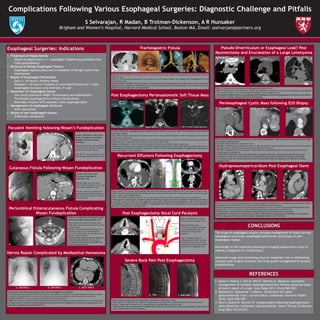
Tracheogastric Fistula
Fig A, B: CT chest (POD 5) in a patient with recurrent pneumonia shows tracheo-esophageal fistula, confirmed on virtual
bronchoscopy.
Fig C, D: The 3D volume rendered pre (C) and post stent (D) images. Patient was managed with esophageal stent and
endotracheal tube.
Teaching point: Potential causes of fistula include dissection during surgery (more likely due to low location of
fistula) and prolonged low intubation.Virtual bronchoscopy and 3D reformations further help to characterize the
fistula and plan endoscopic stent placement.
Post Esophagectomy Perianastomotic Soft Tissue Mass
Recurrent Effusions Following Esophagectomy
Fig A: Contrast CT in patient post esophagectomy shows large bilateral pleural effusions which were chylous on pleural tap.
Fig B: Conventional lymphangiogram demonsted leak, treated with thoracic duct embolization with resolution of chyle
leak on follow up CT (Fig D and E).
Fig C: The normal anatomy of thoracic duct is extremely variable resulting in high incidence of surgical injury.
Teaching point: Attenuation of pleural effusions following surgery may not be diagnostic. High index of clinical
suspicion in patients with recurrent effusions, appropriate imaging (lymphangiogram) as well as thoracentesis
are needed to make an accurate diagnosis. Triglyceride levels may not be elevated in the early post operative
period (1-3 weeks) due to fasting state.
Post Esophagectomy Vocal Cord Paralysis
Fig A: Preoperative CT shows
normal position of vocal cords.
Fig B: Immediate post
operatively, patient developed
vocal cord paralysis that was
medialized with gel foam
injection.
Fig C: Schematic diagram
showing course of the recurrent
laryngeal nerves which may be
damaged during cervical surgical
dissection for esophagectomy.
A B C D
67-year-old woman, esophagectomy for adenocarcinoma of the GE junction in 2007 with enlarging peri-anastomotic
soft tissue nodule.
Fig A: PET CT of peri-anastomotic soft tissue nodule shows only minimal FDG uptake similar to background and no other
sites of abnormal uptake.
Fig B, C: Chest MRI T2-weighted images show central fluid signal and post contrast subtraction scans shows peripheral
enhancing walls. imaging findings consistent with complex cystic mass.
Differential diagnosis – tumor with necrosis, indolent infection, granulation tissue, nodal mass.
Teaching point: Endoscopy revealed no evidence of tumor and the mass is now stable after 24 months. Not all peri-
anastomotic soft masses represent recurrence. As seen in this case granulomatous or inflammatory masses can
occur and biopsy is key to diagnosis.
Severe Back Pain Post Esophagectomy
Post-op course complicated by mediastinitis, presented 1 month later to ER.
Fig A: Sagittal CT demonstrates endplate irregularity and disc space loss, epidural gas collection (yellow arrow).
Fig B, C: Sagittal MRI reveals discitis, osteomyelitis and epidural abscess (red arrows).
Teaching point: Due to postoperative mediastinitis, and the close proximity of the neo-esophagus to the thoracic
spine, osteomyelitis may be a delayed.
Pseudo‐Diverticulum or Esophageal Leak? Post
Myomectomy and Enucleation of a Large Leiomyoma
Fig A: CT demonstrates large lobulated esophageal mass (yellow arrow).
Fig B: CT following oral contrast reveals irregular gas collection (red arrow) in continuity with esophagus. A mediastinal
drain was placed due to fever, with subsequent drainage of pus and defervescence.
Teaching point: Differentiation of outpouching of the esophageal wall from an esophageal perforation can be difficult.
The paitent’s clinical symptoms determines management.
Periesophageal Cystic Mass following EUS Biopsy
New onset back pain following EUS Biopsy of Cystic Periesophageal Mass – differentials include duplication Cyst, Node
or Leiomyoma with Necrosis.
Fig A NCCT: High attenuation retrocrural mass .
Fig B CECT: Peripheral rim enhancement of retrocrural mass which is closely associated with the aorta and contrast filled
esophagus (arrows), differential includes abscess complicating hematoma.
Fig C CECT: repeated due to increasing pain, shows large saccular pseudoaneurym arising anteriorly with associated
enlarging periaortic hematoma.
Teaching Point: Because of close proximity of the esophagus to the aorta, endoscopic procedures may cause
aortic injury. In this case, distal thoracic aorta pseudoaneurysm caused by infected esophageal duplication
cyst developed and was treated with aortic resection and replacement by homograft and removal of residual
esophageal cyst.
Hydropneumopericardium Post Esophageal Stent
Fig A: Pre stent CT s/p partial esophagectomy and esophagogastric anastomosis for esophageal caracinoma shows dilated
esophagus with air-fluid (white arrows) level proximal to anastomosis (green arrow).
Fig B, C: Esophageal stent (red arrow) placed across malignant stricture, note new moderate hydropneumopericardium
(yellow arrows). E.coli was found in pericardial fluid suggesting an esophageal pericardial fistula.
Teaching point: Post-operative complicated hydropneumopericardium should raise concern for
pyopneumopericardium which may be due to surgical complication from gastropericardial fistula or bacteremia
Patients may present with pericarditis cardiac tamponade with fever as in this case.
CONCLUSIONS
The scope of esophageal surgery includes management of hiatal hernias,
esophageal carcinoma, esophageal perforation and biopsy of peri-
esophageal masses.
Knowledge of the expected postsurgical imaging appearance is key to
making a diagnosis of complications.
Advanced image post processing plays an important role in delineating
complex post surgical anatomy and helps guide management of surgical
complications.
REFERENCES
1. Hama Y, Hahira J, Emi M, Kite R, Shimizu K, Okada M. Successful
management of multiple esophagorespiratory fistulas using two types
of stent: report of a case. Surg Today 2011; 41(4):560-562.
2. Katsanos K, Sabharwal T, Adam A. Stenting of the upper
gastrointestinal tract: current status. Cardiovasc Intervent Radiol
2010; 33(4):690-705.
3. Paul S, Bueno R. Section VI: complications following esophagectomy—
early detection, treatment, and prevention. Semin Thorac Cardiovasc
Surg 2003;15:210–215.
A. Pre-Stent B. Post Stent C. Post Stent
A. NCCT day 1 B. CT day 1(oral+) IV(+) C. CECT 1 month later
A.Pre-op CT B C
A B. T2W C. Post GAD
A
B C
A B C
D
E
A. PET CT 2010 B. MRI T2W 2011 C. MRI T1W Post contrast subtraction
B