
Recommended
More Related Content
Similar to histopathology of dental caries .pdf
Similar to histopathology of dental caries .pdf (20)
Recently uploaded
Recently uploaded (20)
histopathology of dental caries .pdf
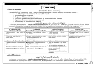
- 1. Dr. Ahmed El_Nashar 4. Histopathology of dental caries 1. Enamel caries 1. Smooth surface caries ( examined by electron microscope ) - Permeation of acids within enamel causes a series of submicroscopic changes , recorded by electron microscope as follows ….. 1. Loss of the interprismatic or inter-rod substances of the enamel. 2. Increased prominence of the rods. 3. Roughening of the ends of the enamel rods. 4. Degradation of the mucopolysaccharides present in the interprismatic organic substance. 5. Appearance of transverse striations of the enamel rods. 6. Accentuation of the incremental striae of Retzius. ( examined by ground section and transmitted light ) - As the caries process advances, a triangle or a cone shaped lesion with the apex toward DEJ and the base toward the surface of the tooth. Several zones can be distinguished before actual destruction , Working from backward forward zones appears in the following arrangement …… 1. Translucent zone 2. Dark zone 3. Body of the lesion. 4. Surface zone initial demineralization Demineralization and remineralization. greatest demineralization. greater degree of remineralization • Volume of Pores is 1 % of volume of enamel. • Volume of Pores is 2-4% of volume of enamel. • Volume of Pores is 5 – 25 % of volume of enamel. • Volume of Pores is 1 % of volume of enamel. • The pores are larger than that in normal enamel. • Some of the pores are large but others are smaller than those of the translucent zone. • pores are larger than those found in sound enamel. • In this zone 1.2% of minerals are lost • In this zone 6% of minerals are lost • In this zone 24% of minerals are lost. • Ii appears translucent with transmitted light. • This zone is sometimes missing or only present along a part of the lesion. • it appears dark with transmitted light (light brown in color). • dark zone is narrow in acute caries and wide in slowly advancing caries • forms the largest area in the carious lesion • The lost minerals are replaced by unbound water and organic material , derived from saliva and microorganisms • This area is recognizable clinically as brown spot • It appears relatively unaffected. It is about 40 µm thick. 1. pits and fissure caries א א - As the caries process advances, a triangle or a cone shaped lesion with the apex toward the surface of the tooth and the base toward the DEJ . Several zones can be distinguished before actual destruction , Working from backward forward zones appears in the following arrangement ……
- 2. Dr. Ahmed El_Nashar 2. Dentin caries 1, Caries in primary dentin Early dentinal changes Advanced dentinal changes Fatty degeneration Dentinal sclorosis Zone of decalcification Bacterial invasion Decomposed dentine Fatty degeneration of Tome's fibers of the dentinal tubules. • a calcification of the dentinal tubules that tends to seal them off against further penetration by microorganisms • formed as a result of reaction of vital dentinal tubules and vital pulp. • Initial decalcification involves the walls of the tubules by diffusion of acids. • then, the dentine matrix is destroyed progressively by proteolysis. • No bacteria present in this zone. 1. Pure forms of MOs penetrate D.T., There are 2 waves of MOs.. 1st wave p acidogenic MOs. 2nd wave p Proteolytic MOs. 2. The decalcification of the walls of the denlinal tubules leads to their confluence. • complete destruction. • The presence of considerable amount of globular dentine accounts for the rapid spread of caries in so- called malacotic or soft teeth. 3. ch. ch. by prescence of …. Beaded dentinal tubules Liquefaction foci Transverse cleft • thickening and swelling of the walls of dentinal tubules at irregular intervals and increase the diameter of the dentinal tubules due to packing of the tubules by MOs • They are tubules and filled with necrotic debris, which tend to increase in size by expansion • They are transverse clefts are extend at right angles to dentinal tubules • May be due to extension of caries 1.along the lateral branches of the tubules 2.along the incremental lines of dentine. 3.along the matrix fibers which run in this direction. 2. Caries in secondary dentin • the same as primary dentin but somewhat slower because the dentinal tubules are fewer in number and more irregular in their course. • Caries may spread laterally at the junction of the primary and secondary dentine and produces a separation of the two layers. • Causes of lateral spread of caries at D.E.J may be due to one of the flowing causes…… 1. The high amount of organic content of dentino-enamel junction. 2. Branching of the dentinal tubules, enamel tufts and enamel spindles, 3. Direction of the enamel rods.