
Recommended
More Related Content
Similar to digestive system.ppt
Similar to digestive system.ppt (20)
More from AderawAlemie
More from AderawAlemie (20)
Recently uploaded
Recently uploaded (20)
digestive system.ppt
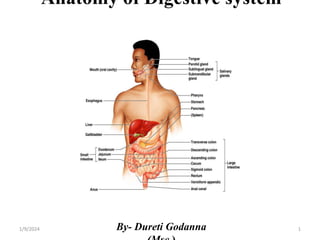
- 1. Anatomy of Digestive system By- Dureti Godanna 1 1/9/2024
- 2. Objectives At the end of this session you are expected to List organs of digestive system Locate all organs of digestive system anatomically. Understand the anatomy of digestive organs in relation to other organ s Understand the Vasculatures and innervation of digestive organs 2 1/9/2024
- 4. 4 1/9/2024
- 5. Organs of digestive system Two groups of organs The gastrointestinal (GI) tract Accessory digestive organs 5 1/9/2024
- 6. 6 1/9/2024
- 7. GI tract and Accessory digestive organs GI tract Approximately 9m long Extends from the mouth to the anus. Oral cavity Stomach Pharynx Small intestine, Esophagus Large intestine. . Accessory organs Teeth Liver Tongue Gallbladder Salivary glands Pancreas 7 1/9/2024
- 8. Layers of the GI Tract The wall of the GI tract from the lower esophagus to the anal canal has the same basic, four-layered arrangement of tissues. The four layers of the tract from deep to superficial. Mucosa Sub mucosa Muscularis Serosa/adventitia 8 1/9/2024
- 9. Mouth It also known as the oral or buccal cavity . It is formed by the cheeks, lips, hard and soft palates, tongue. The oral cavity is divided into Vestibule And Mouth proper. The vestibule is the area b/n the cheeks and lips externally and the gums and teeth internally. The mouth proper is the space bounded by the teeth. 1/9/2024 9
- 10. 10 1/9/2024
- 11. Tongue Occupies the floor of the mouth and fills the oral cavity when mouth is closed Functions Mixing food with saliva and forming the bolus Initiation of swallowing, and speech Note: Lingual frenulum secures the tongue to the floor of the mouth 11 1/9/2024
- 12. 12 1/9/2024
- 13. Lingual papillae Superior surface bears three types of papillae Filiform - give the tongue roughness and provide friction Fungiform - scattered widely over the tongue and give it a reddish hue Circumvallate - V-shaped row in back of tongue 13 1/9/2024
- 14. 14 1/9/2024
- 15. Teeth Humans as other mammals have hetrodont dentition. i.e. the teeth vary structurally and are adapted to handle food in different ways. An adult human has four types of teeth: Incisors Canines (cupids) Premolars (bicuspids) Molars (six pairs) 15 1/9/2024
- 16. Teeth The teeth, are accessory digestive organs located in sockets of the al veolar processes of the mandible and maxillae. The alveolar processes are covered by the gingivae ( gums). Which extend slightly into each socket. The sockets are lined by the periodontal ligament (periodontal mem brane) Which consists of dense fibrous connective tissue that anchors the te eth to the socket walls and acts as a shock absorber during chewing. 16 1/9/2024
- 17. Dentitions Humans have two dentitions i.e., two sets of teeth develop in a person’s life time. Deciduous (milk) teeth Begin to develop in each jaw before birth. The first teeth usually erupt at 6 to 8 monthly after birth beginning with the incisor. Eruption of these teeth have completed by 2 ½ years. 20 in number Formula for deciduous dentition of humans I-2/2, C -1/1 , DM -2/2 = 10 x 2 = 20 (DM = deciduous molar) 17 1/9/2024
- 18. 18 1/9/2024
- 19. Permanent teeth Replace the deciduous teeth at predictable sequence from age 6 to 17. The third molars, or wisdom teeth, are the last to erupt between the ages of 17 to 25. 32 in number Formula for permanent dentition I- 2/2, C- 1/1, P- 2/2, m-3/3 = 16x2 = 32 1/9/2024 19
- 20. 20 1/9/2024
- 21. Parts of teeth A tooth consists of a crown and one or more roots. The crown Functional part that is visible above the gum. The root is the unseen portion that supports and fastens the tooth in the jawbone. The root - attached to the tooth-bearing bone the alveolar processes of the jaws by a fibrous ligament called the periodontal ligament. 21 1/9/2024
- 22. 22 1/9/2024
- 23. 23 1/9/2024
- 24. Salivary Glands Secret saliva. Saliva has various functions: • Serves as a solvent • Cleansing the teeth • Dissolving food chemicals so that they can be tasted • Contains enzymes which digest starch • Contains mucous which lubricates the pharynx to facilitate swallowing 24 1/9/2024
- 25. Salivary Glands….. It is a gland that releases a secretion called saliva into the oral cavity . The mucous membrane of the mouth and tongue contains many sma ll salivary glands. Open directly or indirectly via short ducts to the oral cavity. These glands include labial, buccal and palatal glands in the lips, ch eeks, and palate, respectively. Lingual glands in the tongue, all of which make a small contributio n to saliva. 25 1/9/2024
- 26. Salivary Glands Most saliva is secreted by the major salivary glands. Lie beyond the oral mucosa, into ducts that lead to the oral cavity. The parotid gland Largest salivary glands Found between the skin and the masseter muscle. Located anterior and inferior to the auricle. The parotid duct, which is about 5cm long drains into the oral cavity opposite the 2nd upper molar. 26 1/9/2024
- 27. 27 1/9/2024
- 28. Parotid gland …… Each secretes saliva into the oral ca vity via a parotid duct Pierces the Buccinator muscle to op en into the vestibule opposite the se cond maxillary (upper) molar tooth. 28 1/9/2024
- 29. 29 1/9/2024
- 30. 30 1/9/2024
- 31. The pharynx It is funnel-shaped tube that extends fro m the internal nares to the esophagus p osteriorly and to the larynx anteriorly. The pharynx consists of external and internal muscles. The external include the superior, middle inferior muscles Which constrict the pharynx during swallowing. 31 1/9/2024
- 32. Pharynx…… These muscles elevate the larynx and pharynx in swallowing and during speaking. The inferior constrictor muscle prevents air from entering the esophagus during breathing 32 1/9/2024
- 33. Esophagus This is a collapsible muscular tube (25cm). Connects the pharynx to the stomach. Located posterior to the trachea. It passes through the diaphragm is an opening called esophageal hiatus. 33 1/9/2024
- 34. Esophagus The upper third of the esophagus contains skeletal muscle. The middle third contains both skeletal and smooth muscle, and the terminal portion contains only smooth muscle. The terminal portion of the esophagus is slightly narrowed due to the presence of the lower esophageal (gastro esophageal) sphincter. This prevents regurgitation of stomach contents into the esophagus. 34 1/9/2024
- 35. 35 1/9/2024
- 36. 36 1/9/2024
- 37. 1/9/2024 37
- 38. Peritoneum The peritoneum is the largest serous membrane of the body. It consists of a layer of simple squamous epithelium with an underlyi ng supporting layer of areolar connective tissue. The peritoneum is divided into the parietal and visceral peritoneu m Parietal peritoneum lines the wall of the abdominal cavity. Visceral peritoneum covers some of the organs in the cavity and is their serosa 38 1/9/2024
- 39. Peritoneal cavity The slim space containing lubricating s erous fluid. Found between the parietal and viscer al portions of the peritoneum. In certain diseases, the peritoneal cavit y may become distended by the accum ulation of several liters of fluid, a cond ition called ascites 39 1/9/2024
- 40. Stomach The stomach is a J-shaped pouch and is the most distensible part of the GIT. Found directly inferior to the diaphragm in the abdomen. The stomach connects the esophagus to the duodenum (the first part of the small intestine) The functions of the stomach are: Store food as it is mechanically churned with gastric secretions Initiate the digestion of proteins Move food into the small intestine as a chyme (pasty material) Secretion of gastric juice 40 1/9/2024
- 41. Anatomy of the Stomach The stomach has four main regions: 1. Cardia: surrounds the opening of th e esophagus into the stomach. 2. Fundus: Rounded portion superior t o and to the left of the Cardia 3. Body: Inferior to the fundus is the la rge central portion of the stomach. 4. Pyloric part 41 1/9/2024
- 42. Pyloric part It is divided in to three regions The first region is pyloric antrum, connects to the body of the stomach. The second region is pyloric canal The pylorus which in turn connect s to the duodenum. When the stomach is empty, the m ucosa lies in large folds or rugae. 42 1/9/2024
- 43. 43 1/9/2024
- 44. 44 1/9/2024
- 45. Anatomy of the Stomach….. The concave medial border of the stomach is called the lesser curv ature. The convex lateral border is called the greater curvature. 45 1/9/2024
- 46. 1/9/2024 46
- 47. 1/9/2024 47
- 48. Histology of the Stomach The gastric glands contain three types of exocrine gland cells Secrete their products into the stomach lu men: Mucous neck cells , Chief cells and Parie tal cells mucous neck cells secrete mucus Parietal cells produce intrinsic factor (ne eded for absorption of vitamin B12) and hydrochloric acid. The chief cells secrete pepsinogen and ga stric lipase. 48 1/9/2024
- 49. Vasculature of the stomach The right and left gastric arteries run along the lesser curvature while the right and left gastro-omental arteries run along the greater curvature. The fundus and upper body of stomach receive blood from the short and posterior gastric arteries branches of the splenic artery. 49 1/9/2024
- 50. 1/9/2024 50
- 51. 51 1/9/2024
- 53. Small intestine Most digestion and absorption of nutrients occur in a long tube called the small intest ine. Its length alone provides a large surface ar ea for digestion and absorption. The area is further increased by circular fo lds, villi, and microvilli. The small intestine begins at the pyloric sp hincter of the stomach. Opens into the large intestine. 53 1/9/2024
- 54. Small intestine It is the portion of the GIT b/n the pyloric sphincter of the stomach and the ileocecal valve opening into the large intestine. It is the site where digestion is completed and nutrients are absorbed. 54 1/9/2024
- 55. Functions of small intestine Reception of the secretions from the liver and pancreas Chemically breakdown of chyme Absorption of nutrients Transportation of the remaining undigested material to the large intestine. The small intestine is divided into three regions Duodenum Jejunum Ileum 55 1/9/2024
- 56. The duodenum It is a relatively fixed C- shaped tube (25cm long) Extend from the pyloric sphincter to the duodeno jejunal flexure. Its left concave surface receives bile secretions through the common bile duct from the liver and gallbladder, and pancreatic secretions through the duct of the pancreas. Both ducts unite to form a common entry into the duodenum called the hepatopancreatic ampulla (or ampulla of Vater). 56 1/9/2024
- 57. 57 1/9/2024
- 58. The blood supply to the duodenum It comes from two sources. The first and second part are supplied by the gastro duodenal arte ry. The third and fourth parts are supplied by SMA. Duodenal veins, follow the arteries and drain into the portal vein. 1/9/2024 58
- 59. The jejunum (1m long) extends from the duodenum to the ileum. It has a slightly larger lumen and more internal folds than the ileum. The ileum (2m long) makes up the remaining part of the small intestine. The terminal portion of the ileum empties into the medial side of the cecum through the ileocecal valve. The walls of the ileum have an abundance of lymphatic tissue aggregated into nodules called mesenteric (payer’s) patches 59 1/9/2024
- 60. Characteristics of Jejunum & ileum Characteristics Jejunum ileum color Deeper red Paler pink caliber 2-4cm 2-3cm wall Thick and heavy Thin and light Vascularity Greater Lesser Fat in mesentery less more 60 1/9/2024
- 61. Blood supplies The jejunum and ileum have: Arterial supply from the (superior mesenteric artery) SMA. The SMA runs between the layers of the mesentery and sends many branches to the jejunum and ileum.. Venous drainage from the (superior mesenteric vein) SMV . The SMV lies anterior and to the right of the SMA in the root of the mesentery. 61 1/9/2024
- 62. Large Intestine The large intestine is the terminal portion of the GI tract. It is about 1.5m long begins at the terminal end of the ileum and te rminates at the anus Has 6.5 cm in diameter in living humans The large intestine has little or no digestive functions. It functions Absorb water and electrolytes from the remaining chyme Forms stores Expels feces from the body. 62 1/9/2024
- 63. Large Intestine The large intestine is structurally divided into o Cecum o Colon o Rectum o Anal canal. The cecum, the first part of the large intestine that is continuous with the ascending colon, has a blind intestinal pouch(appendix) in the right lower quadrant. The transverse colon, the largest and most mobile part of the large intestine Crosses the abdomen from the right colic flexure to the left colic fle ure. 63 1/9/2024
- 64. 64 1/9/2024
- 65. Large Intestine Ascending colon ascends on the right side of the abdomen. Reaches the inferior surface of the liver. Turns abruptly to the left to form the right colic (hepatic) flexure. 65 1/9/2024
- 66. 66 1/9/2024
- 67. The rectum It is about 15 cm in length Lies anterior to the sacrum and coccyx. The terminal 2–3 cm of the large intestine is called the anal canal. The opening of the anal canal to the exterior called the anus. It is guarded by an internal anal sphincter of smooth muscle (involu ntary) and an external anal sphincter of skeletal muscle (voluntary). Normally these sphincters keep the anus closed except during the eli mination of feces. 67 1/9/2024
- 68. 68 1/9/2024
- 69. 1/9/2024 69
- 70. Accessory digestive organs These organs aid in the chemical break down of food. These are Liver Gallbladder pancreas The liver and pancreas function as exocrine glands . Because their secretions are transported to the lumen of the GIT via ducts. 70 1/9/2024
- 71. Liver The liver is the largest internal organ of the body Weighing about 1.3 kg in an adult. It is reddish-brown in color because of its great vascularity. The liver has two major lobes 1. The right lobe 2. The left lobe 71 1/9/2024
- 72. The liver carries out numerous functions: osynthesis, storage, and release of glycogen osynthesis of blood proteins oPhagocytosis of old red blood cells oDetoxify toxic substances oproduction of bile. 72 1/9/2024
- 73. Liver…. The liver is inferior to the diaphragm Occupies most of the right hypochondriac and part of the epigastric regions of the abdominopelvic cavity 73 1/9/2024
- 74. 74 1/9/2024
- 75. Gallbladder This is a pear-shaped sac that is located in a depression of the posteri or surface of the liver. It is 7–10 cm long and typically hangs from the postero inferior mar gin of the liver. 75 1/9/2024
- 76. Gall bladder It is a sac like organ attached to the inferior surface of the liver. It stores and concentrates bile, which drains to it from the liver Bile is a yellowish-green fluid Bile is continuously produced by the liver and drains through the hepatic and common bile ducts to the duodenum for the emulsification and absorption of fats. 76 1/9/2024
- 77. 1/9/2024 77
- 78. 78 1/9/2024
- 79. 79 1/9/2024
- 80. Plain abdominal X-ray abdomen 80 1/9/2024