2. diuretics, implanted with an internal cardiac defibrillator, and
discharged on carvedilol, lisinopril, and furosemide with un-
known compliance. Electrocardiogram did not meet the
criteria for cardiac resynchronization therapy. Following
discharge, she continued to have progressive anasarca and
was admitted 1 month after discharge. She required a dobu-
tamine infusion in addition to a paracentesis for ascites.
Echocardiogram showed diffuse LV hypokinesis with EF
20%. She was discharged on diuretics, and antiretroviral
therapy was continued. However, 1 month antecedent to
transfer to the NIH, she was readmitted to an outside facility
with decompensated HF, associated congestive hepatopathy,
and progressive renal dysfunction. Diuretic therapy was
unsuccessful, prompting IV dobutamine again to augment
perfusion. Clinical status remained tenuous, and dobutamine
was discontinued, and the patient was advised to transition
to hospice care for end-stage HF. However, she desired
continued aggressive medical care, and the NIH was contacted.
She was transferred to the NIH CC while receiving
carvedilol, spironolactone, furosemide, and an antiretroviral
regimen of tenofovir disoproxil fumarate/emtricitabine,
atazanavir, and ritonavir. Physical examination was notable
for blood pressure 96/70 mmHg, pulse 83 b.p.m., O2
saturation 96% on room air, anasarca, and cool peripheral
extremities. Notable admission labs included sodium
120 mmol/L and pro-brain natriuretic peptide 8358 pg/mL.
At the NIH CC, she was treated with intravenous furose-
mide; however, this was held for hypotension and acute
oliguria. She was transferred to the intensive care unit for
inodilator support. An echocardiogram demonstrated
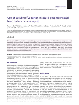
dilation of all cardiac chambers with LVEF 11% (normal >
53%), severe mitral regurgitation (Figure 1A), septal
flattening suggesting right ventricular (RV) volume overload
(Figure 1B), grade III diastolic dysfunction, and lateral E/e’ ra-
tio 23, suggesting an elevated LV filling pressure (Figure 1C,
D). The tricuspid annular plane systolic excursion (not shown)
was consistent with reduced RV systolic function.
Right heart haemodynamics revealed secondary
pulmonary hypertension with mean pulmonary artery pres-
sure 34 mmHg, volume overload with pulmonary capillary
wedge pressure 20 mmHg, vasoconstriction with a high
systemic vascular resistance (SVR) at 1599 dyn·s·cm 5
,
and a low cardiac output state with cardiac index (CI)
1.3 L/min/m2
and mixed venous oxygen saturation (SVO2)
44%. She was started on dobutamine (2.5 μg/kg/min)
and nitroglycerin (20 μg/min). Spironolactone (25 mg)
was continued and carvedilol discontinued. Dobutamine
was titrated to 7.5 μg/kg/min guided by haemodynamics.
Her urine output increased to 200–300 mL/h; SVR de-
creased to 987 dyn·s·cm 5
; and CI and SVO2 improved to
2.6 L/min/m2
and 64%, respectively (Figure 2). She
achieved a fluid balance of 4.6 L by Day 3.
Figure 1 Transthoracic echocardiogram on admission. Imaging showed severe mitral regurgitation (A) and diastolic septal flattening suggestive of
right-sided volume overload (B). Doppler imaging on admission showed a restrictive mitral inflow pattern (C) and diminished tissue Doppler
velocities (D).
A B
C D
Sacubitril/valsartan in ADHF 185
ESC Heart Failure 2018; 5: 184–188
DOI: 10.1002/ehf2.12219
3. Oral afterload reduction was initiated with captopril (Day
4), which was titrated to 50 mg t.i.d., and hydralazine (HYZ)
and isosorbide dinitrate (ISDN) both at 10 mg t.i.d. (Day 5).
On this regimen, nitroglycerin was discontinued (Day 6),
and dobutamine was weaned to 2.5 μg/kg/min (Day 6).
However, this resulted in a decrease in CI from 2.6 to 2.2
L/min/m2
. (Figure 2). HYZ and ISDN were increased to 50
and 20 mg, respectively. On Day 8, her total fluid balance
was 19 L, and she had lost 15 kg. Haemodynamics
revealed mean pulmonary artery pressure 31 mmHg, pul-
monary capillary wedge pressure 16 mmHg, SVR
486 dyn·s·cm 5
, CI 4.4 L/min/m2
, and SVO2 73%. Dobuta-
mine was weaned off (Day 9); however, within 10 h,
she developed anuria with a drop in CI to 1.8 L/min/m2
(Figure 2). Dobutamine was resumed, and HYZ and ISDN
were increased to 75 and 40 mg t.i.d., respectively. The
decision was made to trial sacubitril/valsartan following a
48 h captopril washout period.
On Day 12, sacubitril/valsartan was initiated at the mid-
range dose of 49/51 mg b.i.d. Concerns over tachyphylaxis
prompted a transition from dobutamine to milrinone (Day
12) titrated to 0.375 μg/kg/min. Because she was off dobuta-
mine and was euvolemic, metoprolol tartrate was added (Day
13). Milrinone was successfully weaned off (Day 14) once
sacubitril/valsartan approached a steady-state dose. The
patient maintained adequate blood pressure and urine out-
put. Subsequent changes to her medication regimen included
increasing sacubitril/valsartan to the maximum dose of
97/103 mg b.i.d. (Day 15) and the addition of digoxin
0.25 mg o.d. (Day 15). She displayed no clinical or laboratory
signs of infection for the duration of her intensive care unit
admission.
The patient was transferred to the ward (Day 17) and
transitioned to metoprolol succinate 100 mg o.d. with di-
goxin discontinued. Upon discharge, her pro-brain
natriuretic peptide decreased to 788 pg/mL. An echocardio-
gram performed 41 days after discharge revealed an
increase in LVEF to 35%, improved LV diastolic dimension
(from 64 to 56 mm), and systolic dimension (from 58 mm
to 42 mm), and markedly decreased mitral regurgitation
(Figure 3A). There was no further evidence of septal
flattening or LV diastolic dysfunction (Figure 3B), and there
was normalization of LV filling pressures with a lateral E/e’
ratio of 9 (Figure 3C, D). The tricuspid annular plane
systolic excursion (not shown) suggested improved RV
systolic function. At 10 months after discharge, she has
not been readmitted and on clinic visits has been without
signs of decompensated HF.
Figure 2 Hospital course. Abbreviations: IV, intravenous; PO, by mouth; sPAP, systolic pulmonary artery pressure; dPAP, diastolic pulmonary artery
pressure; mPAP, mean pulmonary artery pressure; PCWP, pulmonary capillary wedge pressure; CO, cardiac output; CI, cardiac index; SVO2, mixed
venous oxygen saturation; SVR, systemic vascular resistance; MAP, mean arterial pressure; PVR, pulmonary vascular resistance; CVP, central venous
pressure.
186 T.D. Bell et al.
ESC Heart Failure 2018; 5: 184–188
DOI: 10.1002/ehf2.12219
4. Discussion
In a patient with HIV-associated cardiomyopathy and stage D
HFrEF, we describe the successful use of sacubitril/valsartan
combined with goal-directed medical therapy to liberate the
patient from inodilator dependence. To our knowledge, this
is the first reported use of sacubitril/valsartan for this
indication. Sacubitril/valsartan was shown to be superior to
enalapril in reducing mortality and HF hospitalizations in
patients with chronic HFrEF.5
Sacubitril/valsartan also
demonstrated fewer rates of inodilator agents (31% risk
reduction) and MCS or HT (22% risk reduction).7
These
secondary outcomes were evident within 30 days of random-
ization, suggesting early effects of sacubitril/valsartan.
Despite optimization of medical therapy and
haemodynamics,ourattemptstoweandobutaminewerecom-
plicatedbyhypoperfusion.Sacubitril/valsartanenabledthedis-
continuationofinodilatorsaftertraditionalafterloadreduction
methods with an angiotensin-converting enzyme inhibitor
failed. The neprilysin inhibitor’s ability to increase levels of en-
dogenous vasoactive peptides may have provided additional
benefits towards maintaining improved organ perfusion once
off inodilator support. However, it is important to mention that
the medication’s potency in reducing SVR and blood pressure
may also be a limitation to its use in advanced HF, particularly
if blood pressures are marginal. Clinical trials are needed to
assess the efficacy and limitations of sacubitril/valsartan in pa-
tients with advanced HF. One such study under enrollment is
the Entresto in Advanced Heart Failure (LIFE Study).
During end-stage HF, it can be challenging to wean
inotropes. Chronic inotrope infusions may be the only option
for stage D HFrEF patients optimally treated with goal-
directed medical therapy and not candidates for MCS or HT.
If home inotropes are chosen, a discussion must take place
with the patient regarding the palliative nature and
potentially harmful consequences, including increased risk
of death.2
Given the long-term favourable findings of
sacubitril/valsartan and our experience with this patient,
sacubitril/valsartan may be a potential cost-effective option
for inotrope-dependent patients who are not candidates for
MCS and HT.
Acknowledgements
The authors would like to recognize and thank the NIH Clinical
Center Critical Care nursing, social work, and respiratory ther-
apy staff for their expert care of this patient; Brad Moriyama,
Pharm.D, for his assistance with medication interactions; Kelly
Byrne for her assistance in manuscript preparation and sub-
mission; and Matthew A. Adan and Dr. Andrew Catanzaro for
participating in the clinical care of this patient.
Figure 3 Transthoracic echocardiogram after discharge from the hospital. Imaging showed trace mitral regurgitation (A) and no evidence of septal
flattening (B). Doppler imaging showed normalization of the mitral inflow pattern (C) and similar tissue Doppler velocities (D).
A B
D
C
Sacubitril/valsartan in ADHF 187
ESC Heart Failure 2018; 5: 184–188
DOI: 10.1002/ehf2.12219
5. Conflict of interest
None declared.
Funding
National Institutes of Health Clinical Center.
References
1. Mozaffarian D, Benjamin EJ, Go AS,
Arnett DK, Blaha MJ, Cushman M, Das
SR, de Ferranti S, Despres JP, Fullerton
HJ, Howard VJ, Huffman MD, Isasi CR,
Jimenez MC, Judd SE, Kissela BM,
Lichtman JH, Lisabeth LD, Liu S, Mackey
RH, Magid DJ, McGuire DK, Mohler ER
3rd, Moy CS, Muntner P, Mussolino ME,
Nasir K, Neumar RW, Nichol G,
Palaniappan L, Pandey DK, Reeves MJ,
Rodriguez CJ, Rosamond W, Sorlie PD,
Stein J, Towfighi A, Turan TN, Virani SS,
Woo D, Yeh RW, Turner MB. Heart
Disease and Stroke Statistics—2016
update: a report from the American Heart
Association. Circulation 2016; 133:
e38–360.
2. Yancy CW, Jessup M, Bozkurt B, Butler J,
Casey DE Jr, Drazner MH, Fonarow GC,
Geraci SA, Horwich T, Januzzi JL,
Johnson MR, Kasper EK, Levy WC,
Masoudi FA, McBride PE, McMurray JJ,
Mitchell JE, Peterson PN, Riegel B, Sam
F, Stevenson LW, Tang WH, Tsai EJ,
Wilkoff BL. 2013 ACCF/AHA guideline
for the management of heart failure:
executive summary: a report of the
American College of Cardiology
Foundation/American Heart Association
Task Force on practice guidelines. Circula-
tion 2013; 128: 1810–1852.
3. Cruden NL, Fox KA, Ludlam CA, Johnston
NR, Newby DE. Neutral endopeptidase
inhibition augments vascular actions of
bradykinin in patients treated with
angiotensin-converting enzyme inhibi-
tion. Hypertension 2004; 44: 913–918.
4. Wilkinson IB, McEniery CM, Bongaerts
KH, MacCallum H, Webb DJ, Cockcroft
JR. Adrenomedullin (ADM) in the human
forearm vascular bed: effect of neutral en-
dopeptidase inhibition and comparison
with proadrenomedullin NH2-terminal
20 peptide (PAMP). Br J Clin Pharmacol
2001; 52: 159–164.
5. McMurray JJ, Packer M, Desai AS, Gong
J, Lefkowitz MP, Rizkala AR, Rouleau JL,
Shi VC, Solomon SD, Swedberg K, Zile
MR. Angiotensin-neprilysin inhibition
versus enalapril in heart failure. N Engl J
Med 2014; 371: 993–1004.
6. Yancy CW, Jessup M, Bozkurt B, Butler J,
Casey DE Jr, Colvin MM, Drazner MH,
Filippatos G, Fonarow GC, Givertz MM,
Hollenberg SM, Lindenfeld J, Masoudi
FA, McBride PE, Peterson PN, Stevenson
LW, Westlake C. 2016 ACC/AHA/HFSA
focused update on new pharmacological
therapy for heart failure: an update of
the 2013 ACCF/AHA guideline for the
management of heart failure: a report of
the American College of Cardiology/
American Heart Association Task Force
on clinical practice guidelines and the
Heart Failure Society of America. J Am
Coll Cardiol 2016; 68: 1476–1488.
7. Packer M, McMurray JJ, Desai AS, Gong
J, Lefkowitz MP, Rizkala AR, Rouleau
JL, Shi VC, Solomon SD, Swedberg K, Zile
M, Andersen K, Arango JL, Arnold JM,
Belohlavek J, Bohm M, Boytsov S,
Burgess LJ, Cabrera W, Calvo C, Chen
CH, Dukat A, Duarte YC, Erglis A, Fu M,
Gomez E, Gonzalez-Medina A, Hagege
AA, Huang J, Katova T, Kiatchoosakun
S, Kim KS, Kozan O, Llamas EB, Martinez
F, Merkely B, Mendoza I, Mosterd A,
Negrusz-Kawecka M, Peuhkurinen K,
Ramires FJ, Refsgaard J, Rosenthal A,
Senni M, Sibulo AS Jr, Silva-Cardoso J,
Squire IB, Starling RC, Teerlink JR,
Vanhaecke J, Vinereanu D, Wong RC.
Angiotensin receptor neprilysin inhibition
compared with enalapril on the risk of
clinical progression in surviving patients
with heart failure. Circulation 2015;
131: 54–61.
188 T.D. Bell et al.
ESC Heart Failure 2018; 5: 184–188
DOI: 10.1002/ehf2.12219