2. 2 Child Kidney Dis • 2019;23:1-6 www.chikd.org
ofchildren.GlomerularlesionsassociatedwithINSinclude
multiple histologic types, such as minimal change NS
(MCNS), focal segmental glomerulosclerosis, membrano
proliferative glomerulonephritis, and membranous ne
phropathy1,5)
. All these types of morphology share a com
mon feature with podocyte foot process effacement and
structural disorganization of the glomerular filtration bar
rier leading to proteinuria. These ultrastructural abnor
malities typically resolve with corticosteroid-induced cli
nicalremissions5)
.However,themechanismsofthisacquired
and reversible abnormality in glomerular ultrastructure
and filtration barrier permeability are incompletely under
stood.
Shalhoub, in 1974, proposed that the increased glome
rular permeability to plasma protein in MCNS is due to a
circulating factor released by T-cells; however, until date
the search for the circulating factor has been unsuccessful.
In addition, advances in the knowledge of podocyte patho
biology in the mechanism underlying proteinuria have led
to NS being recognized as a podocytopathy. Based on this
knowledge, several hypotheses have been proposed to
explain the role of podocytes and related molecules in the
mechanism underlying proteinuria in INS. Although the
pathophysiology of NS has not been completely explained
until now, it is considered a complex multifactorial disease
with an immunological component. In this review, we dis
cussrecentresearchfindingsinthepathogenesisofMCNS.
Pathogenesis of MCNS
Podocyte foot process effacement is the ultrastructural
hallmark in MCNS, however, the pathogenesis leading to
podocyteeffacementisnotclear.Systemicfactors,immune
mediated or circulating, and local factors can contribute
to podocyte effacement, but there is no single unifying
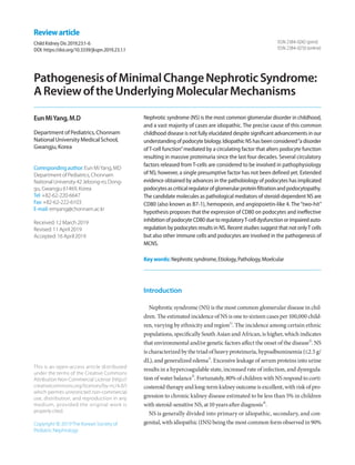
hypothesis (Fig 1).
Immune dysregulation
As immunosuppression with corticosteroids is the main
stay of NS therapy, it is logical to suspect immune dysregu
lation as a pathogenic factor in disease development. In
1974,ShalhoubpostulatedthatMCNSisadisorderofT-cell
function, resulting in increased plasma levels of lympho
Minimal Change Nephrotic Syndrome
Systemic Glomerular
T and B cell dysfunction Podocyte dysfunction
Angplt4, CD80
Cytokines (e.g. IL-8, IL-13)
and
other circulating factors
(e.g. hemopexin, Angplt4)
Glomerular filtration barrier damage
(Podocyte foot process effacement, GBM charge loss etc.)
Treg
Fig 1. Pathogenetic pathways leading to minimal change nephrotic syndrome
Fig 1. Pathogenetic pathways leading to minimal change nephrotic syndrome. Angplt4, angio
poitin-like 4;Treg, regulatoryT-cell;GBM, glomerular basement membrane.
3. Yang EM • Pathogenesis of MCNS 3
www.chikd.org
cyte-derived permeability factor6)
. This hypothesis was
based on the following clinical observations: [1] remission
is commonly accompanied by measles infection whereby
cell-mediated immunity is suppressed; [2] MCNS is asso
ciated with Hodgkin’s disease, which is a known T-cell dis
order; [3] patients show good response to corticosteroids
and cyclophosphamide, which are inhibitors of T-cell func
tion; [4] humoral component deposition (immunoglobulin
and components) is absent in glomeruli, which is unlike
that in other glomerular disorders. Therefore, the massive
proteinuria and hypoalbuminemia that characterize NS
were thought to result from increased glomerular capillary
wall permeability due to T-cell activation triggered by
several stimuli, such as viral infection or allergens.
1.Roleofcytokines
Investigators have made attempts, based on Shalhoub’s
hypothesis,toidentifythecirculatingfactorsreleasedfrom
T-cells that increase glomerular permeability to serum
proteins, and some studies have confirmed that capillary
permeability factor is detectable in patients with NS7,8)
. Of
the various factors presumed to increase glomerular per
meability to serum proteins, the most likely pathogenic
factors are considered to be cytokines, which are small pro
teins secreted by the cells of both the innate and adaptive
immune systems that transfer information within the im
mune system9,10)
. Patients who suffered relapses were found
to have elevated serum or urine levels of various cytokines,
including interleukin (IL)-211,12)
, soluble IL-2 receptor11,13)
,
interferon-gamma11,12)
, IL-814-16)
, IL-1317-19)
, tumor necrosis
factor-α20)
, and vascular endothelial growth factor21)
. Of
the many known cytokines, IL-8 and IL-13 in particular
have been proposed to be most likely to be circulating
factors. IL-8 may play a role in proteinuria by affecting the
metabolism of glomerular basement membrane (GBM)
components15)
. Additionally, urinary IL-8 levels were
higher in patients who suffered relapses and had a positive
correlation with the degree of proteinuria16)
. However, rat
podocytes incubated in vitro with high concentrations of
human IL-8 showed no difference in GBM permeability22)
.
IL-13 has been revealed to stimulate intracellular podocyte
protein trafficking and proteolysis in vitro17)
. IL-13-trans
fectedratsdevelopedsevereproteinuriaandshowedMCNS-
like nephropathy18)
. Increased IL-13 also induced the over
expression in podocytes of CD80, recently identified as a
possible molecular mechanism underlying proteinuria in
NS23)
. However, not all patient with MCNS present serum
IL-13 elevation during relapse, and serum IL-13 is also
knowntobeincreasedinclinicalconditionsnotassociated
with proteinuria, such as allergy including asthma and
atopy24, 25)
. Studies conducted over the last 40 years have
reported conflicting results regarding the role of cytokines
in MCNS.
2.RoleofregulatoryT-cells(Tregs)andB-cells
Historically, MCNS has been considered a T-cell disease;
however, advances in the study of basic immunology have
contributed to a more articulated understanding of its pa
thogenesistakingintoaccountTregsandB-cells.Normally,
cytokine release by T-cells is transient owing to the activa
tion of Tregs that interact with T effector cells to suppress
cytokine production. Tregs have been suggested to consti
tute a second step in an MCNS cascade, of which the first
remains unclear26,27)
. The induction of Treg led to a marked
reduction in proteinuria in animal models, and most pa
tients with MCNS showed decreased levels of Treg28,29)
.
Shimada et al. reported that abnormal censoring of podo
cyte CD80 expression could underlie Treg dysfunction or
impaired autoregulation by podocytes. Treg dysfunction
could lead to transient massive proteinuria becoming per
sistent, following which podocyte injury, and eventually,
MCNS,occur27)
.UnlikeroleofT-cellsinMCNS,whichhas
been extensively studied, the role of B-cells is currently not
well understood. Clinical trials have been conducted that
demonstratedMCNSremissionafterB-celldepletionusing
the anti-CD20 monoclonal antibody rituximab30,31)
. The
recent successful use of anti-CD20 monoclonal antibodies
forthetreatmentofsteroidsensitiveNSraisesthepossibility
of B-cells either influencing T-cells or themselves being
primaryplayersinNS.CD80isexpressedbybothactivated
B- and T- cells, and increased nitric oxide production by
B-cells observed in NS patients with relapse further sup
ports the possibility of B-cell involvement10,32)
. Altogether,
these results revealed that not only T-cells, but also B-cells
or B-cell products might be implicated in the causal me
chanism of MCNS via the abnormal regulation of T-cell
function by circulating B-cells or by communication bet
ween B- and T-cells33)
. However, information on the role of
4. 4 Child Kidney Dis • 2019;23:1-6 www.chikd.org
B-cells is currently limited.
Systemic circulating factors
1.Hemopexin
Hemopexinisanabundantplasmaproteinthateffectively
scavengesheme.Ithasbeenproposedthatvariousisoforms
ofhemopexinexist10)
.Undernormalconditions,circulating
hemopexin is inactive, but under certain conditions, it be
comes activated as a serine protease34)
. The active isoform
of hemopexin has been reported to be implicated in the
pathogenesis of MCNS. Infusion of human plasma hemo
pexin in rats induces reversible proteinuria accompanied
by podocyte foot process effacement and loss of the nega
tive charge of the GBM35)
. Furthermore, hemopexin in
duces nephrin-dependent cytoskeletal rearrangement in
podocytes and affects permeability of the glomerular filtra
tion barrier by reduction in glycocalyx. The effects of he
mopexin were inhibited by pretreatment with normal
human plasma and serine protease inhibitors36)
. Hemo
pexinissuspectedtobeacirculatingfactorcausingMCNS,
although the mechanism of hemopexin activation and the
inhibitory factors that activate hemopexin under normal
conditions is unclear.
2.Angiopoietin-like4(Angptl4)
Angptl4isaglycoproteinsecretedmainlyintheliverand
adipose tissue. Angptl4 is minimally expressed in normal
glomeruli, but it is highly upregulated in the serum and
podocytes in experimental models of MCNS and in the
human disease10,37,38)
. There are two isoforms of podocyte-
secreted Angptl4: the hyposialylated form secreted by po
docytes (podocyte-secreted form), and the sialylated form
secreted by skeletal muscles, heart, and adipose tissue (cir
culating form)39)
. Podocyte-secreted Angtpl4 is glucocorti
coid sensitive and has been proposed to be a mediator of
proteinuria. It was observed that its upregulation induced
massive proteinuria, loss of negative charge in the GBM,
and foot process effacement in vivo models of NS, and that
its conversion into sialylated Angptl437)
. In addition, circu
lating Angptl4 was secreted in response to an elevated
plasma free fatty acid to albumin ratio when proteinuria
reached the nephrotic range, subsequently resulting in
hypertriglyceridemia38)
. This indicates that Angptl4 can
be developed as a biomarker of MCNS in feature studies.
Co-stimulatory molecule CD80
CD80,alsoknownasB7-1,isatransmembranemolecule
present on the surface of both antigen presenting cells and
activated B-cells, and acts as a co-stimulatory signal for T-
cell activation10)
. CD80, present on the surface of antigen
presentingcells,bindsCD28oneffectorT-cellsorcytotoxic
T lymphocyte-associated protein 4 (CTLA4) in regulator
T-cells, determining T-cell activation (CD28) or inhibition
(CTLA4)40)
.In2004,Reiseretal.reportedthatundercertain
conditions,podocytescanexpressCD80,anditsexpression
results in the development of a proteinuric condition23)
.
Proteinuria was not induced in CD80 knockout mice by
lipopolysaccharides administration, but it was induced in
SCID mice, which are deficient in T- and B-cell functions,
showing that CD80 plays a key role independent of T- and
B-cells23)
. Increased CD80 levels in urine are observed in
patients with MCNS with relapse compared to those in
remission and with other glomerular diseases (lupus, focal
segmental glomerulosclerosis)41,42)
. A recent study has re
ported that high urinary CD80 excretion might be a bio
marker for steroid responsiveness and a predictor for good
prognosis in NS43)
. In addition, polyinosinic:polycytidylic
acid(polyI:C),aligandofToll-likereceptor3whichmimics
viral infection, promotes podocyte CD80 expression44)
.
This offers a possible reasoning to explain the frequent re
lapse of MCNS after upper respiratory virus infection.
Because PolyI:C induces only transient proteinuria, im
paired regulatory mechanisms after CD80 induction were
postulated as a second hit cause of MCNS27)
. Suppression
of CD80 expression could be a novel therapeutic strategy
for MCNS; however, more evidence is required to support
this idea.
Conclusions
The pathophysiology of NS is still far from being fully
explained, although recent advances in podocyte biology
haveprovidednovelinsightsintothepossibilityofNSbeing
5. Yang EM • Pathogenesis of MCNS 5
www.chikd.org
a podocytopathy. MCNS is regarded as a multifactorial
disease. It is hypothesized that MCNS is a podocytopathy
and that CD80 or other circulatory factors are the triggers
for proteinuria. Recent and future research will lead to
new therapeutic targets in MCNS.
Conflict of interest
This study was funded by Individual Basic Science &
Engineering Research Program through the Ministry of
Education of the Republic of Korea and National Research
Foundation of Korea (NRF-2016R1D1A1B03933207)
ORCID
Eun Mi Yang http://orcid.org/0000-0001-9410-5855
References
1. Noone DG, Iijima K, Parekh R. Idiopathic nephrotic syndrome in
children.Lancet2018;392:61-74.
2. Banh TH, Hussain-Shamsy N, Patel V, Vasilevska-Ristovska J,
Borges K, Sibbald C, et al. Ethnic Differences in Incidence and
Outcomes of Childhood Nephrotic Syndrome. Clin J AM Soc
Nephrol2016;11:1760-8.
3. Kaneko K, Tsuji S, Kimata T, Kitao T, Yamanouchi S, Kato S. Patho
genesisofchildhoodidiopathicnephroticsyndrome:aparadigm
shiftfromT-cellstopodocytes.WorldJPediatr2015;11:21-8.
4. Mendonca AC, Oliveira EA, Fróes BP, Faria LD, Pinto JS, Nogueira
MM, et al. A predictive model of progressive chronic kidney dis
ease in idiopathic nephrotic syndrome. Pediatr Nephrol 2015;30:
2011-20.
5. Pais Pariya, Avner ED. Nephrotic syndrome. In: Klegman RM,
Stanton BF, Geme III JWS, editros. Nelson textbook of pediatrics,
20thed.Philadelphia:Elsevier,2016:2521-6
6. Shalhoub RJ. Pathogenesis of lipoid nephrosis: a disorder of T-
cellfunction.Lancet1974;2:556-60.
7. Koyama A, Fujisaki M, Kobayashi M, Igarashi M, Narita M. A glo
merular permeability factor produced by human T cell hybrido
mas.KidneyInt1991;40:453-60.
8. Savin VJ, Sharma R, Sharma M, McCarthy ET, Swan SK, Ellis E, et
al. Circulating factor associated with increased glomerular per
meability to albumin in recurrent focal segmental glomerulosc
lerosis.NEnglJMed1996;334:878-83.
9. Araya CE, Wasserfall CH, Brusko TM, Mu W, Segal MS, Johnson RJ,
et al. A case of unfulfilled expectations. Cytokines in idiopathic
minimal lesion nephrotic syndrome. Pediatr Nephrol 2006;21:
603-10.
10. Kaneko K. Molecular mechanism in the pathogenesis of idio
pathicnephroticsyndrome..1sted.Tokyo:Springer,2016.
11. Daniel V, Trautmann Y, Konrad M, Nayir A, Schärer K. T-lympho
cyte populations, cytokines and other growth factors in serum
and urine of children with idiopathic nephrotic syndrome. Clin
Nephrol.1997;47:289-97.
12. Neuhaus TJ, Wadhwa M, Callard R, Barratt TM. Increased IL-2, IL-4
and interferon-gamma (IFN-gamma) in steroid-sensitive ne
phroticsyndrome.ClinExpImmunol1995;100:475-9.
13. MandreoliM,BeltrandiE,Casadei-MaldiniM,ManciniR,Zucchelli
A, Zucchelli P. Lymphocyte release of soluble IL-2 receptors in
patients with minimal change nephropathy. Clin Nephrol 1992;
37:177-82.
14. Garin EH, Blanchard DK, Matsushima K, Djeu JY. IL-8 production
by peripheral blood mononuclear cells in nephrotic patients.
KidneyInt1994;45:1311-7.
15. Garin EH, West L, Zheng W. Effect of interleukin-8 on glomerular
sulfated compounds and albuminuria. Pediatr Nephrol 1997;11:
274-9.
16. Souto MF, Teixeira AL, Russo RC, Penido MG, Silveira KD, Teixeira
MM, et al. Immune mediators in idiopathic nephrotic syndrome:
evidence for a relation between interleukin 8 and proteinuria.
PediatrRes2008;64:637-42.
17. VanDenBergJG,AtenJ,AnninkC,RaveslootJH,WeberE,Weening
JJ. Interleukin-4 and -13 promote basolateral secretion of H(+)
and cathepsin L by glomerular epithelial cells. Am J Physiol Renal
Physiol2002;282:F26-33.
18. Lai KW, Wei CL, Tan LK, Tan PH, Chiang GS, Lee CG, et al. Over
expression of interleukin-13 induces minimal-change-like ne
phropathyinrats.JAmSocNephrol2007;18:1476-85.
19. Tain YL, Chen TY, Yang KD. Implications of serum TNF-beta and
IL-13inthetreatmentresponseofchildhoodnephroticsyndrome.
Cytokine.2003;21:155-9.
20. Suranyi MG, Guasch A, Hall BM, Myers BD. Elevated levels of
tumor necrosis factor-alpha in the nephrotic syndrome in hu
mans.AmJKidneyDis1993;21:251-9.
21. Matsumoto K, Kanmatsuse K. Elevated vascular endothelial
growthfactorlevelsintheurineofpatientswithminimal-change
nephroticsyndrome.ClinNephrol2001;55:269-74.
22. Cho MH, Lee HS, Choe BH, Kwon SH, Chung KY, Koo JH, et al.
Interleukin-8 and tumor necrosis factor-alpha are increased in
minimal change disease but do not alter albumin permeability.
AmJNephrol2003;23:260-6.
23. Reiser J, von Gersdorff G, Loos M,Oh J,Asanuma K,Giardino L,et
al. Induction of B7-1 in podocytes is associated with nephrotic
syndrome.JClinInvest2004;113:1390-7.
24. Gandhi NA, Pirozzi G, Graham NMH. Commonality of the IL-4/IL-
13 pathway in atopic diseases. Expert Rev Clin Immunol 2017;13:
6. 6 Child Kidney Dis • 2019;23:1-6 www.chikd.org
425-37.
25. Corren J. Role of interleukin-13 in asthma. Curr Allergy Asthma
Rep2013;13:415-20.
26. Bertelli R, Bonanni A, Di Donato A, Cioni M, Ravani P, Ghiggeri
GM. Regulatory T cells and minimal change nephropathy: in the
midstofacomplexnetwork.ClinExpImmunol2016;183:166-74.
27. Shimada M, Araya C, Rivard C, Ishimoto T, Johnson RJ, Garin EH.
Minimalchangedisease:a"two-hit"podocyteimmunedisorder?
PediatrNephrol2011;26:645-9.
28. Le Berre L, Bruneau S, Naulet J, Renaudin K, Buzelin F, Usal C, et al.
Induction of T regulatory cells attenuates idiopathic nephrotic
syndrome.JAmSocNephrol2009;20:57-67.
29. Araya C, Diaz L, Wasserfall C, Atkinson M, Mu W, Johnson R, et al.
T regulatory cell function in idiopathic minimal lesion nephrotic
syndrome.PediatrNephrol2009;24:1691-8.
30. Tellier S, Brochard K, Garnier A, Bandin F, Llanas B, Guigonis V, et
al. Long-term outcome of children treated with rituximab for
idiopathicnephroticsyndrome.PediatrNephrol2013;28:911-8.
31. Iijima K, Sako M, Kamei K, Nozu K. Rituximab in steroid-sensitive
nephrotic syndrome: lessons from clinical trials. Pediatr Nephrol
2018;33:1449-55.
32. Iharada A, Kaneko K, Tsuji S, Hasui M, Kanda S, Nishiyama T. In
creased nitric oxide production by T- and B-cells in idiopathic
nephrotic syndrome. PediatrNephrol2009;24:1033-8.
33. ElieV,FakhouryM,DeschenesG,Jacqz-AigrainE.Physiopathology
of idiopathic nephrotic syndrome: lessons from glucocorticoids
andepigeneticperspectives.PediatrNephrol2012;27:1249-56.
34. BakkerWW,vanDaelCM,PierikLJ,vanWijkJA,NautaJ,Borghuis
T, et al. Altered activity of plasma hemopexin in patients with
minimal change disease in relapse. Pediatr Nephrol 2005;20:
1410-5.
35. Cheung PK, Klok PA, Baller JF, Bakker WW. Induction of experi
mental proteinuria in vivo following infusion of human plasma
hemopexin.KidneyInt2000;57:1512-20.
36. Lennon R, Singh A, Welsh GI, Coward RJ, Satchell S, Ni L, et al.
Hemopexin induces nephrin-dependent reorganization of the
actin cytoskeleton in podocytes. J Am Soc Nephrol 2008;19:
2140-9.
37. ClementLC,Avila-CasadoC,MacéC,SoriaE,BakkerWW,Kersten
S, et al. Podocyte-secreted angiopoietin-like-4 mediates prote
inuria in glucocorticoid-sensitive nephrotic syndrome. Nat Med
2011;17:117-22.
38. Clement LC, Macé C, Avila-Casado C, Joles JA, Kersten S, Chugh
SS. Circulating angiopoietin-like 4 links proteinuria with hypertri
glyceridemiainnephroticsyndrome.NatMed2014;20:37-46.
39. BertelliR,BonanniA,CaridiG,CanepaA,GhiggeriGM.Molecular
and Cellular Mechanisms for Proteinuria in Minimal Change Dis
ease.FrontMed2018;5:170.
40. Abbas AK, Sharpe AH. T-cell stimulation: an abundance of B7s.
NatMed1999;5:1345-6.
41. Garin EH, Diaz LN, Mu W, Wasserfall C, Araya C, Segal M, et al.
Urinary CD80 excretion increases in idiopathic minimal-change
disease.JAmSocNephrol2009;20:260-6.
42. Garin EH, Mu W, Arthur JM, Rivard CJ, Araya CE, Shimada M, et al.
Urinary CD80 is elevated in minimal change disease but not in
focalsegmentalglomerulosclerosis.KidneyInt2010;78:296-302.
43. Ling C, Liu X, Shen Y, Chen Z, Fan J, Jiang Y, et al. Urinary CD80
excretion is a predictor of good outcome in children with pri
marynephroticsyndrome.PediatrNephrol2018;33:1183-7.
44. Ishimoto T, Shimada M, Gabriela G, Kosugi T, Sato W, Lee PY, et al.
Toll-like receptor 3 ligand, polyIC, induces proteinuria and glo
merular CD80, and increases urinary CD80 in mice. Nephrol Dial
Transplantation2013;28:1439-46.