Cancer Research: Effects of Insulin-like Factor -2 (IGF-2), Collagen, and Fibronectin on the Proliferation and α5-Integrins Expression of the Rhabdomyosarcoma-derived (RD) Cell Line
•Download as PPT, PDF•
1 like•378 views

Cancer Research: Effects of Insulin-like Factor -2 (IGF-2), Collagen, and Fibronectin on the Proliferation and α5-Integrins Expression of the Rhabdomyosarcoma-derived (RD) Cell Line

Recommended
More Related Content
What's hot
What's hot (20)
Viewers also liked
Similar to Cancer Research: Effects of Insulin-like Factor -2 (IGF-2), Collagen, and Fibronectin on the Proliferation and α5-Integrins Expression of the Rhabdomyosarcoma-derived (RD) Cell Line
Similar to Cancer Research: Effects of Insulin-like Factor -2 (IGF-2), Collagen, and Fibronectin on the Proliferation and α5-Integrins Expression of the Rhabdomyosarcoma-derived (RD) Cell Line (20)
More from Raul Soto
More from Raul Soto (19)
Recently uploaded
Recently uploaded (20)
Cancer Research: Effects of Insulin-like Factor -2 (IGF-2), Collagen, and Fibronectin on the Proliferation and α5-Integrins Expression of the Rhabdomyosarcoma-derived (RD) Cell Line
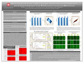
- 1. IGF-2 (ng/m l)IGF-2 (ng/m l) In this experiment we examined the proliferation and 5- integrin surface expression of the rhabdomyosarcoma (RD) cell line when cultured on collagen and fibronectin substrates. We also examined the impact of insulin-likeα growth factor 2 (IGF-2) on proliferation and 5- integrin surface expression. After culturing RD cells for 0, 3, and 7 days on collagen and fibronectin 2D matrices at concentrations of 5µg/mL and 10µg/mL per condition, ourα results indicate that the 10µg/mL fibronectin matrix facilitated the optimal proliferation. Interestingly, using FACS analysis, we identified a 20X increase in 5- integrin surface expression when cultured in both collagen andα fibronectin when compared to their respective controls. We continued our research by studying the effect of IGF-2 on RD cell proliferation and 5- integrin expression using a concentration of 100ng/mL. We found that there was no statistically significant impact on proliferation orα 5-integrin expression when compared to their respective controls. This leads us to believe there is a positive effect on 5- integrin surface expression considering when cultured with collagen and fibronectin alone, there is aα α decrease in surface expression. Confocal microscopy showed rapid and robust proliferation from day 0 to day 3 and day 7. Cell morphology was consistent with the material sheets being elongated and mutlinucleated. Fluorescence was uneven in several photos which we hypothesize to be attributed to an insufficient amount of Live/Dead stain to the amount of RD cells per culture. Future experiments should be performed to confirm and optimize this issue. We noticed standard deviations varying greatly when incubated with this stain and placed into a plate reader. Further optimization assays will be conducted in the future to address these issues. Future experiments include culturing RD cells with various concentrations of IGF-2 on collagen and fibronectin substrates. We would then continue studying its proliferative effects as well as it’s impact on 5- integrin surfaceα expression. We hope to expand this research by looking at overall integrin expression as indicated by ß1- integrin antibodies. Effects of Insulin-like Factor -2 (IGF-2), Collagen, and Fibronectin on the ProliferationEffects of Insulin-like Factor -2 (IGF-2), Collagen, and Fibronectin on the Proliferation andand αα5-Integrins Expression of the5-Integrins Expression of the Rhabdomyosarcoma-derived (RD) Cell LineRhabdomyosarcoma-derived (RD) Cell Line Burke, K.*, Siu, V.*, Soto-Velez, R. * • Tawil, B, PhD, Instructor • Biomedical Engineering 502 : SUMMER 2012Burke, K.*, Siu, V.*, Soto-Velez, R. * • Tawil, B, PhD, Instructor • Biomedical Engineering 502 : SUMMER 2012 * California State University Channel Islands • One University Drive • Camarillo, CA 93012* California State University Channel Islands • One University Drive • Camarillo, CA 93012 RD cells (from ATCC) cultured in RD media (DMEM w/ 10% FBS), washed with sterile Phosphate Buffered Saline (PBS) and trypsinized for 5 minutes at room temp (RT), post media-aspiration, were centrifuged (200rcf for 5 minutes) prior to resuspension of cell pellet, in 1 mL of RD media. Cell concentrations were calculated using measurements obtained from the Nexcelom T4 Cellometer. Incubation of RD cells onto 24-well plates were then performed first in 2D, then 3D, collagen and fibronectin (SIGMA ALDRICH) matrices. Incubation (37°C) was maintained for Days 0, 3, and 7. Proliferation was measured utilizing 150µL of live/dead Cell Viability assay (Invitrogen) added to each well and incubated for 20 minutes at RT. Confocal images were obtained using an Olympus IX-71 Inverted Microscope and QCapture pro software as well as reading average fluorescence intensity (494 nm ex / 517 nm em) over each well using a Molecular Devices F5 plate reader. FilterMax F5 Multi-mode Plate Reader Software (Molecular Devices) provided raw fluorescent read data. Raw data were analyzed and evaluated using Microsoft Excel. 300µL of thrombin was then added to each well for 3D matrices. 400uL of RD media was added to each well after 30 minutes, for a total amount of 1mL per well. -5 integrin expression was initiated with RD cellα culture in collagen and fibronectin, incubated in 5mL 0.25% trypsin-EDTA for 5min at RT, neutralized with RD media, centrifuged and decanted, with the pellet resuspended in RD media to 4x10⁴ /mL cell stock. 500uL of cell stock used to seed wells in 24-well plate to 2x10² cells/well, incubated for days 0 (1hr) and 7. Preparation for FACS analysis protocol (Guava EasyCyte Mini flow cytometer) began with aspirating wells, post incubation, adding 500µL 0.25% trypsin-EDTA, incubating for 5 min at RT and adding 500µL RD media, transferring cell solution to 1.5mL conical tube, and centrifuging. A single PBS wash during centrifugation was performed prior to resuspending in 100uL anti 5α -antibody solution (1-10 dilution of anti α5 Ab in PBS) and incubating in ice for 60 min. Three total PBS washes during centrifugation were performed, with a final cell pellet re-suspension in 300µL PBS. Experiments were repeated with the addition of IGF-2 to look at its effects on proliferation and a5-integrin surface expression. The addition of IGF-2 (100 ng/mL) to RD cells within a 2D collagen matrix versus a 2D fibronectin matrix, required five 24-well plates, each with collagen and fibronectin, measured at days 0, 3, & 7 for proliferation, and days 0 and 7 for FACS. MiniTab 14 software was used produce histograms, 2-sample t-tests, and response-surface computational models. Materials and MethodsMaterials and Methods Tumor metastasis, which involves the migration of cancer cells from the primary tumor to other organs, is responsible for a majority of cancer-related deaths. Identification of the growth factors which promote cancer cell proliferation, and of the integrins used by cancer cells for proliferation and motility, are key aspects in cancer research; as it is the first step in the development of compounds with the ability to block the activity or inhibit the expression of these integrins and growth factors. The purpose of this experiment is to determine if expression of 5- integrins is up-regulated in RD cells, and if IGF-2 has aα significant effect on RD cells proliferation. In some cancers, the interaction of 5- integrins with ECM proteins such as fibronectin are known to be involved inα proliferation, migration, motility, and metastasis, and in helping cancer cells survive chemotherapy. Up-regulation of growth factor signaling promotes cell proliferation and motility in soft tissue sarcomas, such as RMS. Insulin-like growth factor (IGF)- family proteins are involved in the initiation and maintenance of the cancer phenotype in many types of adult and childhood cancer. In particular, IGF-2 is overexpressed in RMS cells; and inhibition of its receptor IGF-2R with antibodies inhibits tumor cell growth. RD is a human cell line derived from RMS. RD cells secrete IGF-2 endogenously as an autocrine factor to enable proliferation and motility. This makes IGF-family proteins potential targets for anti-cancer therapies. IntroductionIntroduction AbstractAbstract Rhabdomyosarcoma (RMS) is a rare type of soft tissue sarcoma, more common in children. In order to develop treatments, our research aims to identify which integrins RMS cells use for motility, and which growth factors increase or decrease its proliferation. In this study, RD cells, derived from human RMS, were cultured in 2D fibronectin and collagen matrices. The effect of insulin- like growth factor-2 (IGF-2) in RD cell proliferation was observed using fluorescent microscopy, and measured using a Live/Dead stain. Response surface computational methods were used to construct a mathematical model of RD cell growth as a function of the concentration of IGF-2, and the concentration of protein substrate. Expression of 5- integrins was measuredα using flow cytometry using an anti- 5 antibody.α Experimental results indicate that IGF-2 upregulates RD cell proliferation cultured in vitro in 2D fibronectin matrices, but not when cultured in vitro in 2D collagen matrices. Results also show that the collagen concentration in the 2D matrix has a significant and positive effect on RD cell proliferation, while the fibronectin concentration does not. Finally, it was found that the expression of 5- integrins is up-regulated in RD cells treated with IGF-2α . Figure 1. Effect of IGF-2 in RD Cell Proliferation for Various Collagen Constructs - (a) Experimental results show that the presence of IGF-2 growth factor (100 ng/ml) did not have a significant effect on the proliferation of RD cells when grown in 2D collagen constructs. Graph is standardized at 100% fluorescence against Day 0. Experimental results also show that RD cell proliferation was greater at the lower concentration of collagen. (b) Response surface computational model for RD cell proliferation, as a function of both collagen concentration and IGF-2 concentration. Figure 4. Fluorescent micrographs depicting changes in cell density as a function of collagen concentration and presence / absence of IGF-2 growth factor, for three timepoints. * = equal contributor ResultsResults ConclusionsConclusions Figure 5. Fluorescent micrographs depicting changes in cell density as a function of fibronectin concentration and presence / absence of IGF- 2 growth factor, for three timepoints. Figure 2. Effect of IGF-2 in RD Cell Proliferation for Various Fibronectin Constructs - (a) Experimental results show that the presence of IGF-2 growth factor (100 ng/ml) had a significant effect on the proliferation of RD cells when grown in a 2D fibronectin construct. Graph is standardized at 100% fluorescence against Day 0. Experimental results also show that the concentration of fibronectin did not have a statistically significant effect on RD cell proliferation. (b) Response surface computational model for RD cell proliferation, as a function of both fibronectin concentration and IGF-2 concentration. (a) (b) (a) (b) Figure 3. 5 integrin expression in RD cells treated with IGF-2 growth factor (100 ng/ml) increases after 7 days, inα both collagen and fibronectin 2D constructs. Experimental GroupsExperimental Groups and Controlsand Controls 2D RD cells in Collagen or Fibronectin 2D matrix Cells /well ConditionsConditions Sample typeSample type 1x10⁴ Collagen 5µg/mL & 10µg/mL EXPERIMENTAL Fibronectin 5µg/mL & 10µg/mL No Collagen or Fibronectin CONTROL 0 Collagen or Fibronectin 3D Fibrinogen (F) + Thrombin (T) at various concentrations in 3D matrix Cells /well ConditionsConditions Sample typeSample type 2x10⁴ F10+T20 EXPERIMENTAL F10+T40 F20+T20 F20+T40 0 Same as above four conditions CONTROL Integrin Collagen and Fibronectin with the addition of 5 integrin antibodyα Cells /well ConditionsConditions Sample typeSample type 2x10² Collagen 5µg/mL & 10µg/mL EXPERIMENTAL Fibronectin 5 and 10 µg/mL No Collagen or Fibronectin, with Ab CONTROL No Collagen or Fibronectin, without Ab IGF-2 Collagen and Fibronectin with the addition of IGF-2 Growth Factor Cells /well ConditionsConditions Sample typeSample type 2.5x10⁴ Collagen 5µg/mL & 10µg/mL EXPERIMENTAL Fibronectin 5 and 10 µg/mL 0 No Collagen or Fibronectin without IGF-II CONTROL Fibronectin (µg/ml) IGF-2 up-regulates expression of 5 integrinsα