ONLY THE LAST QUESTION IS THE POINT OF POST. THE OTHER PAGES ARE B.pdf
Final Poster
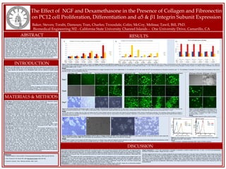
1. RESULTS
The Effect of NGF and Dexamethasone in the Presence of Collagen and Fibronectin
on PC12 cell Proliferation, Differentiation and α5 & β1 Integrin Subunit Expression
Baker, Steven; Torabi, Damoun; Tran, Charles; Trousdale, Colin; McCoy, Melissa; Tawil, Bill, PhD.
Biomedical Engineering 502 - California State University Channel Islands – One University Drive, Camarillo, CA
DISCUSSION
PC12 cells are derived from the rat adrenal medulla with an embryonic origin, and in the presence of Normal Growth Factor (NGF) will cause the cells to
differentiate creating long processes known as neurite varicosites (Figure 4). Conversely, dexamethasone will cause PC12 cells to differentiate into
chromaffin-like cells with shorter extensions. Our research tested the difference between these two compounds plated with either fibronectin or collagen
at concentrations of 5ug and 10ug. The experiment was carried out in both a 2D and 3D medium to look at all aspects of proliferation, differentiation and
integrin extensions.
Proliferation: 1. Cells treated with NGF and Dexamethasone on a collagen medium proliferated by day 4 in all conditions except collagen 5ug/mL and
NGF. From day 4 to 7 there was shown to be a decrease in proliferation in all conditions except collagen 5ug/mL plus NGF and collagen 10ug/mL plus
Dexamethasone. (Figure 1A). 2. Cells coated with fibronectin showed a decrease in proliferation on day 4 except for the condition of fibronectin at
5ug/mL and dexamethasone and uncoated NGF. On day 7 all cells showed an increase in proliferation. (Figure 1B). 3) In a 3D fibrin construct cells
proliferated from day 1 to 4 and a drastic decrease in proliferation from day 4 to day 7. (Figure 1C).
Differentiation: 1. Collagen was shown to have the greatest extensions on Day 7 on collagen 5ug/mL with NGF. (Figure 2A). 2. Fibronectin exhibited
no extensions on any of the condition (Figure 2B). 3. In a 3D construct no differentiation was observed (Figure 3).
Integrin Expression: 1. PC12 cells demonstrate a decrease in expression of Alpha-5 integrin from day 1 to day 7. 2. PC12 cells demonstrate a
decrease in expression of Beta-1 integrin from day 1 to day 7.
The data suggests that PC12 cells on collagen proliferate the best sometime before day 7, and past 7 days on fibronectin. Future studies should check
time points before and after day 4 to access proliferation on collagen. Fibronectin studies should extend the experiment past 7 days in order to determine
the maximum time of proliferation. Das and colleagues determined that cell differentiation in NGF will be at a maximum after 6 days time (2004).
However, our results showed the greatest differentiation on collagen at day 7. This conflict needs to be verified by observing the cells at day 6 and day 8.
The images of this experiment depict no extensions on fibronectin, future studies should verify the possibility of extensions on fibronectin (Rossino,
1990). Future studies will look at proliferation, differentiation, and integrin expression of PC12 cells in a fibrin 3D construct while influenced by NGF and
Dexamethasone.
Proliferation and differentiation are the most important steps when beginning any experiment involving
the use of living cells whether it be in vitro drug testing, genetic study or immunohistochemistry and in
many cases requires the utilization of an extracellular matrix and the influence of specific growth
factors.
The PC12 suspension cell line has been known to differentiate based upon the growth factor involved,
developing neurite varicosities when exposed to Nerve Growth Factor (NGF). PC12 cells have also
been shown to have a greater number of extensions when treated with cellular gangliosides (Li, 1998).
When treated with Dexamethasone PC12 cells differentiate chromaffin-like structures. However,
Jameson has shown this condition inhibits proliferation and neuriite extension. In an extracellular
matrix of fibronectin or an environment lacking an extracellular matrix in which it becomes difficult for
the PC12 cells integrins to attach, an introduced growth factor has been shown to have the function of
limiting the proliferation of cells with no evidence of differentiation of the cells. A collagen extracellular
matrix has been shown to be a greater candidate for cellular attachment of PC12 cells, most obviously
when exposed to NGF and showing neurite growth.
Immunohistochemistry is used to observe the secretion of collagen and fibronectin from PC12 cells
using collagen and fibronectin primary and secondary antibodies. FACS analysis techniques are used
to show Alpha-5 and Beta-1 integrin subunit antibodies to observe the expression of Alpha and Beta
integrin subunits in PC12 cells.
INTRODUCTION
ABSTRACT
PC12 is a cell line derived from the pheochromocytoma of the rat adrenal medulla, that have an
embryonic origin from the neural crest that has a mixture of neuroblastic cells and eosinophilic cells.
We observed proliferation vs differentiation ratio in fibronectin and collagen matrix in presence and
absence of Nerve Growth Factor (NGF) and Dexamethasone at Day 1, Day 4, and Day 7. A
constant concentration of NGF (50 ng/ml) and Dexamethasone (5µM) were used during the
experiment but for each collagen or fibronectin matrix, we had variations of 5µg/ml or 10µg/ml
coated wells and also uncoated wells. There is no evidence of differentiation in presence of NGF
and Dexamethasone in fibronectin coated wells. However, in collagen coated wells, the proliferation
clearly decreases and we can see an obvious differentiation of PC12 cells even in Day 4. NGF
alters neurite growth and Dexamethasone causes formation of chromaffin-like cells. The
proliferation rate was also measured in a 3D matrix environment. We also so observed the
expression of Alpha-5 and Beta-1 integrin subunits in PC12 Cells in 2D environment on Day 1
versus 3D environment on Day 7 by FACS analysis. Using the Immunohistochemistry technique to
observe the secretion of fibronectin and collagen by PC12 cells did not give us a clear result.
40Fibrinogen: 20Thrombin20Fibrinogen: 20Thrombin
Figure 3. PC12 Cells proliferation microscopy on day 1,
4 and 7 in 3D Fibrin construct PC12 cells seeded in a 3D
fibrin construct at 40mg Fibrinogen (F):20mg Thrombin (T)
or 20mg F:20mg T. Proliferation was imaged via inverted
microscope at Day 1, 4, and 7.
Results
1. No differentiation observed in either construct condition.
5ug Collagen + NGF 10ug Collagen + NGF 5ug Collagen+Dexamethasone 10ug Collagen+Dexamethasone 5ug Fibronectin + NGF 10ug Fibronectin + NGF 5ug Fibronectin+Dexamethasone 10ug Fibronectin+Dexamethasone
Day1
Day4
Day7
Figure 2. PC12 Cells proliferation microscopy on day 1, 4 and 7 on Collagen, Fibronectin and 3D Fibrin construct A) PC12 cells seeded on 5µg or 10µg collagen coated wells treated with either NGF at 50ng or Dexamethasone at 5mM. Proliferation was
imaged via inverted microscope at Day 1, 4, and 7. B) PC12 cells seeded on 5µg or 10µg fibronectin coated wells treated with either NGF 50ng or Dexamethasone at 5mM. Proliferation was imaged via inverted microscope at Day 1, 4, and 7.
Results
1. PC12 cells cultured on collagen (5µg and 10µg) and treated with NGF at 50ng exhibited extensive neurite growth while treatment with dexamethasone (5mM) induced chromaffin-like morphology. This morphology change indicates an increase in
differentiation which in turn decreases the proliferation rate. 2. PC12 cells cultured on fibronectin (5µg and 10µg) showed an increase in proliferation. Additionally, no neurite growth or chromaffin-like morphology was observed when treated with NGF at 50ng or
dexamethasone (5mM) respectively.
Figure 5. FACS analysis of α-5 and β-1 integrin subunit expression in PC12 cells in Day 1
(2D) medium and Day 7 (3D) medium.
Results
1. Both α -5 and β -1 expression decreases after 7 days in 3D fibrin construct.
β-1
Day1
Day7
MATERIALS & METHODS
Cell Culture: Pheochromocytoma (PC12) cells were grown in Roswell Park Memorial Institute
(RPMI) 1640 medium containing 10% heat inactivated horse serum and 5% FBS.
2D Proliferation: In the 2D proliferation assay, cells were seeded in 24-well plates initially coated
with 5ng or 10ng of fibronectin or collagen at an initial density of 10,000 cells and stored at 37oC.
After 1, 4, and 7 days, the cells were then extracted from the wells, placed into microfuge tubes, and
centrifuged for 3 minutes at 13.3k rpm. After centrifugation, the supernatant was discarded and the
cells were washed in 500µL PBS and centrifuged again at the same settings. Following this, the PBS
supernatant was discarded, the cells were resuspended in 150µL live-dead stain, and then allowed to
incubate at room temperature in the dark for 15 minutes. Fluorescent samples were quantified using
the FilterMax F5 multimode micro plate reader and imaged using Cy3 filter on an inverted
microscope fitted with QImaging camera and QCapture Pro imaging software. NGF and
Dexamethasone stock solutions were diluted in RPMI to obtain 30mL samples of 50ng NGF and 2µg
dexamethasone. The cells were then diluted into the NGF and dexamethasone solutions to obtain a
concentration of 60,000 cells/mL. 500µL of each solution was added to 24-well plates initially coated
with 5ng and 10ng of collagen or fibronectin to reach a seeding density of 10,000 cells per well. Cells
were then treated and analyzed after 1, 4, and 7 days of incubation as previously described.
3D Proliferation: In the 3D proliferation assay, cells were seeded in 24-well plates initially coated
with 20mg or 10mg Fibrin at an initial density of 10,000 cells and stored at 37oC. After 1, 4, and 7
days, wells were treated and analyzed as previously described.
Flow Cytometry: On Day 1, PC12 cells were removed from the incubator, transferred to a 15mL
falcon tube and centrifuged at 13.3k rpm for 3 minutes. The supernatant was discarded after
centrifugation and the cells were resuspended in trypsin. Fresh media was added to neutralize the
trypsin. 20µL of the cell solution was used to obtain the cell density of 5.9x10⁶ cells/mL. 25.42µL of
the cell solution was then added to 574µL of cold PBS. The cell/PBS solution was aliquot into 6 1.5
microfuge tubes at a volume of 100µL. 2.5µL of Alpha-5 antibody was added to the first two tubes
with the same volume of Beta-1 antibody being added to the next two tubes. The tubes were then
placed on ice and allowed to incubate in the dark for 1 hour. At this point, the fibrin clot for Day 7
analysis was made by adding 25.42µL cell solution to 874.58µL fibrinogen. 150µL of this
cell/fibrinogen solution was added to a 12-well plate along with 150µL thrombin and allowed to sit at
room temperature for 1 hour. 500µL of plain media was added to each well. After the 1 hour of
incubation with antibodies, the FACS analysis of each tube was conducted using the Guava
easyCyte apparatus. On Day 7, the cells were transferred to 1.5mL microfuge tubes and centrifuged
at 500 rpm to separate the cells from residual fibrin while preserving the integrin expression. The
media supernatant was then discarded and the cells were washed twice with 500µL PBS and
centrifuged at the same settings in between each wash. Steps regarding the addition of antibodies
and quantifying via flow cytometer were performed as described above.
Collagen 5+NGF Collagen 5+Dex Collagen 10+NFG Collagen 10+Dex Uncoated+NGF Uncoated+Dex
Day 0 100 100 100 100 100 100
Day 4 89.19342699 120.8813901 250.7410636 276.3739763 215.3933947 665.2361544
Day 6 114.3564977 78.16670864 174.7311828 368.7079163 111.5700121 288.5878686
-50
50
150
250
350
450
550
650
750
Proliferation of PC12 cells on collagen in the presence of NGF and Dexamethsone
Day 0
Day 4
Day 6
Fibronectin 5ug/mL+NGFFibronectin 5ug/mL+DexFibronectin 10ug/mL+NFGFibronectin 10ug/mL+Dex Uncoated+NGF Uncoated+Dex
Day 0 100 100 100 100 100 100
Day 4 79.39317954 376.893717 58.71634083 20.77693557 245.987977 88.5742362
Day 6 559.6339017 2183.763946 1332.725931 383.1009746 666.8191323 768.2597803
-500
0
500
1000
1500
2000
2500
Proliferation of PC12 cells on fibronectin in the presence of NGF and Dexamethsone
Day 0
Day 4
Day 6
Fibrin40 + Cells Fibrin40 - Cells Fibrin20 + Cells Fibrin20 - Cells Cell+Media Media
Day0 100 100 100 100 100 100
Day4 255.9037641 250.5743799 252.3017789 246.5465376 104.0298226 215.5080406
Day7 34.2879317 64.88969468 7.396482028 66.09679821 17.42945384 54.36498295
-50
0
50
100
150
200
250
300
CellProliferation
PC12 Cell Proliferation in 3D Fibrin
Day0
Day4
Day7
Figure 1. PC12 Cells proliferation data on day 1, 4 and 7 on Collagen, Fibronectin and in3D Fibrin construct. A). PC12 cells seeded on 5µg or 10µg collagen coated wells treated with either NGF at 50ng or Dexamethasone at 5mM. Proliferation was measured via microplate reader at Day 1, 4, and 7. B) PC12
cells seeded on 5µg or 10µg fibronectin coated wells treated with either NGF 50ng or Dexamethasone at 5mM. Proliferation was measured via microplate reader at Day 1, 4, and 7. C) PC12 cells seeded in a 3D fibrin construct at 40mg Fibrinogen (F):20mg Thrombin (T) or 20mg F:20mg T. Proliferation was measured
via microplate reader at Day 1, 4, and 7.
Results
1. The proliferation of PC12 cells treated with NGF and Dexamethasone on collagen decreased by Day 7 due to differentiation. 2. The proliferation of PC12 cells treated with NGF and dexamethasone on fibronectin increased by day 7. 3. The proliferation of untreated PC12 cells on fibrin clot increases on Day 4 and a
decrease on day 7 due to the breakdown of the clot
A) B) C)
Figure 4 Brightfield images comparing differentiation of PC12 cells on collagen treated with NGF versus dexamethasone. A&B) PC12 cells treated with NGF at 50ng and cultured on wells coated with
collagen exhibit neurite growth. C&D) PC12 cells treated with dexamethasone at 2mM on collagen exhibits chromaffin-like morphology.
Results
1. PC12 cells in images A and B treated with NGF (50ng) and grown on collagen (5ug) coated well displayed neurite extensions on day 7 and 4 respectively. 2. PC12 cells treated with dexamethasone (5mM)
and grown on collagen (5ug) coated well become chromaffin-like by day 7 (image C and D at 100x and 200x respectively)
A) B) C) D)
References
1. Jameson,Seidler FJ,Qiao D,Slotkin TA Neuropsychopharmacology. 2006 Aug;31(8):1647-58
2. Das, Freudenrich TM, Mundy WR. 2004 Neurotoxicol Teratol. 26(3) 397-406.
3. Rossino P, Gavazzi, Timpl. 1990 Exp Cell Res. 189(1) 100-8.
α-5
Day1
Day7