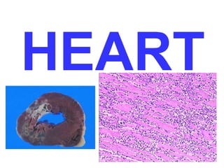
SlideShare a Scribd company logo
1 of 130
HEART
THE HEART
• Normal
• Pathology
– Heart Failure: L, R
– Heart Disease
•
•
•
•
•
•
•
•

Congenital: LR shunts, RL shunts, Obstructive
Ischemic: Angina, Infarction, Chronic Ischemia, Sudden Death
Hypertensive: Left sided, Right sided
Valvular: AS, MVP, Rheumatic, Infective, Non-Infective,
Carcinoid, Artificial Valves
Cardiomyopathy: Dilated, Hypertrophic, Restrictive, Myocarditis,
Other
Pericardium: Effusions, Pericarditis
Tumors: Primary, Effects of Other Primaries
Transplants
NORMAL Features
•
•
•
•
•

6000 L/day
250-300 grams
40% of all deaths (2x cancer)
Wall thickness ~ pressure
(i.e., a wall is only as thick as it has to be)
– LV=1.5 cm
– RV= 0.5 cm
– Atria =.2 cm

• Systole/Diastole
• Starling’s Law
TERMS
• CARDIO”MEGALY”
– DILATATION, any chamber, or all
– HYPERTROPHY, and chamber, or all
STRIATIONS

NUCLEUS

DISCS

SARCOLEMMA

SARC. RETIC.

MITOCHONDRIA

ENDOTHELIUM

FIBROBLASTS

GLYCOGEN

A.N.P.
S.A. NodeAV NodeBundle of HIS L. Bundle, R. Bundle
AX

Anterior
Lateral
Posterior
Septal

VLA

HLA
VALVES
• AV:
– TRICUSPID
– MITRAL

13 cm
11 cm

• SEMILUNAR:
– PULMONIC
– AORTIC

8 cm
6 cm
CARDIAC AGING
Chambers

Epicardial Coronary
Arteries

Increased left atrial cavity size

Tortuosity

Decreased left ventricular cavity size

Increased cross-sectional luminal area

Sigmoid-shaped ventricular septum

Calcific deposits
Atherosclerotic plaque

Myocardium
Valves
Aortic valve calcific deposits
Mitral valve annular calcific deposits
Fibrous thickening of leaflets
Buckling of mitral leaflets toward the left atrium

Increased mass
Increased subepicardial fat

Brown atrophy
Lipofuscin deposition

Basophilic degeneration (glyc.)
Amyloid deposits
CARDIAC AGING
Aorta
Dilated ascending aorta with rightward shift
Elongated (tortuous) thoracic aorta
Sinotubular junction calcific deposits
Elastic fragmentation and collagen accumulation
Atherosclerotic plaque
BROWN
ATROPHY, HEART

LIPOFUCSIN
Pathologic Pump Possibilities
• Primary myocardial failure
(MYOPATHY)
• Obstruction to flow (VALVE)
• Regurgitant flow (VALVE)
• Conduction disorders (CONDUCTION
SYSTEM)
• Failure to contain blood (WALL
INTEGRITY)
• DEFINITION
• TRIAD

CHF

– 1) TACHYCARDIA
– 2) DYSPNEA
– 3) EDEMA
• FAILURE of Frank Starling mechanism
• HUMORAL FACTORS
– Catecholamines (nor-epinephrine)
– ReninAngiotensionAldosterone
– Atrial Natriuretic Polypeptide (ANP)

• HYPERTROPHY and DILATATION
HYPERTROPHY
• PRESSURE OVERLOAD (CONCENTRIC)
• VOLUME OVERLOAD (CHF)
• LVH, RVH, atrial, etc.
• 2X normal weight ischemia
• 3X normal weight HTN
• >3X normal weightMYOPATHY, aortic
regurgitation
CHF: Autopsy Findings
• Cardiomegaly
• Chamber Dilatation
• Hypertrophy of myocardial fibers,
BOXCAR nuclei
Left Sided Failure
• Low output vs. congestion
• Lungs
– pulmonary congestion and edema
– heart failure cells

• Kidneys
– pre-renal azotemia
– salt and fluid retention
• renin-aldosterone activation
• natriuretic peptides

• Brain: Irritability, decreased attention,
stuporcoma
Left Heart Failure Symptoms
• Dyspnea
– on exertion
– at rest

• Orthopnea
– redistribution of peripheral edema fluid
– graded by number of pillows needed

• Paroxysmal Nocturnal Dyspnea (PND)
LEFT Heart Failure
Dyspnea

Orthopnea
PND (Paroxysmal Nocturnal
Dyspnea)
Blood tinged sputum
Cyanosis
Elevated pulmonary “WEDGE”
pressure (PCWP) (nl = 2-15 mm Hg)
Right Sided Heart Failure
• Etiology
– left heart failure
– cor pulmonale

• Symptoms and signs
– Liver and spleen
• passive congestion (nutmeg liver)
• congestive spleenomegaly
• ascites

– Kidneys
– Pleura/Pericardium
• pleural and pericardial effusions
• transudates

– Peripheral tissues
RIGHT Heart Failure
FATIGUE
“Dependent” edema
JVD
Hepatomegaly (congestion)
ASCITES, PLEURAL EFFUSION
GI
Cyanosis
Increased peripheral venous pressure
(CVP) (nl = 2-6 mm Hg)
HEART DISEASE
• CONGENITAL (CHD)
•
•
•
•

ISCHEMIC (IHD)
HYPERTENSIVE (HHD)
VALVULAR (VHD)
MYOPATHIC (MHD)
CONGENITAL HEART
DEFECTS

• Faulty embryogenesis (week 3-8)
• Usually MONO-morphic (i.e., SINGLE
lesion) (ASD, VSD, hypo-RV, hypo-LV)
• May not be evident until adult life
(Coarctation, ASD)
• Overall incidence 1% of USA births
• INCREASED simple early detection via
non invasive methods, e.g., US, MRI,
CT, etc.
Incidence per Million Live
Births

%

4482

42

1043

10

Pulmonary stenosis

836 

8 

Patent ductus arteriosus

781 

7 

Tetralogy of Fallot

577 

5 

Coarctation of aorta

492 

5 

Atrioventricular septal defect
Aortic stenosis

396 

4 

388 

4 

Transposition of great arteries
Truncus arteriosus
Total anomalous pulmonary venous connection
Tricuspid atresia

388 

4 

136 

1 

120 

1 

Malformation
Ventricular septal defect
Atrial septal defect
GENETICS
• Gene abnormalities in only 10% of CHD
• Trisomies 21, 13, 15, 18, XO
• Mutations of genes which encode for
transcription factorsTBX5ASD,VSD
 NKX2.5ASD
• Region of chromosome 22 important in
heart development, 22q11.2
deletionconotruncus, branchial arch,
face
ENVIRONMENT
• RUBELLA
• TERATOGENS
CHD

• LR SHUNTS: all “D’s” in their names
– NO cyanosis
– Pulmonary hypertension
– SIGNIFICANT pulmonary hypertension is
IRREVERSIBLE

• RL SHUNTS: all “T’s” in their names
– CYANOSIS (i,.e., “blue” babies)
– VENOUS EMBOLI become SYSTEMIC
“paradoxical”
• OBSTRUCTIONS: aorta or pulomnary
artery
• ASD
• VSD
• ASVD
• PDA

LR

NON CYANOTIC

IRREVERSIBLE
PULMONARY
HYPERTENSION
IS THE MOST
FEARED
CONSEQUENCE
ASD
• NOT patent foramen ovale
• Usually asymptomatic until adulthood
• SECUNDUM (90%): Defective fossa
ovalis
• PRIMUM (5%): Next to AV valves, mitral
cleft
• SINUS VENOSUS (5%): Next to SVC
with anomalous pulmonary veins
draining to SVC or RA
VSD
•
•
•
•
•

By far, most common CHD defect
Only 30% are isolated
Often with TETRALOGY of FALLOT
90% involve the membranous septum
If muscular septum is involved, likely to
have multiple holes
• SMALL ones often close spontaneously
• LARGE ones progress to pulmonary
hypertension
PDA
• 90% isolated
• HARSH, machinery-like murmur
• LR, possibly RL as pulmonary
hypertension approaches systemic
pressure
• Closing the defect may be life saving
• Keeping it open may be life saving
(Prostaglandin E1). Why? Ans: TGA,
TA, TAPVC
AVSD

• Associated with defective,
inadequate AV valves
• Can be partial, or COMPLETE
(ALL 4 CHAMBERS FREELY
COMMUNICATE)
RL
• Tetralogy of Fallot
• Transposition of great arteries
• Truncus arteriosus
• Total anomalous pulmonary venous
connection

• Tricuspid atresia
RL SHUNTS
• TETRALOGY of FALLOT most COMMON
– 1) VSD, large
– 2) OBSTRUCTION to RV flow
– 3) Aorta OVERRIDES the VSD
– 4) RVH
– SURVIVAL DEPENDS on SEVERITY of
SUBPULMONIC STENOSIS
– Can be a “PINK” tetrology if pulmonic
obstruction is small, but the greater the
obstruction, the greater is the RL shunt
TGA (TRANSPOSITION
of GREAT ARTERIES)
• NEEDS a SHUNT for
survival, obviously
– PDA or PFO (65%),
“unstable” shunt
– VSD (35%), “stable” shunt
– RV>LV in thickness
– Fatal in first few months
– Surgical “switching”
TRUNCUS ARTERIOSIS
TRICUSPID ATRESIA
• Hypoplastic RV
• Needs a shunt, ASD, VSD, or PDA
• High mortality
Total Anomalous Pulmonary
Venous Connection (TAPVC)
• PULMONARY VEINS do NOT go into
LA, but into L. innominate v. or
coronary sinus
• Needs a PFO or a VSD
• HYPOPLASTIC LA
OBSTRUCTIVE CHD
• COARCTATION of aorta
• Pulmonary stenosis/atresia
• Aortic stenosis/atresia
COARCTATION of AORTA
•
•
•
•
•

M>F
But XO’s frequently have it
INFANTILE FORM (proximal to PDA) (SERIOUS)
ADULT FORM (CLOSED DUCTUS, i.e., NO PDA)
Bicuspid aortic valve 50% of the time
PULMONIC
STENOSIS/ATRESIA
• If 100% atretic, hypoplastic RV with ASD
• Clinical severity ~ stenosis severity
AORTIC
STENOSIS/ATRESIA
• VALVULAR
– If severe, hypoplastic LVfatal

• SUB-valvular (subaortic)
– Aortic wall THICK BELOW cusps

• SUPRA-valvular
– Aortic wall THICK ABOVE cusps in
ascending aorta
HEART DISEASE
• CONGENITAL (CHD)

• ISCHEMIC (IHD)
• HYPERTENSIVE (HHD)
• VALVULAR (VHD)
• MYOPATHIC (MHD)
SYNDROMES of IHD
•
•
•
•

Angina Pectoris: Stable, Unstable
Myocardial Infarction (MI, AMI)
Chronic IHD CHF (CIHD)
Sudden Cardiac Death (SCD)

• “Acute” Coronary Syndromes:
– UNSTABLE ANGINA
– AMI
– SCD (Sudden Cardiac Death)
IHD RISK
• Number of plaques
• Distribution of plaques
• Size, structure of plaques
ACUTE CORONARY SYNDROMES
• “The acute coronary syndromes are
frequently initiated by an
unpredictable and abrupt conversion
of a stable atherosclerotic plaque to
an unstable and potentially lifethreatening atherothrombotic lesion
through superficial erosion,
ulceration, fissuring, rupture, or deep
hemorrhage, usually with
superimposed thrombosis.”
EPIDEMIOLOGY
(same as atherosclerosis)

• ½ million die of IHD yearly in USA
• 1 million in 1963. Why?
– Prevention of control controllable risk factors
– Earlier, better diagnostic methods
– PTCA, CABG, arrythmia control

• 90% of IHD patients have
ATHEROSCLEROSIS (no surprise here)
ACUTE CORONARY
SYNDROME FACTORS
• ACUTE PLAQUE CHANGE *******
• Inflammation
• Thrombus
• Vasoconstriction
******* MOST IMPORTANT
ACUTE PLAQUE CHANGE
• Rupture/Refissuring
• Erosion/Ulceration, exposing ECM
• Acute Hemorrhage

NB: Plaques do NOT have to be severely stenotic to
cause acute changes, i.e., 50% of AMI results from
thromboses of plaques showing LESS THAN 50%
stenosis
INFLAMMATION
• Endothelial cells release CAMs,
selectins
• T-cells release TNF, IL-6, IFN-gamma to
stimulate and activate endothelial cells
and macrophages
• CRP (α-2 globulin) predicts the
probability of damage in angina
patients
THROMBUS
• Total occlusion
• Partial
• Embolization
VASOCONSTRICTION
• Circulating adrenergic agonists, i.e., α
• Platelet release products, e.g., ADP
• Endothelially released factors, such as
endothelin
Coronary Artery Pathology in Ischemic Heart Disease
Syndrome

Plaque
Stenoses Disruption

Stable angina

>75%

No

No

Unstable angina

Variable

Frequent

Nonocclusive, often with thromboemboli

Transmural
myocardial infarction

Variable

Frequent

Occlusive

Subendocardial
myocardial infarction

Variable

Variable

Widely variable, may be absent,
partial/complete, or lysed

Sudden death

Usually
severe

Frequent

Often small platelet aggregates or thrombi
and/or thromboemboli

Plaque-Associated Thrombus
ANGINA PECTORIS
•
•
•
•
•

Paroxysmal (sudden)
Recurrent
15 sec.15 min.
Reduced perfusion, but NO infarction
THREE TYPES
– STABLE: relieved by rest or nitro
– PRINZMETAL: SPASM is main feature, responds to
nitro, S-T elevation.
Often younger with
people not much atherosclerotic narrowing.
– UNSTABLE (crescendo, PRE-infarction, Q-wave
angina): perhaps some thrombosis, perhaps some
non transmural necrosis, perhaps some
embolization, but DISRUPTION of PLAQUE is
universally agreed upon
MYOCARDIAL INFARCTION
• Transmural vs. Subendocardial (inner 1/3)
• DUH! EXACT SAME risk factors as
atherosclerosis
• Most are TRANSMURAL, and MOST are
caused by coronary artery occlusion
• In the 10% of transmural MIs NOT associated
with atherosclerosis:
– Vasospasm
– Emboli, e.g., mural thrombus
– UNexplained
MYOCARDIAL RESPONSE
Feature

Time

Onset of ATP depletion

Seconds

Loss of contractility

<2 min

ATP reduced
to 50% of normal  

10 min

to 10% of normal  

40 min

Irreversible cell injury

20–40 min

Microvascular injury

>1 hr
PROGRESSION OF NECROSIS
TIMING of Gross and Microscopic Findings

GROSS

MICROSCOPIC

½–4 hr

None

Usually none; variable waviness of fibers at border

4–12 hr

Occasionally dark mottling

Beginning coagulation necrosis; edema; hemorrhage


12–24 hr

Dark mottling

Ongoing coagulation necrosis; pyknosis of nuclei;
myocyte hypereosinophilia; marginal contraction
band necrosis; beginning neutrophilic infiltrate


1–3 days

Mottling with yellow-tan
infarct center

Coagulation necrosis, with loss of nuclei and striations;
interstitial infiltrate of neutrophils


3–7 days

Hyperemic border; central
yellow-tan softening

Beginning disintegration of dead myofibers, with dying
neutrophils; early phagocytosis of dead cells by
macrophages at infarct border


7–10
days

Maximally yellow-tan and
soft, with depressed redtan margins

Well-developed phagocytosis of dead cells; early
formation of fibrovascular granulation tissue at
margins


10–14
days

Red-gray depressed infarct
borders

Well-established granulation tissue with new blood
vessels and collagen deposition


2–8 wk

Gray-white scar, progressive
from border toward core
of infarct

Increased collagen deposition, with decreased
cellularity


>2 mo

Scarring complete

Dense collagenous scar
1 day (pyknosis, “waviness”)
3-4 days (neutrophils)
7 days (macrophages)
Weeks (organization)
Months (fibrosis)
RE-PERFUSION

• Thrombolysis
• PTCA
• CABG

• Reperfusion CANNOT restore necrotic
or dead fibers, only reversibly injured
ones, and prevent further necrosis.
• REPERFUSION “INJURY”
– Free radicals
– Interleukins
AMI DIAGNOSIS
• SYMPTOMS
• EKG 1) Q-waves, 2) T-wave inversion, 3)

ST-T

elevation

• DIAPHORESIS
• (10% of MIs are “SILENT” with Qwaves)
• CKMB gold standard enzyme
• Troponin-I, Troponin-T better
• CRP predicts risk of AMI in angina
patients
COMPLICATIONS
•
•
•
•
•
•
•
•
•

Wall motion abnormalities
Arrhythmias
Rupture (4-5 days)
Pericarditis
RV infarction
Infarct extension
Mural thrombus
Ventricular aneurysm
Papillary muscle dysfunction
(regurgitation)
• CHF
CIHD, aka, ischemic
“cardiomyopathy”
• Progress to CHF often with no
pathologic or clinical evidence of
localized infarction
– Extensive atherosclerosis
– No infarct
– Hypertrophy & Dilatation present
SUDDEN CARDIAC DEATH
• 350,000 in USA yearly from atherosclerosis
• NON-atherosclerotic sudden cardiac death includes:

– Congenital coronary artery disease
– Aortic stenosis
– MVP, i.e., mitral valve prolapse
– Myocarditis
– Cardiomyopathy (sudden death in young athletes)
– Pulmonary hypertension
– *Conduction defects
– *HTN, hypertrophy of UNKNOWN etiology
AUTOPSY findings in SCD
• >75% narrowing of 1-3 vessels
• Healed infarcts 40%
• “ARRHYTHMIA” is often a very
convenient conclusion when no
anatomic findings are present, i.e.,
“wastebasket” diagnosis
HEART DISEASE
• CONGENITAL (CHD)
• ISCHEMIC (IHD)

• HYPERTENSIVE (HHD)
• VALVULAR (VHD)
• MYOPATHIC (MHD)
HHD (Left)
• DEFINITION: Hypertrophic
adaptive response of the heart,
which can progress:
– Myocardial dysfunction
– Cardiac dilatation
– CHF
– Sudden death
NEEDED for DIAGNOSIS:
• LVH (LV>2.0 and/or Heart>500 gm.)
• HTN (>140/90)
PREVALENCE:
• WHAT % of USA people have
hypertension?
PREVALENCE:
• WHAT % of USA people have
hypertension?

•

Answer:

25%
HISTOPATHOLOGY
• INCREASED FIBER (MYOCYTE)
THICKNESS

• INCREASED nuclear size with
increased “blockiness” (boxcar nucleus)
CLINICAL
• EKG in LVH
Summary of LVH Criteria
1) R-I + S-III >25 mm
2) S-V1 + R-V5 >35 mm
3) ST-T depr. in L lead
4) R-wave in L lead >11 mm
5) LAE + other criteria
Positive Criteria: 1=possible
2=probable 3=definite

ATRIAL FIBRILLATION

Why?*

CHF, cardiac dilatation, pulmonary
venous congestion and dilatation
COURSE:
• NORMAL longevity, death from
other causes
• Progressive IHD
• Progressive renal damage,
hemorrhagic CVA (Which arteries?)
• CHF
HHD (Right) = COR PULMONALE
• ACUTE: Massive PE (No RVH)
• CHRONIC: COPD, CRPD,
Pulmonary artery disease, chest
wall motion impairment (RVH)
Diseases of the Pulmonary
Parenchyma
Chronic obstructive pulmonary disease

Disorders Affecting Chest Movement
Kyphoscoliosis
Marked obesity (pickwickian syndrome)
Neuromuscular diseases

Diffuse pulmonary interstitial fibrosis
Pneumoconioses
Cystic fibrosis
Bronchiectasis

Disorders Inducing Pulmonary Arterial
Diseases of the Pulmonary Vessels
Constriction
Metabolic acidosis
Recurrent pulmonary thromboembolism
Hypoxemia
Primary pulmonary hypertension
Chronic altitude sickness
Extensive pulmonary arteritis (e.g., Wegener  Obstruction to major airways
Idiopathic alveolar hypoventilation
granulomatosis)
Drug-, toxin-, or radiation-induced vascular 
obstruction
Extensive pulmonary tumor microembolism
HEART DISEASE
• CONGENITAL (CHD)
• ISCHEMIC (IHD)
• HYPERTENSIVE (HHD)

• VALVULAR (VHD)
• MYOPATHIC (MHD)
V HD
alvular

• Opening problems:
•

Stenosis
Closing problems: Regurgitation
or Incompetence or “insufficiency” (as
opposed to coronary “insufficiency”)
• AS

70% of all VHD

– Calcification of a deformed valve
– “Senile” calcific AS
– Rheum, Heart Dis.

• MS
–Rheumatic Heart Disease
AORTIC STENOSIS
2X gradient pressure
LVH (but no hypertension), ischemia
Cardiac decompensation, angina, CHF
50% die in 5 years if angina present
50% die in 2 years if CHF present
MITRAL ANNULAR
CALCIFICATION
• Calcification of the
mitral “skeleton”
• Usually NO
dysfunction
• Regurgitation
usually, but
Stenosis possible
• F>>M
REGURGITATIONS

• AR

– Rheumatic
– Infectious
– Aortic dilatations
• Syphilis
• Rheumatoid Arthritis
• Marfan

• MR

–MVP
–
–
–
–

Infectious
Fen-Phen
Papillary muscles, chordae tendinae
Calcification of mitral ring (annulus)
Mitral Valve Prolapse (MVP)
• MYXOMATOUS degeneration of the
mitral valve
• Associated with connective tissue
disorders
• “Floppy” valve

• 3% incidence, F>>M
• Easily seen on echocardiogram
MVP: CLINICAL FEATURES
•
•
•
•
•
•

Usually asymptomatic
Mid-systolic “click”
Holosystolic murmur if regurg. present
Occasional chest pain, dyspnea
97% NO untoward effects
3% Infective endocarditis, mitral
insufficiency, arrythmias, sudden death
RHEUMATIC Heart Disease
• Follows a group A strep infection, a
few weeks later
• DECREASE in “developed” countries
• PANCARDITIS: 1) Endocarditis,
2) Myocarditis, 3) Pericarditis
ACUTE:
-Inflammation
-Aschoff bodies
-Anitschkow cells
-Pancarditis
-Vegetations on
chordae tendinae at
leaflet junction

CHRONIC:
THICKENED VALVES
COMMISURAL FUSION
THICK, SHORT,
CHORDAE TENDINAE
CLINICAL FEATURES
•
•
•
•
•

Migratory Polyarthritis
Myocarditis
Subcutaneous nodules
Erythema marginatum
Sydenham chorea
INFECTIOUS ENDOCARDITIS
• Microbes
–

Usually

strep viridans

– Often Staph aureus in IVD users
– Enterococci

– HAČEK (normal oral flora, gram - , in children)
•
•
•
•
•

Hemophilus influenzae
Actinobacillus
Cardiobacterium
Eikenella
Kingella

– Fungi, rickettsiae, chlamydia
INFECTIOUS ENDOCARDITIS
• Acute: 50% mortality (course=days)

• SUB-acute: LOW mortality (course=weeks)
VEGETATIONS
• INFECTIVE >5mm
• NON-Infective <5mm
DIAGNOSIS=MMm, Mmmm, mmmmm
• MAJOR
•
•
•

•
•
•
•
•
•
•

Positive blood culture(s) indicating characteristic organism or persistence of unusual organism
Echocardiographic findings, including valve-related or implant-related mass or abscess, or
partial separation of artificial valve
New valvular regurgitation

minor
Predisposing heart lesion or intravenous drug use
Fever
Vascular lesions, including arterial petechiae, subungual/splinter hemorrhages, emboli, septic
infarcts, mycotic aneurysm, intracranial hemorrhage, Janeway lesions
Immunologic phenomena, including glomerulonephritis, Osler nodes, Roth spots, rheumatoid
factor
Microbiologic evidence, including single culture showing uncharacteristic organism
Echocardiographic findings consistent with but not diagnostic of endocarditis, including new
valvular regurgitation, pericarditis
NON-infective VEGETATIONS
• <5 mm
• PE
• Trousseau syndrome (migratory
thrombophlebitis with malignancies)
• s/p Swan-Ganz
• Libman-Saks with SLE (both sides of
valve)
•
•
•
•

Carcinoid Syndrome
Episodic skin flushing
Cramps
Nausea & Vomiting
Diarrhea

• ↑serotonin, ↑ 5HIAA in urine
• FIBROUS INTIMAL THICKENING
– RV, Tricuspid valve, Pulmonic valve (all RIGHT side)
– Similar to what Fen-Phen does on the LEFT side
ARTIFICIAL VALVES
• Mechanical
• Xenografts (porcine)
• 60% have complications within
10 years
HEART DISEASE
• CONGENITAL (CHD)
• ISCHEMIC (IHD)
• HYPERTENSIVE (HHD)
• VALVULAR (VHD)

• MYOPATHIC (MHD)
• PERICARDIAL DISEASE
CARDIOMYOPATHIES
•
•
•
•
•
•
•

Inflammatory
Immunologic
Metabolic
Dystrophies
Genetic
Idiopathic
Toxic

• DILATED (DCM)
– SY-stolic dysfunction

• HYPERTROPHIC (HCM)
– DIA-stolic dysfunction

• RESTRICTIVE (RCM)
– DIA-stolic dysfunction
Functional
Pattern
Dilated

LVEF
<40%

Mechanisms of
Heart Failure
Impairment of

contractility
(systolic
dysfunction)

Hypertrop
hic

50–

Impairment of

80%

compliance
(diastolic
dysfunction)

Restrictive 45–

90%

Impairment of

compliance
(diastolic
dysfunction)

Causes

Indirect
Myocardial
Dysfunction (Not
Cardiomyopathy)

Idiopathic; alcohol;
peripartum; genetic;
myocarditis;
hemochromatosis;
chronic anemia;
doxorubicin
(Adriamycin);
sarcoidosis

Ischemic heart
disease; valvular
heart disease;
hypertensive
heart disease;
congenital heart
disease

Genetic; Friedreich
ataxia; storage
diseases; infants of
diabetic mothers

Hypertensive
heart disease;
aortic stenosis

Idiopathic;
amyloidosis;
radiation-induced
fibrosis

Pericardial
constriction
Cardiac Infections

Toxins

Metabolic

Viruses: Cox-B, esp.

Alcohol

Hyperthroidism

Chlamydia

Cobalt

Hypothyroidism

Rickettsia

Catecholamines (stress)

Hyperkalemia

Bacteria

Carbon monoxide

Hypokalemia

Fungi

Lithium

Protozoa

Hydrocarbons

Nutritional deficiency (protein,
thiamine, other avitaminoses)

Arsenic

Neuromuscular
Disease
Friedreich ataxia (frataxin)
Muscular dystrophy
Congenital atrophies

Immunologic
Myocarditis (several
forms)
Post-transplant rejection

Hemochromatosis

Cyclophosphamide

Infiltrative

Doxorubicin (Adriamycin)
and daunorubicin

Leukemia
Carcinomatosis

Storage Disorders and Sarcoidosis
Other Depositions
Radiation-induced
Hunter-Hurler syndrome
(Dermatan-Heparan)
Glycogen storage disease
Fabry disease (glyco-lipid)
Amyloidosis

fibrosis
DILATED cardiomyopathy
•
•
•
•
•
•

Chamber thickness (not just LVH)
Adults
Progressively declining LVEF
LVEF ~ prognosis
50% die in 2 years
3 Main causes
– Myocarditis
– ETOH
– Adriamycin
DCM

Path:
4 chamber dilatation
Hypertrophy, also 4 chambers
Interstitial Fibrosis
Arrhythmogenic Right Ventricular Cardiomyopathy
(Arrhythmogenic Right Ventricular Dysplasia)

This is an uncommon dilated cardiomyopathy predominantly RIGHT ventricle.
So is NAXOS syndrome.

Wooly Hair

Palmoplantar keratoderma
HYPERTROPHIC cardiomyopathy
• Also called IHSS, (Idiopathic Hypertrophic
Subaortic Stenosis)
– GENETIC defects involving:

• Beta-myosin heavy chain
• Troponin T
• Alpha-tropomyosin
• Myosin binding protein C

– PATHOLOGY: Massive hypertrophy, Asymmetric
septum, DISARRAY of myocytes, INTERSTITIAL
fibrosis
– CLINICAL:
filling

↓chamber volume, ↓SV, ↓ diastolic
RESTRICTIVE cardiomyopathy
•
•
•
•

(idiopathic)
↓ ventricular compliance
Chiefly affects DIASTOLE
NORMAL chamber size and wall
thickness
• THREE similar diseases affecting
predominantly the
SUBENDOCARDIAL area:
– Endomyocardial Fibrosis (African
children)
– Loeffler Endomyocarditis (eosinophilic
leukemia)
– Endocardial Fibroelastosis (infants)
MYOCARDITIS
• INFLAMMATION of MYOCARDIUM
• Chiefly microbial

–COXACKIE A & B, CMV, HIV
–
–
–
–
–

Trypanosoma cruzi (Chagas dis.), 80%
Trichinosis
Toxoplasmosis
Lyme disease (5%)
Diphtheria

• IMMUNE: Post-viral, rheumatic, SLE, drug
hypersensitivityalpha-methyl dopa, sulfas
LYMPHOCYTIC INFILTRATES are the USUAL pattern of ALL myocarditis, but
eosinophils, giant cells, and even trypanosomes can be seen occasionally
OTHER Myocarditides
•
•
•
•

Adriamycin
Cyclophosphamide
Catecholamines (Pheochromocytomas)
Amyloid, systemic or primary cardiac
– Congo red stain: green birefringence with
polarization

• Amyloid, aging
– Congo red stain: green birefringence with
polarization

• Hemochromatosis (Prussian Blue)

• BOTH

HYPER-, HYPO- -thyroidism
PERICARDIUM
• Normally 30-50 ml clear serous fluid
– Visceral (epicardium)
– Parietal (fibrous pericardium)
– PERICARDIAL EFFUSIONS TAMPONADE
• Ruptured MI
• Traumatic perforation
• Ruptured aortic dissection
PERICARDITIS
• SEROUS: Rheum. Fever (RF), SLE,
scleroderma, tumors, uremia

• FIBRINOUS: MI (Dressler), uremia, radiation,
RF, SLE, s/p open heart surgery

•
•
•
•

PURULENT: infective, bacterial
HEMORRHAGIC: Malignancy, TB
CASEOUS: TB
CHRONIC: (ADHESIVE, CONSTRICTIVE)
TUMORS

• 90% benign “mesenchymal”, i.e., stromal

– MYXOMAS (LEFT ATRIUM MOST
COMMON)
–
–
–
–

FIBROMAS
LIPOMAS
FIBROELASTOMAS (valvular, usually papillary)
RHABDOMYOMA (Most common cardiac tumor in
children)

• 10% SARCOMAS
MYXOMA
Cardiac effects of NON-cardiac tumors
• Direct Consequences of Tumor
– Pericardial and myocardial metastases
– Large vessel obstruction
– Pulmonary tumor emboli

• Indirect Consequences of Tumor
(Complications of Circulating Mediators)
–
–
–
–

Nonbacterial thrombotic endocarditis (Trousseau) (NBTE)
Carcinoid heart disease
Pheochromocytoma-associated heart disease
Myeloma-associated amyloidosis

• Effects of Tumor Therapy
– Chemotherapy
– Radiation therapy
CARDIAC TRANSPLANT
PATHOLOGY
• Most patients are on
immunosuppressives
• 5 year survival >60%
CARDIAC TRANSPLANT
PATHOLOGY

More Related Content

What's hot

20 kidney
20 kidney20 kidney
20 kidneyReach Na
 
Karaci̇ğer patoloji̇si̇ prati̇ği̇ 2014 2015
Karaci̇ğer patoloji̇si̇ prati̇ği̇ 2014 2015Karaci̇ğer patoloji̇si̇ prati̇ği̇ 2014 2015
Karaci̇ğer patoloji̇si̇ prati̇ği̇ 2014 2015g ö
 
Vasculitis classification, secondary forms, mimickers
Vasculitis  classification, secondary forms, mimickersVasculitis  classification, secondary forms, mimickers
Vasculitis classification, secondary forms, mimickersShinjan Patra
 
21 lower ut
21 lower ut21 lower ut
21 lower utReach Na
 
Tubulointerstitial diseases of Kidney
Tubulointerstitial diseases of KidneyTubulointerstitial diseases of Kidney
Tubulointerstitial diseases of KidneyUsman Shams
 
Microangiopathic hemolytic Anemia & Hemolytic Uremic Syndrome
Microangiopathic hemolytic Anemia & Hemolytic Uremic SyndromeMicroangiopathic hemolytic Anemia & Hemolytic Uremic Syndrome
Microangiopathic hemolytic Anemia & Hemolytic Uremic SyndromeArya Anish
 
Congenital heart disease
Congenital heart diseaseCongenital heart disease
Congenital heart diseaseBharat Pokhrel
 
Pathology of The Urinary System
Pathology of The Urinary SystemPathology of The Urinary System
Pathology of The Urinary SystemMuhammad Amir Sohail
 
The boot shaped heart sign
The boot shaped heart signThe boot shaped heart sign
The boot shaped heart signThorsang Chayovan
 
Minarcik robbins 2013_ch18-liver
Minarcik robbins 2013_ch18-liverMinarcik robbins 2013_ch18-liver
Minarcik robbins 2013_ch18-liverElsa von Licy
 
Medullary Carcinoma Thyroid
Medullary Carcinoma ThyroidMedullary Carcinoma Thyroid
Medullary Carcinoma ThyroidAtifa Ambreen
 
Infective Endocarditis- Prevention and Management
Infective Endocarditis- Prevention and ManagementInfective Endocarditis- Prevention and Management
Infective Endocarditis- Prevention and ManagementSmarakranjan Rout
 

What's hot (20)

20 kidney
20 kidney20 kidney
20 kidney
 
Karaci̇ğer patoloji̇si̇ prati̇ği̇ 2014 2015
Karaci̇ğer patoloji̇si̇ prati̇ği̇ 2014 2015Karaci̇ğer patoloji̇si̇ prati̇ği̇ 2014 2015
Karaci̇ğer patoloji̇si̇ prati̇ği̇ 2014 2015
 
Vasculitis classification, secondary forms, mimickers
Vasculitis  classification, secondary forms, mimickersVasculitis  classification, secondary forms, mimickers
Vasculitis classification, secondary forms, mimickers
 
21 lower ut
21 lower ut21 lower ut
21 lower ut
 
Tubulointerstitial diseases of Kidney
Tubulointerstitial diseases of KidneyTubulointerstitial diseases of Kidney
Tubulointerstitial diseases of Kidney
 
Microangiopathic hemolytic Anemia & Hemolytic Uremic Syndrome
Microangiopathic hemolytic Anemia & Hemolytic Uremic SyndromeMicroangiopathic hemolytic Anemia & Hemolytic Uremic Syndrome
Microangiopathic hemolytic Anemia & Hemolytic Uremic Syndrome
 
Congenital heart disease
Congenital heart diseaseCongenital heart disease
Congenital heart disease
 
Renal Pathology Tutorial
Renal Pathology TutorialRenal Pathology Tutorial
Renal Pathology Tutorial
 
Pathology of The Urinary System
Pathology of The Urinary SystemPathology of The Urinary System
Pathology of The Urinary System
 
Renal Pathology Lectures_Ppt Series (4 in 1)
Renal Pathology Lectures_Ppt Series (4 in 1)Renal Pathology Lectures_Ppt Series (4 in 1)
Renal Pathology Lectures_Ppt Series (4 in 1)
 
Haemostasis
HaemostasisHaemostasis
Haemostasis
 
hipovolemik şok resüsitasyonu
hipovolemik şok resüsitasyonuhipovolemik şok resüsitasyonu
hipovolemik şok resüsitasyonu
 
Liver and biliary tract pathology
Liver and biliary tract pathologyLiver and biliary tract pathology
Liver and biliary tract pathology
 
Acute Myeloid Leukemia
Acute Myeloid LeukemiaAcute Myeloid Leukemia
Acute Myeloid Leukemia
 
Pathology Review-Term2
Pathology Review-Term2Pathology Review-Term2
Pathology Review-Term2
 
The boot shaped heart sign
The boot shaped heart signThe boot shaped heart sign
The boot shaped heart sign
 
Heart pathology
Heart pathologyHeart pathology
Heart pathology
 
Minarcik robbins 2013_ch18-liver
Minarcik robbins 2013_ch18-liverMinarcik robbins 2013_ch18-liver
Minarcik robbins 2013_ch18-liver
 
Medullary Carcinoma Thyroid
Medullary Carcinoma ThyroidMedullary Carcinoma Thyroid
Medullary Carcinoma Thyroid
 
Infective Endocarditis- Prevention and Management
Infective Endocarditis- Prevention and ManagementInfective Endocarditis- Prevention and Management
Infective Endocarditis- Prevention and Management
 

Viewers also liked

9. hear pathology; neoplasms
9. hear pathology; neoplasms9. hear pathology; neoplasms
9. hear pathology; neoplasmsKrishna Tadepalli
 
Megaloblastic anaemia
Megaloblastic anaemiaMegaloblastic anaemia
Megaloblastic anaemiaRam Negi
 
12 heart
12 heart12 heart
12 heartReach Na
 
11 vessels
11 vessels11 vessels
11 vesselsReach Na
 
17 gi
17 gi17 gi
17 giReach Na
 
Chapter 7
Chapter 7Chapter 7
Chapter 7Reach Na
 
Chapter 6
Chapter 6Chapter 6
Chapter 6Reach Na
 
24 breast
24 breast24 breast
24 breastReach Na
 
23 female
23 female23 female
23 femaleReach Na
 
14 wbc
14 wbc14 wbc
14 wbcReach Na
 
Megaloblastic anemia
Megaloblastic anemiaMegaloblastic anemia
Megaloblastic anemiaorampo
 

Viewers also liked (12)

9. hear pathology; neoplasms
9. hear pathology; neoplasms9. hear pathology; neoplasms
9. hear pathology; neoplasms
 
Megaloblastic anaemia
Megaloblastic anaemiaMegaloblastic anaemia
Megaloblastic anaemia
 
12 heart
12 heart12 heart
12 heart
 
11 vessels
11 vessels11 vessels
11 vessels
 
17 gi
17 gi17 gi
17 gi
 
Chapter 7
Chapter 7Chapter 7
Chapter 7
 
Chapter 6
Chapter 6Chapter 6
Chapter 6
 
24 breast
24 breast24 breast
24 breast
 
23 female
23 female23 female
23 female
 
Megaloblastic anemia mak
Megaloblastic anemia makMegaloblastic anemia mak
Megaloblastic anemia mak
 
14 wbc
14 wbc14 wbc
14 wbc
 
Megaloblastic anemia
Megaloblastic anemiaMegaloblastic anemia
Megaloblastic anemia
 

Similar to Minarcik robbins 2013_ch12-heart

Valvular heart disease
Valvular heart disease Valvular heart disease
Valvular heart disease Ahmed Adel
 
Cyanotic Heart Diseases
Cyanotic Heart DiseasesCyanotic Heart Diseases
Cyanotic Heart DiseasesThe Medical Post
 
Congenital heart diseases
Congenital heart diseasesCongenital heart diseases
Congenital heart diseasesDavis Kurian
 
Congenital heart disease
Congenital heart diseaseCongenital heart disease
Congenital heart diseaseEngidaw Ambelu
 
Congenital Heart Diseases
Congenital Heart DiseasesCongenital Heart Diseases
Congenital Heart DiseasesThe Medical Post
 
CONGENITAL HEART DISEASE LECTURE NOTES MD3.pptx
CONGENITAL HEART DISEASE LECTURE NOTES MD3.pptxCONGENITAL HEART DISEASE LECTURE NOTES MD3.pptx
CONGENITAL HEART DISEASE LECTURE NOTES MD3.pptxErhardRutakulemberwa
 
Mitral stenosis
Mitral stenosisMitral stenosis
Mitral stenosissahasam
 
Approach to congenital cyanotic heart diseases.pptx
Approach to congenital cyanotic heart diseases.pptxApproach to congenital cyanotic heart diseases.pptx
Approach to congenital cyanotic heart diseases.pptxMedicalSuperintenden19
 
approach to congenital cyanotic heart diseases
approach to congenital cyanotic heart diseasesapproach to congenital cyanotic heart diseases
approach to congenital cyanotic heart diseasesRyanKhan40
 
Approach to congenital cyanotic heart diseases
Approach to congenital cyanotic heart diseases Approach to congenital cyanotic heart diseases
Approach to congenital cyanotic heart diseases Dr.Debasis Maity
 
Eisenmenger Syndrome
Eisenmenger SyndromeEisenmenger Syndrome
Eisenmenger SyndromeNishant Tyagi
 
Valvular heart diseases
Valvular heart diseasesValvular heart diseases
Valvular heart diseasesabelfelege
 
Congestive Cardiac Failure presentation and diagnosis
Congestive Cardiac Failure presentation and diagnosisCongestive Cardiac Failure presentation and diagnosis
Congestive Cardiac Failure presentation and diagnosisShah Abbas
 
Congenital heart defects
Congenital heart defectsCongenital heart defects
Congenital heart defectsUsman Shams
 
Lecture 4-Valvular Heart Diseases (1).ppt
Lecture 4-Valvular Heart Diseases (1).pptLecture 4-Valvular Heart Diseases (1).ppt
Lecture 4-Valvular Heart Diseases (1).pptSuzanM1
 
PA/IVS - Dr. Julius King Kwedhi
PA/IVS - Dr. Julius King KwedhiPA/IVS - Dr. Julius King Kwedhi
PA/IVS - Dr. Julius King KwedhiDr. Julius Kwedhi
 
Introduction to congenital heart disease
Introduction to congenital heart diseaseIntroduction to congenital heart disease
Introduction to congenital heart diseaseBalasingam Balagobi
 
congenital_heart_diseases_in_adults.ppt
congenital_heart_diseases_in_adults.pptcongenital_heart_diseases_in_adults.ppt
congenital_heart_diseases_in_adults.pptPedro Carpio, MD
 

Similar to Minarcik robbins 2013_ch12-heart (20)

Valvular heart disease
Valvular heart disease Valvular heart disease
Valvular heart disease
 
Cyanotic Heart Diseases
Cyanotic Heart DiseasesCyanotic Heart Diseases
Cyanotic Heart Diseases
 
Congenital heart diseases
Congenital heart diseasesCongenital heart diseases
Congenital heart diseases
 
Congenital heart disease
Congenital heart diseaseCongenital heart disease
Congenital heart disease
 
Congenital Heart Diseases
Congenital Heart DiseasesCongenital Heart Diseases
Congenital Heart Diseases
 
CONGENITAL HEART DISEASE LECTURE NOTES MD3.pptx
CONGENITAL HEART DISEASE LECTURE NOTES MD3.pptxCONGENITAL HEART DISEASE LECTURE NOTES MD3.pptx
CONGENITAL HEART DISEASE LECTURE NOTES MD3.pptx
 
Acyanotic heart disease
Acyanotic heart diseaseAcyanotic heart disease
Acyanotic heart disease
 
Mitral stenosis
Mitral stenosisMitral stenosis
Mitral stenosis
 
Approach to congenital cyanotic heart diseases.pptx
Approach to congenital cyanotic heart diseases.pptxApproach to congenital cyanotic heart diseases.pptx
Approach to congenital cyanotic heart diseases.pptx
 
approach to congenital cyanotic heart diseases
approach to congenital cyanotic heart diseasesapproach to congenital cyanotic heart diseases
approach to congenital cyanotic heart diseases
 
Approach to congenital cyanotic heart diseases
Approach to congenital cyanotic heart diseases Approach to congenital cyanotic heart diseases
Approach to congenital cyanotic heart diseases
 
Eisenmenger Syndrome
Eisenmenger SyndromeEisenmenger Syndrome
Eisenmenger Syndrome
 
Valvular heart diseases
Valvular heart diseasesValvular heart diseases
Valvular heart diseases
 
Congestive Cardiac Failure presentation and diagnosis
Congestive Cardiac Failure presentation and diagnosisCongestive Cardiac Failure presentation and diagnosis
Congestive Cardiac Failure presentation and diagnosis
 
Congenital heart defects
Congenital heart defectsCongenital heart defects
Congenital heart defects
 
Lecture 4-Valvular Heart Diseases (1).ppt
Lecture 4-Valvular Heart Diseases (1).pptLecture 4-Valvular Heart Diseases (1).ppt
Lecture 4-Valvular Heart Diseases (1).ppt
 
Aortic stenosis.pptx
Aortic stenosis.pptxAortic stenosis.pptx
Aortic stenosis.pptx
 
PA/IVS - Dr. Julius King Kwedhi
PA/IVS - Dr. Julius King KwedhiPA/IVS - Dr. Julius King Kwedhi
PA/IVS - Dr. Julius King Kwedhi
 
Introduction to congenital heart disease
Introduction to congenital heart diseaseIntroduction to congenital heart disease
Introduction to congenital heart disease
 
congenital_heart_diseases_in_adults.ppt
congenital_heart_diseases_in_adults.pptcongenital_heart_diseases_in_adults.ppt
congenital_heart_diseases_in_adults.ppt
 

More from Elsa von Licy

Styles of Scientific Reasoning, Scientific Practices and Argument in Science ...
Styles of Scientific Reasoning, Scientific Practices and Argument in Science ...Styles of Scientific Reasoning, Scientific Practices and Argument in Science ...
Styles of Scientific Reasoning, Scientific Practices and Argument in Science ...Elsa von Licy
 
Strategie Decisions Incertitude Actes conference fnege xerfi
Strategie Decisions Incertitude Actes conference fnege xerfiStrategie Decisions Incertitude Actes conference fnege xerfi
Strategie Decisions Incertitude Actes conference fnege xerfiElsa von Licy
 
Rainville pierre
Rainville pierreRainville pierre
Rainville pierreElsa von Licy
 
Neuropsychophysiologie
NeuropsychophysiologieNeuropsychophysiologie
NeuropsychophysiologieElsa von Licy
 
L agressivite en psychanalyse (21 pages 184 ko)
L agressivite en psychanalyse (21 pages   184 ko)L agressivite en psychanalyse (21 pages   184 ko)
L agressivite en psychanalyse (21 pages 184 ko)Elsa von Licy
 
C1 clef pour_la_neuro
C1 clef pour_la_neuroC1 clef pour_la_neuro
C1 clef pour_la_neuroElsa von Licy
 
Hemostase polycop
Hemostase polycopHemostase polycop
Hemostase polycopElsa von Licy
 
Vuillez jean philippe_p01
Vuillez jean philippe_p01Vuillez jean philippe_p01
Vuillez jean philippe_p01Elsa von Licy
 
Spr ue3.1 poly cours et exercices
Spr ue3.1   poly cours et exercicesSpr ue3.1   poly cours et exercices
Spr ue3.1 poly cours et exercicesElsa von Licy
 
Plan de cours all l1 l2l3m1m2 p
Plan de cours all l1 l2l3m1m2 pPlan de cours all l1 l2l3m1m2 p
Plan de cours all l1 l2l3m1m2 pElsa von Licy
 
M2 bmc2007 cours01
M2 bmc2007 cours01M2 bmc2007 cours01
M2 bmc2007 cours01Elsa von Licy
 
Bioph pharm 1an-viscosit-des_liquides_et_des_solutions
Bioph pharm 1an-viscosit-des_liquides_et_des_solutionsBioph pharm 1an-viscosit-des_liquides_et_des_solutions
Bioph pharm 1an-viscosit-des_liquides_et_des_solutionsElsa von Licy
 
Poly histologie-et-embryologie-medicales
Poly histologie-et-embryologie-medicalesPoly histologie-et-embryologie-medicales
Poly histologie-et-embryologie-medicalesElsa von Licy
 
Methodes travail etudiants
Methodes travail etudiantsMethodes travail etudiants
Methodes travail etudiantsElsa von Licy
 
Atelier.etude.efficace
Atelier.etude.efficaceAtelier.etude.efficace
Atelier.etude.efficaceElsa von Licy
 
There is no_such_thing_as_a_social_science_intro
There is no_such_thing_as_a_social_science_introThere is no_such_thing_as_a_social_science_intro
There is no_such_thing_as_a_social_science_introElsa von Licy
 

More from Elsa von Licy (20)

Styles of Scientific Reasoning, Scientific Practices and Argument in Science ...
Styles of Scientific Reasoning, Scientific Practices and Argument in Science ...Styles of Scientific Reasoning, Scientific Practices and Argument in Science ...
Styles of Scientific Reasoning, Scientific Practices and Argument in Science ...
 
Strategie Decisions Incertitude Actes conference fnege xerfi
Strategie Decisions Incertitude Actes conference fnege xerfiStrategie Decisions Incertitude Actes conference fnege xerfi
Strategie Decisions Incertitude Actes conference fnege xerfi
 
Rainville pierre
Rainville pierreRainville pierre
Rainville pierre
 
Neuropsychophysiologie
NeuropsychophysiologieNeuropsychophysiologie
Neuropsychophysiologie
 
L agressivite en psychanalyse (21 pages 184 ko)
L agressivite en psychanalyse (21 pages   184 ko)L agressivite en psychanalyse (21 pages   184 ko)
L agressivite en psychanalyse (21 pages 184 ko)
 
C1 clef pour_la_neuro
C1 clef pour_la_neuroC1 clef pour_la_neuro
C1 clef pour_la_neuro
 
Hemostase polycop
Hemostase polycopHemostase polycop
Hemostase polycop
 
Antiphilos
AntiphilosAntiphilos
Antiphilos
 
Vuillez jean philippe_p01
Vuillez jean philippe_p01Vuillez jean philippe_p01
Vuillez jean philippe_p01
 
Spr ue3.1 poly cours et exercices
Spr ue3.1   poly cours et exercicesSpr ue3.1   poly cours et exercices
Spr ue3.1 poly cours et exercices
 
Plan de cours all l1 l2l3m1m2 p
Plan de cours all l1 l2l3m1m2 pPlan de cours all l1 l2l3m1m2 p
Plan de cours all l1 l2l3m1m2 p
 
M2 bmc2007 cours01
M2 bmc2007 cours01M2 bmc2007 cours01
M2 bmc2007 cours01
 
Feuilletage
FeuilletageFeuilletage
Feuilletage
 
Chapitre 1
Chapitre 1Chapitre 1
Chapitre 1
 
Biophy
BiophyBiophy
Biophy
 
Bioph pharm 1an-viscosit-des_liquides_et_des_solutions
Bioph pharm 1an-viscosit-des_liquides_et_des_solutionsBioph pharm 1an-viscosit-des_liquides_et_des_solutions
Bioph pharm 1an-viscosit-des_liquides_et_des_solutions
 
Poly histologie-et-embryologie-medicales
Poly histologie-et-embryologie-medicalesPoly histologie-et-embryologie-medicales
Poly histologie-et-embryologie-medicales
 
Methodes travail etudiants
Methodes travail etudiantsMethodes travail etudiants
Methodes travail etudiants
 
Atelier.etude.efficace
Atelier.etude.efficaceAtelier.etude.efficace
Atelier.etude.efficace
 
There is no_such_thing_as_a_social_science_intro
There is no_such_thing_as_a_social_science_introThere is no_such_thing_as_a_social_science_intro
There is no_such_thing_as_a_social_science_intro
 

Recently uploaded

Call Girls Nagpur Just Call 9907093804 Top Class Call Girl Service Available
Call Girls Nagpur Just Call 9907093804 Top Class Call Girl Service AvailableCall Girls Nagpur Just Call 9907093804 Top Class Call Girl Service Available
Call Girls Nagpur Just Call 9907093804 Top Class Call Girl Service AvailableDipal Arora
 
(👑VVIP ISHAAN ) Russian Call Girls Service Navi Mumbai🖕9920874524🖕Independent...
(👑VVIP ISHAAN ) Russian Call Girls Service Navi Mumbai🖕9920874524🖕Independent...(👑VVIP ISHAAN ) Russian Call Girls Service Navi Mumbai🖕9920874524🖕Independent...
(👑VVIP ISHAAN ) Russian Call Girls Service Navi Mumbai🖕9920874524🖕Independent...Taniya Sharma
 
Lucknow Call girls - 8800925952 - 24x7 service with hotel room
Lucknow Call girls - 8800925952 - 24x7 service with hotel roomLucknow Call girls - 8800925952 - 24x7 service with hotel room
Lucknow Call girls - 8800925952 - 24x7 service with hotel roomdiscovermytutordmt
 
(Rocky) Jaipur Call Girl - 9521753030 Escorts Service 50% Off with Cash ON De...
(Rocky) Jaipur Call Girl - 9521753030 Escorts Service 50% Off with Cash ON De...(Rocky) Jaipur Call Girl - 9521753030 Escorts Service 50% Off with Cash ON De...
(Rocky) Jaipur Call Girl - 9521753030 Escorts Service 50% Off with Cash ON De...indiancallgirl4rent
 
Top Rated Bangalore Call Girls Mg Road ⟟ 8250192130 ⟟ Call Me For Genuine Sex...
Top Rated Bangalore Call Girls Mg Road ⟟ 8250192130 ⟟ Call Me For Genuine Sex...Top Rated Bangalore Call Girls Mg Road ⟟ 8250192130 ⟟ Call Me For Genuine Sex...
Top Rated Bangalore Call Girls Mg Road ⟟ 8250192130 ⟟ Call Me For Genuine Sex...narwatsonia7
 
Top Rated Bangalore Call Girls Richmond Circle ⟟ 8250192130 ⟟ Call Me For Gen...
Top Rated Bangalore Call Girls Richmond Circle ⟟ 8250192130 ⟟ Call Me For Gen...Top Rated Bangalore Call Girls Richmond Circle ⟟ 8250192130 ⟟ Call Me For Gen...
Top Rated Bangalore Call Girls Richmond Circle ⟟ 8250192130 ⟟ Call Me For Gen...narwatsonia7
 
Call Girls Varanasi Just Call 9907093804 Top Class Call Girl Service Available
Call Girls Varanasi Just Call 9907093804 Top Class Call Girl Service AvailableCall Girls Varanasi Just Call 9907093804 Top Class Call Girl Service Available
Call Girls Varanasi Just Call 9907093804 Top Class Call Girl Service AvailableDipal Arora
 
Call Girls Aurangabad Just Call 9907093804 Top Class Call Girl Service Available
Call Girls Aurangabad Just Call 9907093804 Top Class Call Girl Service AvailableCall Girls Aurangabad Just Call 9907093804 Top Class Call Girl Service Available
Call Girls Aurangabad Just Call 9907093804 Top Class Call Girl Service AvailableDipal Arora
 
Chandrapur Call girls 8617370543 Provides all area service COD available
Chandrapur Call girls 8617370543 Provides all area service COD availableChandrapur Call girls 8617370543 Provides all area service COD available
Chandrapur Call girls 8617370543 Provides all area service COD availableDipal Arora
 
Night 7k to 12k Navi Mumbai Call Girl Photo 👉 BOOK NOW 9833363713 👈 ♀️ night ...
Night 7k to 12k Navi Mumbai Call Girl Photo 👉 BOOK NOW 9833363713 👈 ♀️ night ...Night 7k to 12k Navi Mumbai Call Girl Photo 👉 BOOK NOW 9833363713 👈 ♀️ night ...
Night 7k to 12k Navi Mumbai Call Girl Photo 👉 BOOK NOW 9833363713 👈 ♀️ night ...aartirawatdelhi
 
Russian Escorts Girls Nehru Place ZINATHI 🔝9711199012 ☪ 24/7 Call Girls Delhi
Russian Escorts Girls  Nehru Place ZINATHI 🔝9711199012 ☪ 24/7 Call Girls DelhiRussian Escorts Girls  Nehru Place ZINATHI 🔝9711199012 ☪ 24/7 Call Girls Delhi
Russian Escorts Girls Nehru Place ZINATHI 🔝9711199012 ☪ 24/7 Call Girls DelhiAlinaDevecerski
 
Call Girls Cuttack Just Call 9907093804 Top Class Call Girl Service Available
Call Girls Cuttack Just Call 9907093804 Top Class Call Girl Service AvailableCall Girls Cuttack Just Call 9907093804 Top Class Call Girl Service Available
Call Girls Cuttack Just Call 9907093804 Top Class Call Girl Service AvailableDipal Arora
 
💎VVIP Kolkata Call Girls Parganas🩱7001035870🩱Independent Girl ( Ac Rooms Avai...
💎VVIP Kolkata Call Girls Parganas🩱7001035870🩱Independent Girl ( Ac Rooms Avai...💎VVIP Kolkata Call Girls Parganas🩱7001035870🩱Independent Girl ( Ac Rooms Avai...
💎VVIP Kolkata Call Girls Parganas🩱7001035870🩱Independent Girl ( Ac Rooms Avai...Taniya Sharma
 
Call Girls Service Surat Samaira ❤️🍑 8250192130 👄 Independent Escort Service ...
Call Girls Service Surat Samaira ❤️🍑 8250192130 👄 Independent Escort Service ...Call Girls Service Surat Samaira ❤️🍑 8250192130 👄 Independent Escort Service ...
Call Girls Service Surat Samaira ❤️🍑 8250192130 👄 Independent Escort Service ...CALL GIRLS
 
VIP Mumbai Call Girls Hiranandani Gardens Just Call 9920874524 with A/C Room ...
VIP Mumbai Call Girls Hiranandani Gardens Just Call 9920874524 with A/C Room ...VIP Mumbai Call Girls Hiranandani Gardens Just Call 9920874524 with A/C Room ...
VIP Mumbai Call Girls Hiranandani Gardens Just Call 9920874524 with A/C Room ...Garima Khatri
 
Call Girls Ludhiana Just Call 9907093804 Top Class Call Girl Service Available
Call Girls Ludhiana Just Call 9907093804 Top Class Call Girl Service AvailableCall Girls Ludhiana Just Call 9907093804 Top Class Call Girl Service Available
Call Girls Ludhiana Just Call 9907093804 Top Class Call Girl Service AvailableDipal Arora
 
Best Rate (Hyderabad) Call Girls Jahanuma ⟟ 8250192130 ⟟ High Class Call Girl...
Best Rate (Hyderabad) Call Girls Jahanuma ⟟ 8250192130 ⟟ High Class Call Girl...Best Rate (Hyderabad) Call Girls Jahanuma ⟟ 8250192130 ⟟ High Class Call Girl...
Best Rate (Hyderabad) Call Girls Jahanuma ⟟ 8250192130 ⟟ High Class Call Girl...astropune
 
Call Girls Siliguri Just Call 9907093804 Top Class Call Girl Service Available
Call Girls Siliguri Just Call 9907093804 Top Class Call Girl Service AvailableCall Girls Siliguri Just Call 9907093804 Top Class Call Girl Service Available
Call Girls Siliguri Just Call 9907093804 Top Class Call Girl Service AvailableDipal Arora
 
Call Girls Ooty Just Call 9907093804 Top Class Call Girl Service Available
Call Girls Ooty Just Call 9907093804 Top Class Call Girl Service AvailableCall Girls Ooty Just Call 9907093804 Top Class Call Girl Service Available
Call Girls Ooty Just Call 9907093804 Top Class Call Girl Service AvailableDipal Arora
 
Kesar Bagh Call Girl Price 9548273370 , Lucknow Call Girls Service
Kesar Bagh Call Girl Price 9548273370 , Lucknow Call Girls ServiceKesar Bagh Call Girl Price 9548273370 , Lucknow Call Girls Service
Kesar Bagh Call Girl Price 9548273370 , Lucknow Call Girls Servicemakika9823
 

Recently uploaded (20)

Call Girls Nagpur Just Call 9907093804 Top Class Call Girl Service Available
Call Girls Nagpur Just Call 9907093804 Top Class Call Girl Service AvailableCall Girls Nagpur Just Call 9907093804 Top Class Call Girl Service Available
Call Girls Nagpur Just Call 9907093804 Top Class Call Girl Service Available
 
(👑VVIP ISHAAN ) Russian Call Girls Service Navi Mumbai🖕9920874524🖕Independent...
(👑VVIP ISHAAN ) Russian Call Girls Service Navi Mumbai🖕9920874524🖕Independent...(👑VVIP ISHAAN ) Russian Call Girls Service Navi Mumbai🖕9920874524🖕Independent...
(👑VVIP ISHAAN ) Russian Call Girls Service Navi Mumbai🖕9920874524🖕Independent...
 
Lucknow Call girls - 8800925952 - 24x7 service with hotel room
Lucknow Call girls - 8800925952 - 24x7 service with hotel roomLucknow Call girls - 8800925952 - 24x7 service with hotel room
Lucknow Call girls - 8800925952 - 24x7 service with hotel room
 
(Rocky) Jaipur Call Girl - 9521753030 Escorts Service 50% Off with Cash ON De...
(Rocky) Jaipur Call Girl - 9521753030 Escorts Service 50% Off with Cash ON De...(Rocky) Jaipur Call Girl - 9521753030 Escorts Service 50% Off with Cash ON De...
(Rocky) Jaipur Call Girl - 9521753030 Escorts Service 50% Off with Cash ON De...
 
Top Rated Bangalore Call Girls Mg Road ⟟ 8250192130 ⟟ Call Me For Genuine Sex...
Top Rated Bangalore Call Girls Mg Road ⟟ 8250192130 ⟟ Call Me For Genuine Sex...Top Rated Bangalore Call Girls Mg Road ⟟ 8250192130 ⟟ Call Me For Genuine Sex...
Top Rated Bangalore Call Girls Mg Road ⟟ 8250192130 ⟟ Call Me For Genuine Sex...
 
Top Rated Bangalore Call Girls Richmond Circle ⟟ 8250192130 ⟟ Call Me For Gen...
Top Rated Bangalore Call Girls Richmond Circle ⟟ 8250192130 ⟟ Call Me For Gen...Top Rated Bangalore Call Girls Richmond Circle ⟟ 8250192130 ⟟ Call Me For Gen...
Top Rated Bangalore Call Girls Richmond Circle ⟟ 8250192130 ⟟ Call Me For Gen...
 
Call Girls Varanasi Just Call 9907093804 Top Class Call Girl Service Available
Call Girls Varanasi Just Call 9907093804 Top Class Call Girl Service AvailableCall Girls Varanasi Just Call 9907093804 Top Class Call Girl Service Available
Call Girls Varanasi Just Call 9907093804 Top Class Call Girl Service Available
 
Call Girls Aurangabad Just Call 9907093804 Top Class Call Girl Service Available
Call Girls Aurangabad Just Call 9907093804 Top Class Call Girl Service AvailableCall Girls Aurangabad Just Call 9907093804 Top Class Call Girl Service Available
Call Girls Aurangabad Just Call 9907093804 Top Class Call Girl Service Available
 
Chandrapur Call girls 8617370543 Provides all area service COD available
Chandrapur Call girls 8617370543 Provides all area service COD availableChandrapur Call girls 8617370543 Provides all area service COD available
Chandrapur Call girls 8617370543 Provides all area service COD available
 
Night 7k to 12k Navi Mumbai Call Girl Photo 👉 BOOK NOW 9833363713 👈 ♀️ night ...
Night 7k to 12k Navi Mumbai Call Girl Photo 👉 BOOK NOW 9833363713 👈 ♀️ night ...Night 7k to 12k Navi Mumbai Call Girl Photo 👉 BOOK NOW 9833363713 👈 ♀️ night ...
Night 7k to 12k Navi Mumbai Call Girl Photo 👉 BOOK NOW 9833363713 👈 ♀️ night ...
 
Russian Escorts Girls Nehru Place ZINATHI 🔝9711199012 ☪ 24/7 Call Girls Delhi
Russian Escorts Girls  Nehru Place ZINATHI 🔝9711199012 ☪ 24/7 Call Girls DelhiRussian Escorts Girls  Nehru Place ZINATHI 🔝9711199012 ☪ 24/7 Call Girls Delhi
Russian Escorts Girls Nehru Place ZINATHI 🔝9711199012 ☪ 24/7 Call Girls Delhi
 
Call Girls Cuttack Just Call 9907093804 Top Class Call Girl Service Available
Call Girls Cuttack Just Call 9907093804 Top Class Call Girl Service AvailableCall Girls Cuttack Just Call 9907093804 Top Class Call Girl Service Available
Call Girls Cuttack Just Call 9907093804 Top Class Call Girl Service Available
 
💎VVIP Kolkata Call Girls Parganas🩱7001035870🩱Independent Girl ( Ac Rooms Avai...
💎VVIP Kolkata Call Girls Parganas🩱7001035870🩱Independent Girl ( Ac Rooms Avai...💎VVIP Kolkata Call Girls Parganas🩱7001035870🩱Independent Girl ( Ac Rooms Avai...
💎VVIP Kolkata Call Girls Parganas🩱7001035870🩱Independent Girl ( Ac Rooms Avai...
 
Call Girls Service Surat Samaira ❤️🍑 8250192130 👄 Independent Escort Service ...
Call Girls Service Surat Samaira ❤️🍑 8250192130 👄 Independent Escort Service ...Call Girls Service Surat Samaira ❤️🍑 8250192130 👄 Independent Escort Service ...
Call Girls Service Surat Samaira ❤️🍑 8250192130 👄 Independent Escort Service ...
 
VIP Mumbai Call Girls Hiranandani Gardens Just Call 9920874524 with A/C Room ...
VIP Mumbai Call Girls Hiranandani Gardens Just Call 9920874524 with A/C Room ...VIP Mumbai Call Girls Hiranandani Gardens Just Call 9920874524 with A/C Room ...
VIP Mumbai Call Girls Hiranandani Gardens Just Call 9920874524 with A/C Room ...
 
Call Girls Ludhiana Just Call 9907093804 Top Class Call Girl Service Available
Call Girls Ludhiana Just Call 9907093804 Top Class Call Girl Service AvailableCall Girls Ludhiana Just Call 9907093804 Top Class Call Girl Service Available
Call Girls Ludhiana Just Call 9907093804 Top Class Call Girl Service Available
 
Best Rate (Hyderabad) Call Girls Jahanuma ⟟ 8250192130 ⟟ High Class Call Girl...
Best Rate (Hyderabad) Call Girls Jahanuma ⟟ 8250192130 ⟟ High Class Call Girl...Best Rate (Hyderabad) Call Girls Jahanuma ⟟ 8250192130 ⟟ High Class Call Girl...
Best Rate (Hyderabad) Call Girls Jahanuma ⟟ 8250192130 ⟟ High Class Call Girl...
 
Call Girls Siliguri Just Call 9907093804 Top Class Call Girl Service Available
Call Girls Siliguri Just Call 9907093804 Top Class Call Girl Service AvailableCall Girls Siliguri Just Call 9907093804 Top Class Call Girl Service Available
Call Girls Siliguri Just Call 9907093804 Top Class Call Girl Service Available
 
Call Girls Ooty Just Call 9907093804 Top Class Call Girl Service Available
Call Girls Ooty Just Call 9907093804 Top Class Call Girl Service AvailableCall Girls Ooty Just Call 9907093804 Top Class Call Girl Service Available
Call Girls Ooty Just Call 9907093804 Top Class Call Girl Service Available
 
Kesar Bagh Call Girl Price 9548273370 , Lucknow Call Girls Service
Kesar Bagh Call Girl Price 9548273370 , Lucknow Call Girls ServiceKesar Bagh Call Girl Price 9548273370 , Lucknow Call Girls Service
Kesar Bagh Call Girl Price 9548273370 , Lucknow Call Girls Service
 

Minarcik robbins 2013_ch12-heart

  • 2. THE HEART • Normal • Pathology – Heart Failure: L, R – Heart Disease • • • • • • • • Congenital: LR shunts, RL shunts, Obstructive Ischemic: Angina, Infarction, Chronic Ischemia, Sudden Death Hypertensive: Left sided, Right sided Valvular: AS, MVP, Rheumatic, Infective, Non-Infective, Carcinoid, Artificial Valves Cardiomyopathy: Dilated, Hypertrophic, Restrictive, Myocarditis, Other Pericardium: Effusions, Pericarditis Tumors: Primary, Effects of Other Primaries Transplants
  • 3. NORMAL Features • • • • • 6000 L/day 250-300 grams 40% of all deaths (2x cancer) Wall thickness ~ pressure (i.e., a wall is only as thick as it has to be) – LV=1.5 cm – RV= 0.5 cm – Atria =.2 cm • Systole/Diastole • Starling’s Law
  • 4. TERMS • CARDIO”MEGALY” – DILATATION, any chamber, or all – HYPERTROPHY, and chamber, or all
  • 5.
  • 7. S.A. NodeAV NodeBundle of HIS L. Bundle, R. Bundle
  • 8.
  • 10. VALVES • AV: – TRICUSPID – MITRAL 13 cm 11 cm • SEMILUNAR: – PULMONIC – AORTIC 8 cm 6 cm
  • 11. CARDIAC AGING Chambers Epicardial Coronary Arteries Increased left atrial cavity size Tortuosity Decreased left ventricular cavity size Increased cross-sectional luminal area Sigmoid-shaped ventricular septum Calcific deposits Atherosclerotic plaque Myocardium Valves Aortic valve calcific deposits Mitral valve annular calcific deposits Fibrous thickening of leaflets Buckling of mitral leaflets toward the left atrium Increased mass Increased subepicardial fat Brown atrophy Lipofuscin deposition Basophilic degeneration (glyc.) Amyloid deposits
  • 12. CARDIAC AGING Aorta Dilated ascending aorta with rightward shift Elongated (tortuous) thoracic aorta Sinotubular junction calcific deposits Elastic fragmentation and collagen accumulation Atherosclerotic plaque
  • 14. Pathologic Pump Possibilities • Primary myocardial failure (MYOPATHY) • Obstruction to flow (VALVE) • Regurgitant flow (VALVE) • Conduction disorders (CONDUCTION SYSTEM) • Failure to contain blood (WALL INTEGRITY)
  • 15. • DEFINITION • TRIAD CHF – 1) TACHYCARDIA – 2) DYSPNEA – 3) EDEMA • FAILURE of Frank Starling mechanism • HUMORAL FACTORS – Catecholamines (nor-epinephrine) – ReninAngiotensionAldosterone – Atrial Natriuretic Polypeptide (ANP) • HYPERTROPHY and DILATATION
  • 16. HYPERTROPHY • PRESSURE OVERLOAD (CONCENTRIC) • VOLUME OVERLOAD (CHF) • LVH, RVH, atrial, etc. • 2X normal weight ischemia • 3X normal weight HTN • >3X normal weightMYOPATHY, aortic regurgitation
  • 17.
  • 18.
  • 19. CHF: Autopsy Findings • Cardiomegaly • Chamber Dilatation • Hypertrophy of myocardial fibers, BOXCAR nuclei
  • 20.
  • 21. Left Sided Failure • Low output vs. congestion • Lungs – pulmonary congestion and edema – heart failure cells • Kidneys – pre-renal azotemia – salt and fluid retention • renin-aldosterone activation • natriuretic peptides • Brain: Irritability, decreased attention, stuporcoma
  • 22. Left Heart Failure Symptoms • Dyspnea – on exertion – at rest • Orthopnea – redistribution of peripheral edema fluid – graded by number of pillows needed • Paroxysmal Nocturnal Dyspnea (PND)
  • 23. LEFT Heart Failure Dyspnea Orthopnea PND (Paroxysmal Nocturnal Dyspnea) Blood tinged sputum Cyanosis Elevated pulmonary “WEDGE” pressure (PCWP) (nl = 2-15 mm Hg)
  • 24. Right Sided Heart Failure • Etiology – left heart failure – cor pulmonale • Symptoms and signs – Liver and spleen • passive congestion (nutmeg liver) • congestive spleenomegaly • ascites – Kidneys – Pleura/Pericardium • pleural and pericardial effusions • transudates – Peripheral tissues
  • 25. RIGHT Heart Failure FATIGUE “Dependent” edema JVD Hepatomegaly (congestion) ASCITES, PLEURAL EFFUSION GI Cyanosis Increased peripheral venous pressure (CVP) (nl = 2-6 mm Hg)
  • 26.
  • 27. HEART DISEASE • CONGENITAL (CHD) • • • • ISCHEMIC (IHD) HYPERTENSIVE (HHD) VALVULAR (VHD) MYOPATHIC (MHD)
  • 28. CONGENITAL HEART DEFECTS • Faulty embryogenesis (week 3-8) • Usually MONO-morphic (i.e., SINGLE lesion) (ASD, VSD, hypo-RV, hypo-LV) • May not be evident until adult life (Coarctation, ASD) • Overall incidence 1% of USA births • INCREASED simple early detection via non invasive methods, e.g., US, MRI, CT, etc.
  • 29. Incidence per Million Live Births % 4482 42 1043 10 Pulmonary stenosis 836  8  Patent ductus arteriosus 781  7  Tetralogy of Fallot 577  5  Coarctation of aorta 492  5  Atrioventricular septal defect Aortic stenosis 396  4  388  4  Transposition of great arteries Truncus arteriosus Total anomalous pulmonary venous connection Tricuspid atresia 388  4  136  1  120  1  Malformation Ventricular septal defect Atrial septal defect
  • 30. GENETICS • Gene abnormalities in only 10% of CHD • Trisomies 21, 13, 15, 18, XO • Mutations of genes which encode for transcription factorsTBX5ASD,VSD  NKX2.5ASD • Region of chromosome 22 important in heart development, 22q11.2 deletionconotruncus, branchial arch, face
  • 32. CHD • LR SHUNTS: all “D’s” in their names – NO cyanosis – Pulmonary hypertension – SIGNIFICANT pulmonary hypertension is IRREVERSIBLE • RL SHUNTS: all “T’s” in their names – CYANOSIS (i,.e., “blue” babies) – VENOUS EMBOLI become SYSTEMIC “paradoxical” • OBSTRUCTIONS: aorta or pulomnary artery
  • 33. • ASD • VSD • ASVD • PDA LR NON CYANOTIC IRREVERSIBLE PULMONARY HYPERTENSION IS THE MOST FEARED CONSEQUENCE
  • 34.
  • 35. ASD • NOT patent foramen ovale • Usually asymptomatic until adulthood • SECUNDUM (90%): Defective fossa ovalis • PRIMUM (5%): Next to AV valves, mitral cleft • SINUS VENOSUS (5%): Next to SVC with anomalous pulmonary veins draining to SVC or RA
  • 36. VSD • • • • • By far, most common CHD defect Only 30% are isolated Often with TETRALOGY of FALLOT 90% involve the membranous septum If muscular septum is involved, likely to have multiple holes • SMALL ones often close spontaneously • LARGE ones progress to pulmonary hypertension
  • 37.
  • 38. PDA • 90% isolated • HARSH, machinery-like murmur • LR, possibly RL as pulmonary hypertension approaches systemic pressure • Closing the defect may be life saving • Keeping it open may be life saving (Prostaglandin E1). Why? Ans: TGA, TA, TAPVC
  • 39. AVSD • Associated with defective, inadequate AV valves • Can be partial, or COMPLETE (ALL 4 CHAMBERS FREELY COMMUNICATE)
  • 40. RL • Tetralogy of Fallot • Transposition of great arteries • Truncus arteriosus • Total anomalous pulmonary venous connection • Tricuspid atresia
  • 41. RL SHUNTS • TETRALOGY of FALLOT most COMMON – 1) VSD, large – 2) OBSTRUCTION to RV flow – 3) Aorta OVERRIDES the VSD – 4) RVH – SURVIVAL DEPENDS on SEVERITY of SUBPULMONIC STENOSIS – Can be a “PINK” tetrology if pulmonic obstruction is small, but the greater the obstruction, the greater is the RL shunt
  • 42.
  • 43. TGA (TRANSPOSITION of GREAT ARTERIES) • NEEDS a SHUNT for survival, obviously – PDA or PFO (65%), “unstable” shunt – VSD (35%), “stable” shunt – RV>LV in thickness – Fatal in first few months – Surgical “switching”
  • 45. TRICUSPID ATRESIA • Hypoplastic RV • Needs a shunt, ASD, VSD, or PDA • High mortality
  • 46. Total Anomalous Pulmonary Venous Connection (TAPVC) • PULMONARY VEINS do NOT go into LA, but into L. innominate v. or coronary sinus • Needs a PFO or a VSD • HYPOPLASTIC LA
  • 47. OBSTRUCTIVE CHD • COARCTATION of aorta • Pulmonary stenosis/atresia • Aortic stenosis/atresia
  • 48. COARCTATION of AORTA • • • • • M>F But XO’s frequently have it INFANTILE FORM (proximal to PDA) (SERIOUS) ADULT FORM (CLOSED DUCTUS, i.e., NO PDA) Bicuspid aortic valve 50% of the time
  • 49. PULMONIC STENOSIS/ATRESIA • If 100% atretic, hypoplastic RV with ASD • Clinical severity ~ stenosis severity
  • 50. AORTIC STENOSIS/ATRESIA • VALVULAR – If severe, hypoplastic LVfatal • SUB-valvular (subaortic) – Aortic wall THICK BELOW cusps • SUPRA-valvular – Aortic wall THICK ABOVE cusps in ascending aorta
  • 51. HEART DISEASE • CONGENITAL (CHD) • ISCHEMIC (IHD) • HYPERTENSIVE (HHD) • VALVULAR (VHD) • MYOPATHIC (MHD)
  • 52. SYNDROMES of IHD • • • • Angina Pectoris: Stable, Unstable Myocardial Infarction (MI, AMI) Chronic IHD CHF (CIHD) Sudden Cardiac Death (SCD) • “Acute” Coronary Syndromes: – UNSTABLE ANGINA – AMI – SCD (Sudden Cardiac Death)
  • 53. IHD RISK • Number of plaques • Distribution of plaques • Size, structure of plaques
  • 54. ACUTE CORONARY SYNDROMES • “The acute coronary syndromes are frequently initiated by an unpredictable and abrupt conversion of a stable atherosclerotic plaque to an unstable and potentially lifethreatening atherothrombotic lesion through superficial erosion, ulceration, fissuring, rupture, or deep hemorrhage, usually with superimposed thrombosis.”
  • 55. EPIDEMIOLOGY (same as atherosclerosis) • ½ million die of IHD yearly in USA • 1 million in 1963. Why? – Prevention of control controllable risk factors – Earlier, better diagnostic methods – PTCA, CABG, arrythmia control • 90% of IHD patients have ATHEROSCLEROSIS (no surprise here)
  • 56. ACUTE CORONARY SYNDROME FACTORS • ACUTE PLAQUE CHANGE ******* • Inflammation • Thrombus • Vasoconstriction ******* MOST IMPORTANT
  • 57. ACUTE PLAQUE CHANGE • Rupture/Refissuring • Erosion/Ulceration, exposing ECM • Acute Hemorrhage NB: Plaques do NOT have to be severely stenotic to cause acute changes, i.e., 50% of AMI results from thromboses of plaques showing LESS THAN 50% stenosis
  • 58.
  • 59. INFLAMMATION • Endothelial cells release CAMs, selectins • T-cells release TNF, IL-6, IFN-gamma to stimulate and activate endothelial cells and macrophages • CRP (Îą-2 globulin) predicts the probability of damage in angina patients
  • 60. THROMBUS • Total occlusion • Partial • Embolization
  • 61. VASOCONSTRICTION • Circulating adrenergic agonists, i.e., Îą • Platelet release products, e.g., ADP • Endothelially released factors, such as endothelin
  • 62.
  • 63. Coronary Artery Pathology in Ischemic Heart Disease Syndrome Plaque Stenoses Disruption Stable angina >75% No No Unstable angina Variable Frequent Nonocclusive, often with thromboemboli Transmural myocardial infarction Variable Frequent Occlusive Subendocardial myocardial infarction Variable Variable Widely variable, may be absent, partial/complete, or lysed Sudden death Usually severe Frequent Often small platelet aggregates or thrombi and/or thromboemboli Plaque-Associated Thrombus
  • 64. ANGINA PECTORIS • • • • • Paroxysmal (sudden) Recurrent 15 sec.15 min. Reduced perfusion, but NO infarction THREE TYPES – STABLE: relieved by rest or nitro – PRINZMETAL: SPASM is main feature, responds to nitro, S-T elevation. Often younger with people not much atherosclerotic narrowing. – UNSTABLE (crescendo, PRE-infarction, Q-wave angina): perhaps some thrombosis, perhaps some non transmural necrosis, perhaps some embolization, but DISRUPTION of PLAQUE is universally agreed upon
  • 65. MYOCARDIAL INFARCTION • Transmural vs. Subendocardial (inner 1/3) • DUH! EXACT SAME risk factors as atherosclerosis • Most are TRANSMURAL, and MOST are caused by coronary artery occlusion • In the 10% of transmural MIs NOT associated with atherosclerosis: – Vasospasm – Emboli, e.g., mural thrombus – UNexplained
  • 66. MYOCARDIAL RESPONSE Feature Time Onset of ATP depletion Seconds Loss of contractility <2 min ATP reduced to 50% of normal   10 min to 10% of normal   40 min Irreversible cell injury 20–40 min Microvascular injury >1 hr
  • 68. TIMING of Gross and Microscopic Findings GROSS MICROSCOPIC ½–4 hr None Usually none; variable waviness of fibers at border 4–12 hr Occasionally dark mottling Beginning coagulation necrosis; edema; hemorrhage   12–24 hr Dark mottling Ongoing coagulation necrosis; pyknosis of nuclei; myocyte hypereosinophilia; marginal contraction band necrosis; beginning neutrophilic infiltrate   1–3 days Mottling with yellow-tan infarct center Coagulation necrosis, with loss of nuclei and striations; interstitial infiltrate of neutrophils   3–7 days Hyperemic border; central yellow-tan softening Beginning disintegration of dead myofibers, with dying neutrophils; early phagocytosis of dead cells by macrophages at infarct border   7–10 days Maximally yellow-tan and soft, with depressed redtan margins Well-developed phagocytosis of dead cells; early formation of fibrovascular granulation tissue at margins   10–14 days Red-gray depressed infarct borders Well-established granulation tissue with new blood vessels and collagen deposition   2–8 wk Gray-white scar, progressive from border toward core of infarct Increased collagen deposition, with decreased cellularity   >2 mo Scarring complete Dense collagenous scar
  • 69. 1 day (pyknosis, “waviness”) 3-4 days (neutrophils) 7 days (macrophages) Weeks (organization) Months (fibrosis)
  • 70. RE-PERFUSION • Thrombolysis • PTCA • CABG • Reperfusion CANNOT restore necrotic or dead fibers, only reversibly injured ones, and prevent further necrosis. • REPERFUSION “INJURY” – Free radicals – Interleukins
  • 71. AMI DIAGNOSIS • SYMPTOMS • EKG 1) Q-waves, 2) T-wave inversion, 3) ST-T elevation • DIAPHORESIS • (10% of MIs are “SILENT” with Qwaves) • CKMB gold standard enzyme • Troponin-I, Troponin-T better • CRP predicts risk of AMI in angina patients
  • 72. COMPLICATIONS • • • • • • • • • Wall motion abnormalities Arrhythmias Rupture (4-5 days) Pericarditis RV infarction Infarct extension Mural thrombus Ventricular aneurysm Papillary muscle dysfunction (regurgitation) • CHF
  • 73. CIHD, aka, ischemic “cardiomyopathy” • Progress to CHF often with no pathologic or clinical evidence of localized infarction – Extensive atherosclerosis – No infarct – Hypertrophy & Dilatation present
  • 74. SUDDEN CARDIAC DEATH • 350,000 in USA yearly from atherosclerosis • NON-atherosclerotic sudden cardiac death includes: – Congenital coronary artery disease – Aortic stenosis – MVP, i.e., mitral valve prolapse – Myocarditis – Cardiomyopathy (sudden death in young athletes) – Pulmonary hypertension – *Conduction defects – *HTN, hypertrophy of UNKNOWN etiology
  • 75. AUTOPSY findings in SCD • >75% narrowing of 1-3 vessels • Healed infarcts 40% • “ARRHYTHMIA” is often a very convenient conclusion when no anatomic findings are present, i.e., “wastebasket” diagnosis
  • 76. HEART DISEASE • CONGENITAL (CHD) • ISCHEMIC (IHD) • HYPERTENSIVE (HHD) • VALVULAR (VHD) • MYOPATHIC (MHD)
  • 77. HHD (Left) • DEFINITION: Hypertrophic adaptive response of the heart, which can progress: – Myocardial dysfunction – Cardiac dilatation – CHF – Sudden death
  • 78. NEEDED for DIAGNOSIS: • LVH (LV>2.0 and/or Heart>500 gm.) • HTN (>140/90)
  • 79. PREVALENCE: • WHAT % of USA people have hypertension?
  • 80. PREVALENCE: • WHAT % of USA people have hypertension? • Answer: 25%
  • 81.
  • 82. HISTOPATHOLOGY • INCREASED FIBER (MYOCYTE) THICKNESS • INCREASED nuclear size with increased “blockiness” (boxcar nucleus)
  • 83. CLINICAL • EKG in LVH Summary of LVH Criteria 1) R-I + S-III >25 mm 2) S-V1 + R-V5 >35 mm 3) ST-T depr. in L lead 4) R-wave in L lead >11 mm 5) LAE + other criteria Positive Criteria: 1=possible 2=probable 3=definite ATRIAL FIBRILLATION Why?* CHF, cardiac dilatation, pulmonary venous congestion and dilatation
  • 84.
  • 85. COURSE: • NORMAL longevity, death from other causes • Progressive IHD • Progressive renal damage, hemorrhagic CVA (Which arteries?) • CHF
  • 86. HHD (Right) = COR PULMONALE • ACUTE: Massive PE (No RVH) • CHRONIC: COPD, CRPD, Pulmonary artery disease, chest wall motion impairment (RVH)
  • 87. Diseases of the Pulmonary Parenchyma Chronic obstructive pulmonary disease Disorders Affecting Chest Movement Kyphoscoliosis Marked obesity (pickwickian syndrome) Neuromuscular diseases Diffuse pulmonary interstitial fibrosis Pneumoconioses Cystic fibrosis Bronchiectasis Disorders Inducing Pulmonary Arterial Diseases of the Pulmonary Vessels Constriction Metabolic acidosis Recurrent pulmonary thromboembolism Hypoxemia Primary pulmonary hypertension Chronic altitude sickness Extensive pulmonary arteritis (e.g., Wegener  Obstruction to major airways Idiopathic alveolar hypoventilation granulomatosis) Drug-, toxin-, or radiation-induced vascular  obstruction Extensive pulmonary tumor microembolism
  • 88. HEART DISEASE • CONGENITAL (CHD) • ISCHEMIC (IHD) • HYPERTENSIVE (HHD) • VALVULAR (VHD) • MYOPATHIC (MHD)
  • 89. V HD alvular • Opening problems: • Stenosis Closing problems: Regurgitation or Incompetence or “insufficiency” (as opposed to coronary “insufficiency”)
  • 90. • AS 70% of all VHD – Calcification of a deformed valve – “Senile” calcific AS – Rheum, Heart Dis. • MS –Rheumatic Heart Disease
  • 91. AORTIC STENOSIS 2X gradient pressure LVH (but no hypertension), ischemia Cardiac decompensation, angina, CHF 50% die in 5 years if angina present 50% die in 2 years if CHF present
  • 92. MITRAL ANNULAR CALCIFICATION • Calcification of the mitral “skeleton” • Usually NO dysfunction • Regurgitation usually, but Stenosis possible • F>>M
  • 93. REGURGITATIONS • AR – Rheumatic – Infectious – Aortic dilatations • Syphilis • Rheumatoid Arthritis • Marfan • MR –MVP – – – – Infectious Fen-Phen Papillary muscles, chordae tendinae Calcification of mitral ring (annulus)
  • 94. Mitral Valve Prolapse (MVP) • MYXOMATOUS degeneration of the mitral valve • Associated with connective tissue disorders • “Floppy” valve • 3% incidence, F>>M • Easily seen on echocardiogram
  • 95. MVP: CLINICAL FEATURES • • • • • • Usually asymptomatic Mid-systolic “click” Holosystolic murmur if regurg. present Occasional chest pain, dyspnea 97% NO untoward effects 3% Infective endocarditis, mitral insufficiency, arrythmias, sudden death
  • 96.
  • 97. RHEUMATIC Heart Disease • Follows a group A strep infection, a few weeks later • DECREASE in “developed” countries • PANCARDITIS: 1) Endocarditis, 2) Myocarditis, 3) Pericarditis
  • 98. ACUTE: -Inflammation -Aschoff bodies -Anitschkow cells -Pancarditis -Vegetations on chordae tendinae at leaflet junction CHRONIC: THICKENED VALVES COMMISURAL FUSION THICK, SHORT, CHORDAE TENDINAE
  • 100.
  • 101. INFECTIOUS ENDOCARDITIS • Microbes – Usually strep viridans – Often Staph aureus in IVD users – Enterococci – HAČEK (normal oral flora, gram - , in children) • • • • • Hemophilus influenzae Actinobacillus Cardiobacterium Eikenella Kingella – Fungi, rickettsiae, chlamydia
  • 102. INFECTIOUS ENDOCARDITIS • Acute: 50% mortality (course=days) • SUB-acute: LOW mortality (course=weeks)
  • 104. DIAGNOSIS=MMm, Mmmm, mmmmm • MAJOR • • • • • • • • • • Positive blood culture(s) indicating characteristic organism or persistence of unusual organism Echocardiographic findings, including valve-related or implant-related mass or abscess, or partial separation of artificial valve New valvular regurgitation minor Predisposing heart lesion or intravenous drug use Fever Vascular lesions, including arterial petechiae, subungual/splinter hemorrhages, emboli, septic infarcts, mycotic aneurysm, intracranial hemorrhage, Janeway lesions Immunologic phenomena, including glomerulonephritis, Osler nodes, Roth spots, rheumatoid factor Microbiologic evidence, including single culture showing uncharacteristic organism Echocardiographic findings consistent with but not diagnostic of endocarditis, including new valvular regurgitation, pericarditis
  • 105.
  • 106. NON-infective VEGETATIONS • <5 mm • PE • Trousseau syndrome (migratory thrombophlebitis with malignancies) • s/p Swan-Ganz • Libman-Saks with SLE (both sides of valve)
  • 107. • • • • Carcinoid Syndrome Episodic skin flushing Cramps Nausea & Vomiting Diarrhea • ↑serotonin, ↑ 5HIAA in urine • FIBROUS INTIMAL THICKENING – RV, Tricuspid valve, Pulmonic valve (all RIGHT side) – Similar to what Fen-Phen does on the LEFT side
  • 108.
  • 109. ARTIFICIAL VALVES • Mechanical • Xenografts (porcine) • 60% have complications within 10 years
  • 110. HEART DISEASE • CONGENITAL (CHD) • ISCHEMIC (IHD) • HYPERTENSIVE (HHD) • VALVULAR (VHD) • MYOPATHIC (MHD) • PERICARDIAL DISEASE
  • 111. CARDIOMYOPATHIES • • • • • • • Inflammatory Immunologic Metabolic Dystrophies Genetic Idiopathic Toxic • DILATED (DCM) – SY-stolic dysfunction • HYPERTROPHIC (HCM) – DIA-stolic dysfunction • RESTRICTIVE (RCM) – DIA-stolic dysfunction
  • 112.
  • 113. Functional Pattern Dilated LVEF <40% Mechanisms of Heart Failure Impairment of contractility (systolic dysfunction) Hypertrop hic 50– Impairment of 80% compliance (diastolic dysfunction) Restrictive 45– 90% Impairment of compliance (diastolic dysfunction) Causes Indirect Myocardial Dysfunction (Not Cardiomyopathy) Idiopathic; alcohol; peripartum; genetic; myocarditis; hemochromatosis; chronic anemia; doxorubicin (Adriamycin); sarcoidosis Ischemic heart disease; valvular heart disease; hypertensive heart disease; congenital heart disease Genetic; Friedreich ataxia; storage diseases; infants of diabetic mothers Hypertensive heart disease; aortic stenosis Idiopathic; amyloidosis; radiation-induced fibrosis Pericardial constriction
  • 114. Cardiac Infections Toxins Metabolic Viruses: Cox-B, esp. Alcohol Hyperthroidism Chlamydia Cobalt Hypothyroidism Rickettsia Catecholamines (stress) Hyperkalemia Bacteria Carbon monoxide Hypokalemia Fungi Lithium Protozoa Hydrocarbons Nutritional deficiency (protein, thiamine, other avitaminoses) Arsenic Neuromuscular Disease Friedreich ataxia (frataxin) Muscular dystrophy Congenital atrophies Immunologic Myocarditis (several forms) Post-transplant rejection Hemochromatosis Cyclophosphamide Infiltrative Doxorubicin (Adriamycin) and daunorubicin Leukemia Carcinomatosis Storage Disorders and Sarcoidosis Other Depositions Radiation-induced Hunter-Hurler syndrome (Dermatan-Heparan) Glycogen storage disease Fabry disease (glyco-lipid) Amyloidosis fibrosis
  • 115. DILATED cardiomyopathy • • • • • • Chamber thickness (not just LVH) Adults Progressively declining LVEF LVEF ~ prognosis 50% die in 2 years 3 Main causes – Myocarditis – ETOH – Adriamycin
  • 116. DCM Path: 4 chamber dilatation Hypertrophy, also 4 chambers Interstitial Fibrosis
  • 117. Arrhythmogenic Right Ventricular Cardiomyopathy (Arrhythmogenic Right Ventricular Dysplasia) This is an uncommon dilated cardiomyopathy predominantly RIGHT ventricle. So is NAXOS syndrome. Wooly Hair Palmoplantar keratoderma
  • 118. HYPERTROPHIC cardiomyopathy • Also called IHSS, (Idiopathic Hypertrophic Subaortic Stenosis) – GENETIC defects involving: • Beta-myosin heavy chain • Troponin T • Alpha-tropomyosin • Myosin binding protein C – PATHOLOGY: Massive hypertrophy, Asymmetric septum, DISARRAY of myocytes, INTERSTITIAL fibrosis – CLINICAL: filling ↓chamber volume, ↓SV, ↓ diastolic
  • 119. RESTRICTIVE cardiomyopathy • • • • (idiopathic) ↓ ventricular compliance Chiefly affects DIASTOLE NORMAL chamber size and wall thickness • THREE similar diseases affecting predominantly the SUBENDOCARDIAL area: – Endomyocardial Fibrosis (African children) – Loeffler Endomyocarditis (eosinophilic leukemia) – Endocardial Fibroelastosis (infants)
  • 120. MYOCARDITIS • INFLAMMATION of MYOCARDIUM • Chiefly microbial –COXACKIE A & B, CMV, HIV – – – – – Trypanosoma cruzi (Chagas dis.), 80% Trichinosis Toxoplasmosis Lyme disease (5%) Diphtheria • IMMUNE: Post-viral, rheumatic, SLE, drug hypersensitivityalpha-methyl dopa, sulfas
  • 121. LYMPHOCYTIC INFILTRATES are the USUAL pattern of ALL myocarditis, but eosinophils, giant cells, and even trypanosomes can be seen occasionally
  • 122. OTHER Myocarditides • • • • Adriamycin Cyclophosphamide Catecholamines (Pheochromocytomas) Amyloid, systemic or primary cardiac – Congo red stain: green birefringence with polarization • Amyloid, aging – Congo red stain: green birefringence with polarization • Hemochromatosis (Prussian Blue) • BOTH HYPER-, HYPO- -thyroidism
  • 123. PERICARDIUM • Normally 30-50 ml clear serous fluid – Visceral (epicardium) – Parietal (fibrous pericardium) – PERICARDIAL EFFUSIONS TAMPONADE • Ruptured MI • Traumatic perforation • Ruptured aortic dissection
  • 124. PERICARDITIS • SEROUS: Rheum. Fever (RF), SLE, scleroderma, tumors, uremia • FIBRINOUS: MI (Dressler), uremia, radiation, RF, SLE, s/p open heart surgery • • • • PURULENT: infective, bacterial HEMORRHAGIC: Malignancy, TB CASEOUS: TB CHRONIC: (ADHESIVE, CONSTRICTIVE)
  • 125.
  • 126. TUMORS • 90% benign “mesenchymal”, i.e., stromal – MYXOMAS (LEFT ATRIUM MOST COMMON) – – – – FIBROMAS LIPOMAS FIBROELASTOMAS (valvular, usually papillary) RHABDOMYOMA (Most common cardiac tumor in children) • 10% SARCOMAS
  • 127. MYXOMA
  • 128. Cardiac effects of NON-cardiac tumors • Direct Consequences of Tumor – Pericardial and myocardial metastases – Large vessel obstruction – Pulmonary tumor emboli • Indirect Consequences of Tumor (Complications of Circulating Mediators) – – – – Nonbacterial thrombotic endocarditis (Trousseau) (NBTE) Carcinoid heart disease Pheochromocytoma-associated heart disease Myeloma-associated amyloidosis • Effects of Tumor Therapy – Chemotherapy – Radiation therapy
  • 129. CARDIAC TRANSPLANT PATHOLOGY • Most patients are on immunosuppressives • 5 year survival >60%

Editor's Notes

  1. Just as we said the Blood Vessel chapter was 1) atherosclerosis and 2) everything else We can now say the heart chapter is 1) ischemic heart disease and 2) everything else
  2. This is the chapter outline, fairly logical
  3. Remember 1.5 cm is considered to be the AVERAGE LV wall thickness, RV is 1/3 that, and atria are ½ RV.
  4. This is a section from “Shotgun Histology”, in other words, the terms on the left describe the entire myocardium. Atrial natriuretic peptide (ANP), atrial natriuretic factor (ANF), atrial natriuretic hormone (ANH), or atriopeptin, is a powerful vasodilator, and a protein (polypeptide) hormone secreted by heart muscle cells. It is involved in the homeostatic control of body water, sodium, potassium and fat (adipose tissue). It is released by muscle cells in the upper chambers (atria) of the heart (atrial myocytes), in response to high blood pressure. ANP acts to reduce the water, sodium and adipose loads on the circulatory system, thereby reducing blood pressure.
  5. The specialized myocytes of the heart’s conduction system, Purkinje fibers, running sub-endocardially, have this unique appearance. I do not recall any pathologist ever pinpointing an EKG abnormaility to a specific histopathologic abnormality of a Purkinje fiber.
  6. Whichever artery winds up supplying the posterior interventricular septum is said to be “DOMINANT”. A thrombosis of WHICH coronary artery would usually result in sudden death? Ans: MAIN left coronary artery.
  7. The myocardial perfusion is a good test of coronary artery and myocardial function.
  8. R&gt;L
  9. These features are seen so commonly in autopsies of elderly people no matter what they died from. Also keep in mind that most people who do not die ACUTELY, die in cardiac failure. Also keep in mind that even though atherosclerosis is NOT considered normal aging, many of the atherosclerotic changes cannot be completely separated from “normal” aging, because it is so common.
  10. One very key philosophical question is whether atherosclerosis is part of aging or not. We can leave that for the philosophers.
  11. The pigment which accumulates with age is called lipofucsin, and caused the heart to appear “browner” than normal. This is called “brown” atrophy of the heart. Lipofucsin is another typical example of a golden brown, slightly refractile, INtrinsic pigment, which looks like hemosiderin, melanin, or bile, but accumulates, as a rule, on opposite poles of the myocyte nucleus. It is also called, appropriately, AGING pigmernt. It appears to be the product of the oxidation of unsaturated fatty acids.
  12. This is the same analogy as the “straw” we talked about in the last chapter on blood vessels. You can classify cardiac diseases functionally into these 5 “pump” categories, like we had only 2 categories with the blood vessels described as straws or conduits.
  13.  ANP acts to reduce the water, sodium and adipose loads on the circulatory system, thereby reducing blood pressure
  14. Very FEW hearts of elderly people at autopsy weigh the normal 250-300 gm. Atherosclerotic or CHF hearts weigh twice as much, hypertensive hearts weight three times as much, and cardiomyopathic hearts often weigh more.
  15. LVH, how do you know this is probably NOT a cardiomyopathy? Ans: Thickening limited to LV
  16. A good general diagram.
  17. Note that not only is the FIBER thick, but so are the nuclei. Note squaring off of the nuclei, so called “BOXCAR” effect.
  18. Can you understand why all of these findings can be related to LEFT sided heart failure? Ans: YES, primarily PULMONARY.
  19. Can you understand why all of these findings can be related to RIGHT sided heart failure? Ans: YES, primarily STSTEMIC.
  20. Does this look like it covers all bases? Ans: YES You can always logically remember heart diseases as being in one of these 5 categories.
  21. Do the NAMES of these congenital heart conditions adequately describe the pathology? Ans: YES Why have I highlighted the “D”s and the “T”s? Ans: D = LR shunt, T= RL shunt (cyanosis, or “blue” babies). # kinds of Congenital heart diseases LR shunts RL shunts Stenoses of Aorta or Pulmonary Artery
  22. LEFT to RIGHT SHUNTS, NON-cyanotic
  23. All the RL congenital shunts are CYANOTIC, and have T’s in their names. CYANOSIS means UN-oxygenated blood is BYPASSING the lungs.
  24. CLASSICAL “TETROLOGY” of FALLOT: 1) VSD, large 2) OBSTRUCTION to RV flow 3) Aorta OVERRIDES the VSD 4) RVH, including subpulmonic (causing stenosis to Pulmonary Artery)
  25. In TRUNCUS ARTERIOSUS, the  embryological structure known as the truncus arteriosus fails to properly divide into the pulmonary trunk and aorta
  26. This is our THIRD category of congenital heart diseases after L—R and RL shunts. In this THIRD type, there is NO LR or RL shunting.
  27. Can ATRESIA be thought of, anatomically, as being SEVERE stenosis? Ans: YES
  28. Would you expect LVH in all cases? YES
  29. If you would like to think of this as a spectrum, be my guest.
  30. CHRONIC plaque PLUS acute thrombosis = Acute coronary syndromes.
  31. Back to the inflammation saga?
  32. IHD “spectrum” It is NOT unusual for the stenosis of stable angina to be MORE than the stenosis of an acute coronary syndrome on which there is plaque disruption!
  33. What happens to myocardium when arteries are suddenly occluded, much of which is gotten from animal research. Note that the above changes are way earlier than gross or microscopic changes!
  34. Why does the necrosis spread from the endocardium to the pericardium (i.e., epicardium)? Ans: Because the subendocardium is the LEAST well perfused by the subepicardial arteries. The “furthest away” theory, BOTH furthest away from the artery and furthest away from the base. The APEX of the myocardium is like the FOOT of a human being, it is the most likely to receive the brunt of ischemic and infarctive phenomena, no matter WHERE along the course of the artery the disease occurs. The apex therefore the most common site of wall motion abnormalities, logically.
  35. NOTE: In ischemia, NO gross or microscopic findings are seen, visible changes are seen only with INFARCTION. You cannot see ISCHEMIA!!! When might myocardial rupture occur? Why?
  36. Coagulative necrosis is PALE early. Or purple. Yellow when macrophages chew up the dead tissue. Sometimes red and soft again with organization or neovascularation. White and firm with fibrosis.
  37. In “reperfusion”, not only are you oxygenating dead myocardial cells, but you are oxygenating the often destructive inflammatory process too.
  38. C-reactive protein (CRP) is a protein found in the blood, made by the liver, the levels of which rise in response to inflammation (i.e. C-reactive protein is an acute phase reactant). Its physiological role is to bind to phosphocholine expressed on the surface of dead or dying cells (and some types of bacteria) in order to activate the complement system.
  39. All logical, I hope?
  40. Even though myopathies are regarded as separate from IHD, you can have an ischemic myocardiopathy.
  41. With the exception of the last two* items, all are easily demonstrated by autopsy.
  42. Why might there NOT be an easily demonstrated thrombus or myocardial changes?
  43. A chamber wall is only as thick as it has to be, i.e., more pressure  more thickness. Please remember LVH does NOT CAUSE the hypertension, it is the adaptive REACTION to it! CHF causes HYPERTENSION, and HYPERTENSION causes CHF!
  44. LV should be no more than 1.5 cm. LV should be no more than 1.5 cm. LV should be no more than 1.5 cm. LV should be no more than 1.5 cm. LV should be no more than 1.5 cm. LV should be no more than 1.5 cm. LV should be no more than 1.5 cm. LV should be no more than 1.5 cm. LV should be no more than 1.5 cm.
  45. *Answer: owing to left atrial enlargement (i.e., dilatation, i.e. “stretching”)
  46. Is this LV primarily hypertrophic, or dilated? What is an EDV?
  47. Answer: lenticulostriate in basal ganglia most susceptible to hypertensive CVA HYPERTENSION causes CHF, CHF causes HYPERTENSION, HYPERTENSION causes CHF, CHF causes HYPERTENSION, HYPERTENSION causes CHF, CHF causes HYPERTENSION, HYPERTENSION causes CHF, CHF causes HYPERTENSION, HYPERTENSION causes CHF
  48. As the alveoli EXPAND in COPD, the arterioles NARROW! In which type of cor pulmonale, might there be NO RVH, acute (such as massive PE or “saddle” embolism”), or chronic?
  49. A reasonably logical way of looking at COR PULMONALE, or RIGHT HEART FAILURE
  50. Why do BOTH stenosis and regurgitation cause hypertrophy of the chamber proximal to the valve?
  51. If you have 4 valves and 2 possibilities of each valve (stenosis or regurgitation), then you have 8 possibilities, but these 2/8 cover 70% of all. So that mean, practically, clinically, R&gt;L and Stenosis&gt;Regurgitation Would a stenosis be more likely than a regurg to be chronic? Ans: YES
  52. LVH is almost a reflex knee jerk conclusion to AS, but in this case there may be NO systemic hypertension. The LV extra work is at the aortic level, not the arteriolar level!
  53. Idiopathic
  54. Define: Stenosis? Regurgitation? Insufficiency? Incompetence? Prolapse?
  55. “Myxomatous” degeneration? Describe it.
  56. Granuloma:Giant Cell::Aschoff Body:Anitschkow Cell
  57. A GREAT classical Sydenham chorea (St. Vitus “Dance”) can be seen at www.youtube.com/watch?v=RnxqqW_nH0k
  58. The great BRIDGE between infectious and autoimmune diseases! Also note this is a PAN-carditis, i.e., : 1) Endocardium 2) Myocardiom 3) Pericardium
  59. What is a haček?
  60. What is difference between “ACUTE” and “SUBACUTE” bacterial endocarditis? Ans: RATE and SEVERITY. The histology may be identical.
  61. Vegetations: 1) rheumatoid = small, at chordae tendinae junction, 2) infectious = big (&gt;5 mm), 3) lupus (Libman-Saks) = BOTH sides 4) NBTE = non-bacterial thrombotic endocarditis (&lt;5 mm)
  62. Another diagram which shows “quantification” of a diagnosis
  63. Splinter hemorrhages, Janeway lesions (palms, soles), Osler’s “nodes” (raised), Roth’s spots (eye). Do you think that for every skin lesion you see there may be 10 visceral lesions which you do NOT see? Ans: Yes, I believe so!
  64. Which valves would you expect to see Swan-Ganz vegetations? Ans: RIGHT HEART
  65. Most carcinoids are from the GI tract, but only 10% of carcinoids produce “carcinoid syndrome”. Why are carcinoids selectively nasty to the RIGHT heart intima? ANS: Lungs break down serotonin. NOTE WELL: The CCPP for carcinoids!
  66. Note the six ETIOLOGIC classes on the LEFT, And 3 FUNCTIONAL classes on the RIGHT
  67. A “restrictive” cardiomyopathy is a wall which is NOT thickened or dilated necessarily, but RIGID in diastolic relaxation, i.e., loss of “compliance”. EF=1/EDV
  68. LVEF = (EDV-ESV)/EDV, strictly a PERCENTAGE LVEF usually differentiateds the HYPERTROPHIC from the DILATED cardiomyopathies. EF=1/EDV
  69. I hope as we run down the list all these etiologic explanations seem logical?
  70. DILATED may be a misleading classification, because these hearts are HYPERTROPHIC also.
  71. A genetic disease
  72. How can you have decreased SV, decreased diastolic filling, but HIGH LVEF?
  73. When you think RESTRICTIVE, think ↓↓↓ COMPLIANCE
  74. MYOCARDIOPATHY/MYOCARDITIS spectrum!
  75. The “bread and butter” pericarditis is classically and most often described in uremia or pericardial infections. What is the exudate? Ans: Fibrin “Bread and butter” pericarditis = fibrinous pericarditis.
  76. True neoplasm?
  77. HVG