The Most Attractive Hyderabad Call Girls Kothapet 𖠋 9332606886 𖠋 Will You Mis...
Is The Mediastinum Wide
1. Emerg Med J 2001;18:183–185 183
The mediastinum—Is it wide?
C E Gleeson, R L Spedding, L A Harding, M Caplan
Abstract
Objective—To determine if the 8 cm upper
limit for mediastinal width applies in the
trauma setting of today. To define the
upper limit of normal mediastinal width
for supine chest films.
Methods—A retrospective review of chest
computed tomography scans was con-
ducted to determine the width and posi-
tion of the mediastinum within the supine
chest. Radiographs were performed using
a model that enabled the degree of
mediastinal magnification to be ascer-
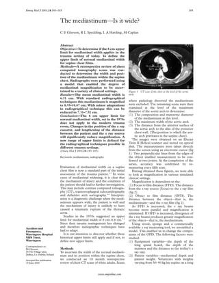
tained in a variety of clinical settings. Figure 1 CT scan of the chest at the level of the aortic
Results—The mean mediastinal width is arch.
6.31 cm. With standard radiographical
techniques this mediastinum is magnified where pathology distorted the mediastinum
to 8.93–10.07 cm. With minor adaptations were excluded. The remaining scans were then
in radiographical technique this can be examined at the level of the maximum
reduced to 7.31–7.92 cm. diameter of the aortic arch to determine:
Conclusion—The 8 cm upper limit for (1) The composition and transverse diameter
normal mediastinal width, set in the 1970s of the mediastinum at this level.
does not apply in the modern trauma (2) The maximum width of the aortic arch.
room. Changes in the position of the x ray (3) The distance from the anterior surface of
cassette, and lengthening of the distance the aortic arch to the skin of the posterior
between the patient and the x ray source chest wall. (The position to which the aor-
will significantly reduce magnification. A tic arch gravitates in the supine chest).
new range of upper limits is defined for The images were obtained on an Elscint
the radiographical techniques possible in Twin II Helical scanner and stored on optical
diVerent trauma settings. disk. The measurements were taken directly
(Emerg Med J 2001;18:183–185) from the screen using an electronic cursor (fig
1). Two perpendicular lines from the edges of
Keywords: mediastinum; radiography the object enabled measurement to be con-
firmed at two points. At the completion of the
series, accuracy was confirmed by re-
Evaluation of mediastinal width on a supine measuring every fifth scan.
chest film is now a standard part of the initial Having obtained these figures, we were able
assessment of the trauma patient.1–3 In some to look at magnification in various simulated
cases of mediastinal widening, it is clear that clinical settings.
the mechanism of injury and the condition of Magnification is dependent on the:
the patient should lead to further investigation. (1) Focus to film distance (FFD). The distance
This may include contrast computed tomogra- from the x ray source (focus) to the x ray film
phy (CT), transoesophageal echocardiography (fig 2).
and definitive arch aortography.4–6 Interpret- (2) Object to film distance (OFD). The
ation is a diagnostic challenge when the medi- distance between the object—that is, the
astinum appears wide, the patient is well and mediastinum—and the x ray film (fig 2).
the mechanism of injury is unlikely to have As FFD is increased, the x ray beams
caused a traumatic rupture of the thoracic become more parallel and magnification is
aorta. minimised. If OFD is increased, divergence of
Studies in the 1970s suggested an upper the x ray beams produces greater magnification
limit for mediastinal width of 8 cm–8.8 cm.7 8 of the object—that is, the mediastinum.
Since then trauma management has changed Using wood, sponge and a commercially
Accident and
and therefore radiographic techniques have available x ray measuring tool, we assembled a
Emergency, had to adapt. model. This enabled us to change the compo-
Warrington Hospital It is our intention to discover whether these nents of the OFD. The following factors were
NHS Trust, historical upper limits still apply and if not, to studied:
Warrington define new upper limits. (1) Equipment variables—the depth of the
Correspondence to:
long spinal board, the depth of the
Dr Gleeson, Methods mattress and the distance to the trolley’s x
25 The Village Gate, To ascertain the width of the normal mediasti- ray tray.
Dalkey, Co Dublin, Ireland
num and its position within the supine chest, (2) Patient variables—mediastinal depth and
Accepted for publication we conducted an 18 month retrospective patient weight. Volunteers with weights
19 June 2000 review of chest CT scans of white adults. Scans varying from 50–90 kg lay supine on a long
www.emjonline.com
2. 184 Gleeson, Spedding, Harding, et al
Typical Optimal Trauma
short 100 cm FFD/short OFD long 180 cm FFD/short OFD shorter FFD/longer OFD
x-ray focus
Divergent x-ray Spinal board
beams Mediastinium x-ray cassette
Wider mediastinal beneath trolley
Wide mediastinal Parallel x-ray image
image x-ray cassette beams
Mattress
Accurate mediastinal
image
Figure 2 How changing the FFD and OFD influences magnification.
Table 1 Components of the mediastinum average aortic diameter was 4.6 cm (SD 0.95).
However, the mean mediastinal width was 6.31
Aorta and:
SVC 197 cm (SD 0.97).
SVC and brachiocephalic 63 The anterior surface of the aorta was
SVC and brachiocephalic and innominate 5 situated at a mean of 15.89 cm (SD 1.69) from
Brachiocephalic 10
Three or more vessels 7 the skin of the posterior chest wall. The average
Total 282 chest diameter was 21.06 cm (SD 2.47).
The average long spinal board depth was 4.5
spine board. The resultant compression of cm. The distance between the spinal board and
the mattress, beneath the board, was the x ray cassette tray beneath six types of
measured to accurately calculate the eVect commercially available trolley varied between
on OFD. 7.1–12.9 cm.
Once the above were quantified radiographs Table 2 shows how the average 6.31 cm
were taken using FFDs of 100, 140, and 180 mediastinum is magnified. In the 1970s supine
cm. chest radiographs were exposed with 100 cm
We then looked at the eVect these variables FFD and the x ray cassette directly beneath the
had on the magnification of a range of medias- patient. This resulted in the mean mediasti-
tinal widths. The geometry was then verified num being magnified to 7.5 cm. Using the
against the verified formula for magnification.9 same FFD of 100 cm, with the longer OFD
commonly used in the modern trauma setting,
Magnification = FFD/FFD−OFD. the average mediastinum is magnified to 9.48
cm.
Results However, by lengthening the FFD to 140 cm
In the 18 month study period, 343 chest CT this can be improved to 8.29 cm. Further
scans were reviewed. Altogether 282 (82%) improvement can be achieved by placing the x
were suitable for inclusion in the study.The ray cassette beneath the spinal board—that is,
other 61 scans were excluded because of shortening the OFD. The resultant mediastinal
pathology in the chest or the abdomen distort- image is narrowed to 7.38 cm.
ing the mediastinum. Data were analysed on a Table 3 shows the upper ranges of mediasti-
SPSS package. nal widths. This range of new upper limits is
At the level of the aortic knuckle, the dependent upon the FFD and the diVering
mediastinum contained the aorta and the other positions for the x ray cassettes. Using the
great vessels (table 1). In all cases, the superior standard used today of 100 cm FFD and long
vena cava (SVC) contributed to the mediasti- OFD, the new upper limit for normal medias-
nal width. The biggest contribution was when tinal width in 95% of the population is 12.4
the SVC lay beside the aorta. cm. By improving radiographic technique, with
The values for the mediastinal and aortic a 140 cm FFD and the x ray cassette under the
diameters were normally distributed. The
Table 3 Magnification of the mediastinum 1 and 2 SD
Table 2 Magnification of the average mediastinum (6.31 cm) in various clinical settings above the mean*
x ray cassette placement x ray cassette placement
Beneath spinal Beneath shallow Beneath Beneath Beneath Beneath deep
FFD (cm) Beneath patient board trolley Beneath deep trolley FFD (cm) patient spinal board shallow trolley trolley
100 7.5 (7.17–7.89)* 7.95 (7.56–8.33) 8.7 (8.26–9.21) 9.48 (8.93–10.07) 100 8.67,9.83 9.18,10.40 10.05,11.39 10.94,12.40
140 7.12 (6.9–7.36) 7.38 (7.31–7.92) 7.86 (7.6–8.15) 8.29 (7.99–8.61) 140 8.22,9.32 8.54,9.68 9.08,10.29 9.56,10.84
180 6.92 (6.76–7.10) 7.45 (7.25–7.65) 7.45 (7.25–7.65) 7.75 (7.54–7.96) 180 8.0,9.07 8.67,9.32 8.60,9.75 8.94,10.14
*Range relates to chest depth. *Mediastinum located at the average AP depth within the chest.
www.emjonline.com
3. The mediastinum 185
spinal board the upper limit is a more FFD and long OFD (table 3). This results
reasonable 9.68 cm. in a greatly magnified mediastinum with
limited clinical usefulness.
Discussion (2) Modifying technique to maximise FFD
The supine chest film is used as a screening and minimise the non-anatomical compo-
tool to detect thoracic aortic rupture (TAR). nent of the OFD. This results in a more
Many authors have discussed the usefulness of useful upper limit (tables 2 and 3 ).
plain film findings.10–12 None is perfect. In his We realise, as have other authors that TAR
review, Woodring found that 7.3% of great ves- can occur within these upper limits and that
sel damage does not result in mediastinal wid- these figures should not replace clinical suspi-
ening.6 Other authors have suggested various cion.6 But by simple adaptations in radio-
techniques for improving plain chest radiologi- graphic technique, more useful information
cal interpretation including M/C ratio or a can be gained from the standard trauma chest
combination of signs.13 14 Most include medias- radiograph.
tinal width as one of the cardinal signs of TAR. In conclusion, using current radiographic
In everyday practice, mediastinal widening is techniques, the 8–8.8 cm upper limit for
still the most commonly sought after sign. Cli- normal mediastinal width does not apply. This
nicians will be familiar with the scenario of the study provides clinicians with a range of upper
chest radiograph revealing an abnormally wide limits applicable to the trauma setting of today.
mediastinum. This may be an unexpected To minimise magnification, we recommend
finding in a patient who is haemodynamically that the x ray plate is placed close to the
stable and has not sustained an injury associ- patient. Ideally this should be directly under
ated with TAR. the spinal board. The FFD should be the
The landmark work of Marsh and Sturm, set maximum achievable in each AED.
the upper limit of normal at 8–8.8 cm.7 8 At
that time trauma management did not rou- We would like to thank student radiographers Andy England
and Nathan de Beer for help with the model, Jackie Johnson
tinely include spinal immobilisation. The (Senior 1 radiographer), the Warrington library staV, the Medi-
patient would have been lifted and the x ray cal Illustration Department at the Royal Liverpool Hospital and
Ann Martin (Senior 1 radiographer) Royal Victoria Hospital
cassette placed directly next to the skin, indeed Belfast for her help with the pilot study.
the patient was often sat upright to have their Funding: this study was provided with funding by the R&D
chest radiograph. The FFD varied widely. budget, Warrington Hospital NHS Trust.
Since the introduction of ATLS, the patient is Conflicts of interest: none.
now pre-packaged on a long spinal board and
placed on a modern trauma trolley, which
includes a thick mattress. Radiographic tech- Contributors
niques have had to adapt. C E Gleeson collected the data, wrote the paper, and acts as
The gold standard chest radiograph devised guarantor. R L Spedding had the original idea, wrote the paper
and supervised the project. L A Harding supervised radio-
for an erect patient has an FFD of 180 cm.9 In graphic techniques and contributed to writing the paper. M
the supine trauma patient, this FFD can be Caplan gave radiology advice and advised on the final draft of
rarely obtained, as the focus of the x ray the paper.
machine cannot be raised high enough above
1 Sandor F. Incidence and significance of traumatic mediasti-
the supine patient. A ceiling suspended system nal haematoma. Thorax 1967;22:43–62.
can achieve longer FFDs than a mobile 2 Wyman AC. Roentgenologic diagnosis of traumatic rupture
of the thoracic aorta. Arch Surg 1953;66:656–63.
machine, but not the recommended 180 cm. 3 American College of Surgeons Committee on Trauma.
Other limitations include the height of the Advanced trauma life support for doctors.Chicago: American
College of Surgeons, 1980.
trolley and, with a mobile system, the reach of 4 Gavant ML, Menke PG, Fabian T, et al. Blunt traumatic
the radiographer. aortic rupture:detection with helical CT of the chest. Radi-
ology 1995;197:125–33.
When placing a x ray cassette directly 5 Saletta S, Lederman E, Fein S, et al. Transoesophageal
beneath the supine patient, the OFD consists echocardiography for the initial evaluation of the widened
mediastinum in trauma patients. J Trauma 1995;39:137–
entirely of the intra-thoracic distance from the 42.
mediastinum to the posterior chest wall. This 6 Woodring JH. The normal mediastinum in blunt traumatic
rupture of the thoracic aorta and brachiocephalic arteries.
component has been quantified for the first J Emerg Med 1990;8:467–76.
time in this study. The ranges in table 2 7 Marsh DG, Sturm JT. Traumatic aortic rupture.roentgeno-
graphic indications for angiography. Ann Thorac Surg
illustrate the contribution of this anatomical 1976;21:337–40.
variable to magnification. 8 Sturm JT, Marsh DG, Kenton CB. Ruptured thoracic aorta:
evolving radiological concepts. Surgery 1979;85:363–7.
The introduction of the spinal board, the 9 Clarke KC. Positioning in ragiography. Vol 1. 10th ed.
thick mattress and the x ray tray beneath the London: Heinemann, 1979.
10 Sefczek SE, Sefczek RJ, Deeb ZL. Radiographic signs of
trolley has lengthened the distance between the acute traumatic rupture of the thoracic aorta. AJR
x ray cassette and the mediastinum. This 1983;141:1259–62.
11 Mirvis SE, Bidwell JK, Buddemeyer EU, et al. Value of chest
longer OFD has increased magnification of all radiography in excluding traumatic aortic rupture. Radiol-
the constituents of the thorax. ogy 1987;163:487–93.
12 Kram HB, Wohlmuth DA, Appel PL, et al. Clinical and
The result of all these changes means that radiographic indications for aortography in blunt chest
for many patients the upper limit of Marsh and trauma. J Vasc Surg 1987;6:168–76.
13 Seltzer SE, D’Orsi C, Kirshner R, et al. Traumatic aortic
Sturm does not apply. To assist AED clini- rupture : plain radiographic findings. AJR 1981;137:1011–
cians, practical options include: 14.
14 Marnocha KE, Maglinte DDT, Woods J, et al. Mediastinal-
(1) Using the new upper limit based on width/chest-width ratio in blunt chest trauma:a reappraisal.
traditional radiographic techniques—short AJR 1984;142:275–7.
www.emjonline.com