
Recommended
More Related Content
Similar to Reduced Radiation Exposure in Dual-Energy Computed Tomography of the Chest: Impact on Image Quality
Similar to Reduced Radiation Exposure in Dual-Energy Computed Tomography of the Chest: Impact on Image Quality (20)
More from MehranMouzam
More from MehranMouzam (10)
Recently uploaded
Recently uploaded (20)
Reduced Radiation Exposure in Dual-Energy Computed Tomography of the Chest: Impact on Image Quality
- 1. 1 Reduced Radiation Exposure in Dual-Energy Computed Tomography of the Chest: Impact on Image Quality ABSTRACT: Objective: This study purports to answer the question: Does a dual-energy CT scan of the chest using reduced radiation result in images of equal or better quality compared to those produced bythe gold standard of care? Methods:With the agreement of the Ethical Review Committee and written informed consent from 32 patients, who received dual-energy CT (DECT) scanof the chest in a dual-source scanner, a second set of images was taken at a reduced radiation dose. On virtual monochromatic images at 40 and 60 keV, three thoracic radiologists evaluated image quality, normal thoracic structures, and pulmonary and mediastinal aberrations. Students analyzed the data using analysis of variance, Kappa statistics, and Wilcoxon signed-rank tests. Results:No irregularities in the scans were missed in the virtual monochrome photographs of all patients at a lower radiation dose, and the images were found to be of sufficient quality. At 40 and 60 keV, standard-of-care pictures produced equal contrast enhancement and lesion detection. Observers were entirely consistent with one another. Among other characteristics, reduced-doseDECT had a CTDIvol of 3.0 ±0.6 mGy, and a size specified doseestimate (SSDE) of 4.0 ±0.6 mGy, a dose-length product(DLP) of 107 ±30 mgy.cm, and an effective dose(ED) of 1.15 ±0.4 mSv. Conclusion: Dual-energy computed tomography of the chest allows for the administration of lower radiation doses (CTDIvol <3 mGy). INTRODUCTION: Dual-energy computed tomography (DECT) was originally a potential alternative that enabled more accurate images, but it required a high doseof radiation and had issues with the two kV image files collected during two consecutive DECT acquisitions not being in the same location at the same time (1). In the 1990s, numerous studies demonstrated that DECT was superior to single-energy CT for
- 2. 2 detecting lung nodules (2). However, according to a study financed by the Fleischner Society, DECT still proved to be unable to distinguish lung nodules toward the decade's end (3). As public awareness of the dangers of CT scan radiation has grown, additional clinical research and technological advancements have been made to minimise CT scan radiation levels (4). According to some experts, DECT can be performed with doses comparable to those used in single-energy CT in multidetector CT (5). In fact, doctors have employed both single-phase and dual-phase DECT scans to detect benign and malignant mediastinal tumours. These tests have identified aneurysms of the aorta, chronic pulmonary embolism, lung emboli, and pulmonary nodules (6,7). Few persons who use chest DECT have considered reducing their daily radiation exposure. Recent studies have demonstrated that DECT scans are effective for examining lung parenchyma and pulmonary embolisms. This is accomplished through the use of material separation techniques and the creation of virtual monochrome images (8,9). As mentioned above, the goal of this study is to determine whether chest dual- energy CT can be administered at lower doses than the current gold standard of care and still produceviable images. METHODOLOGY: The voxel standard deviation was used to assess the number of CT counts and the amount of noise in DECT images (defined as the standard deviation of voxel values). Chest phantoms were used in this study. Double-sided tape was applied to the surface of test tubes containing iopamidol 370 mg/mL diluted contrast material at 1:20 and 1:40 concentrations. Two scans of the phantom were performed using standard-of-care and low-dose DECT procedures. Changing this has no effect on the amount of time required to scan a file or on the size of the file scanned. CT scans and standard deviations (SDs) were calculated for each region at ten locations on the right upper lung and chest wall of the phantom. The diluted contrast solution test tubes were scanned using CT scanners and their standard deviations were recorded twice.
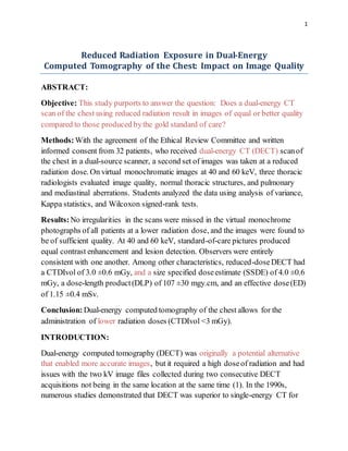
- 3. 3 Figure1: Imageof the anthropomorphicphantom'schest in the transverse plane from a CT scan. Participants in the Study Our Institutional Review Board's Human Research Committee approved this study after everyone signed an informed consent form to participate. To ensure that the research was conducted properly, HIPPAA regulations were strictly adhered to.
- 4. 4 The researchers searched the Radiology Information System (RIS) for patients who needed a conventional chest CT with contrast enhancement. They discovered that there were individuals. To participate in the study, participants needed to be alert, have stable blood pressure, and be at least 56 years old. A CT scan was not performed if the patient could not hold their breath for more than 10 seconds orif the patient was allergic to the contrast used. Certain individuals were unable to participate in the study due to their BMI being greater than 32 kg/m2. Individuals who had previously undergone two CT scans at our clinic — one in the prone position and another in the supine position — were also denied participation due to a history of interstitial lung disease. Forty-five individuals participated in the study. Eight patients stated that they did not wish to be treated after signing an informed consent form. Five persons were forced to abandon the experiment for health reasons after an accident involving a single-energy CT scanner. As a result, there were finally 32 adults participating: 12 men and 20 women. The average age was 71 ±6 years in general. On average, the men were 70 ±5 years old, and the women were 72 ±7 years old, with a range of 56 to 87 years. The 32 individuals who underwent clinical CT scans included those with lung cancer, pneumonia, and other non-pulmonary ailments. For each CT scan performed, the patient received 65mL of IV contrast material. This was achievable in this circumstance because the CT scanner was a multidetector CT scanner with a zooming focus point. It took 35 seconds for the individual to be scanned in preparation for the contrastmedia injection. They were scanned at a rate of 2.5 mL per second. Each scan lasted nearly the same length of time (approximately 3 s). Patients could see how much of their bodywas covered by both standard-of-care and low-dose imaging because they would eventually get a chest CT scan. CT scans with a standard dosetook longer than scans with a low dose. The decreased- dosage technique reduced the quality reference mAs to approximately half that of the standard of care (10). Between the two scans, all other scanning parameters remained constant. It was not included in the set of low-dose images. Each photo set's CTDIvol and dose-length products (DLP) were determined. We were able to determine the water-equivalent diameter (WED) and size-specific dose estimate (SSDE) for each person by utilising software called the Radmetrics Enterprise
- 5. 5 Platform (11). This meant that each chest CT image was estimated because it took 0.14 of a second to convert each image to an EDT. This was the DLP's operation. It resembled a map (12). Images of normal and lower doses were created using Healthcare's proprietary technology. The images were created with the industry-standard medium-smooth soft-tissue reconstruction kernel, a proprietary iterative reconstruction technique (I30f). There were no patients with interstitial lung disease in this research. As a result, we used a soft-tissue kernel for all of the images in order to improve spatial resolution and the ability to see edges (which provides optimal image contrast at the costof spatial resolution). This is how transversely orientated pictures (80/Sn140 kV) were created. Each slice was separated by a 2mm gap. Virtual monochromatic images at 40 and 60 keV, as well as images of blood flow and images with no contrast, were created using dedicated computers. Because virtual monochromatic low-energy images had the same or less noise and additionally might aid in making iodine more visible, they were the best choice (13–16). The images taken at 40 keV were chosen for the investigation becausethey best fit the K-edge of iodine (33 keV). A 60 keV resolution image has better contrastthan a 65-70 keV resolution image due to the absenceof noise. This is why they were chosen for investigation (8, 17). Before the investigators examined the photographs, no scan settings nor patient information were associated with individual participants. Board-certified thoracic radiologists viewed and analyzed the chest CT images using a DICOM-compliant image viewer. Radiologists were unaware of how much radiation was emitted during each imaging series, since they did not administer the tests. Each radiologist examined standard-of-care and low-dose images privately without collaboration. A side-by-side comparison was required to determine whether the image quality was the same for both treatments and whether noise obscured the lesions. Radiologists graded the appearance of images of mediastinal and lung lesions, as well as normal bodycomponents, using a two-point scale. The lesions included lung fissures and the sub-segmental bronchial wall, the pericardium, and sub-centimeter mediastinal lymph nodes. This study examined a variety of factors that influence the appearance of an image (18).
- 6. 6 Two radiologists conducted this research examining the effect of low-dose DECT images on diagnostic information, but they were unable to view what was occurring in the lung parenchyma or mediastinum due to their “blindness.” At 40 and 60 keV, two monochromatic pictures were created (images illustrating standard-of-care methods). An outstanding grade indicated that anomalous structures could be observed clearly in this example due to excellent delineation. If images received a high score, they were perceived as having some blurriness or ambiguity over how to assess them. If they received a low grade, they were considered invisible. Monochromatic 40 keV images were superior to standard-of- care images due to the reduced doseof DECT utilized to increase contrast. The image noise and signal data for the tracheal lumen, mid-thoracic vertebral body, and paraspinal muscle ROIs were gathered concurrently, with CT numbers expressed in HUs. Additionally, CT scans of the right pulmonary artery were performed to determine its appearance on the CT. One researcher created circular ROIs on virtual monochromatic images at 60 and 40 keV using standard-of-care datasets. These took up a lot of space. They occupied an area of between 0.5 and 0.8 cm2 , respectively. The signal to noise ratio (SNR) and contrast to noise ratio (CNR) were also calculated (20). The CNR was determined using tracheal ROIs. Due to the ten-second delay between the standard-of-care and reduced-dose photographs, it was difficult to determine the amount of iodine in blood flow photographs. StatisticalAnalysis: SPSS Statistics, version 25.0, was used to examine the data and determine an appropriate method of analysis. Students employed a t-test to assess the quantity of image noise and the number of CT scans performed in the right pulmonary artery between two groups of individuals. The study used the Wilcoxon signed-rank test to evaluate the subjective image quality of standard of care with low-dose DECT. Cohen's kappa statistic was used to determine the degree to which two radiologists might agree. According to Cohen, those with a scoreof less than 0.20 could claim only “slight agreement,” whereas those individuals with a scoreof more than 0.60 could claim "substantial agreement." As illustrated in the image, a one-way analysis of variance was used to compare HU values between persons who
- 7. 7 received standard of care and those who received less care in a phantom study. We considered this to be significant because our p value was 0.05. RESULTS: The demographics of our patients are summarized in the table below (Table 1). Between standard-of-care and reduced-doseDECT scans, soft tissue and lung parenchyma CT values did not differ substantially (p >0.05, respectively). According to our findings, there was no significant difference in picture noise between standard-of-care and reduced-dosetreatments (p >0.05). (Table 2) Table 1: Characteristics Males Females No. of participants 12 20 Age (years)(Mean ±SD) 70 ± 5 72 ± 7 Weight (kilogram) 80 ± 12 66 ± 10 BMI (Mean ±SD) 25.5 ± 3.7 24.7 ± 3.5 BMI (According to sub categories) ≤ 20 kilogram/meter2 1 2
- 8. 8 20-25 kilogram/meter2 6 9 25-30 kilogram/meter2 4 8 30-32 kilogram/meter2 1 1 Table 2: Standard-of-Care Dual- Energy CT Scan Reduced-Dose Dual- Energy CT Scan Region (HU ±SD) (HU ±SD) Soft tissue (60 keV) 31.4 ±14.6 32.1 ± 4.1 Soft tissue (40 keV) 32.4 ±26.1 30.2 ±28.3 Lung parenchyma (60 keV) -795.8 ±10.7 -792.4 ±12.6
- 9. 9 Lung parenchyma (40 keV) -780.2 ±18.1 -777.8 ±21.5 Contrastmedia 1:20 (40 keV) 1495.1 ±49.2 1487.6 ±53.4 Contrastmedia 1:20 (60 keV) 673.5 ±20.7 667.3 ±23.6 Contrastmedia 1:40 (40 keV) 780.6 ±35.6 767.4 ±34.3 Contrastmedia 1:40 (60 keV) 346.5 ±13.3 339.3 ±19.4 The scans were carried out at an energy level of 60 keV in order to comparetwo monochromatic pictures side by side. DECT images taken at standard and decreased doses indicated normal lung fissures, small lymph nodes, and the pericardium. There were no visible distinctions between these two sorts of photographs. All 39 lesions were identified on virtual monochromatic images in the standard-of-care DECT, but were also visible in the reduced-doseDECT. Non-calcified solid nodules with a diameter of less than one centimeter were found in 27 of the participants. Only one patient had solid nodules that were non-calcified and measured more than one centimetre in diameter. The overall subjective image quality was assessed to be satisfactory in all 32 cases (kappa = 1) when monochromatic 60 keV monochrome images were used in the reduced-doseDECT approach. The detection of lesions can be aided by monochromatic monochrome
- 10. 10 images with a resolution of 40 keV. Despite the dosage reduction, the number and kind of mediastinal lesions were identical in the standard-of-care (60 keV) and reduced-dose(40 keV) pictures. Because of the perfect interobserver agreement (kappa = 1), the reduced-dosepicture sequence inspired a lot of trust. Lung nodules as small as 2 mm in diameter can be detected using this approach. Both intraobserver (kappa = 0.73) and interobserver (kappa = 0.66) agreement were rated as acceptable. There was a considerable improvement in interobserver agreement when noncalcified lung nodules less than 5 mm in diameter were omitted from the analysis. Three patients received four points for having a high level of diagnostic confidence. There were 28 patients who had a high level of diagnostic certainty. Table 3 summarizes the results of the objective image quality evaluation. Noise in the tracheal lumen was significantly reduced (12%) in the reduced-doseimaging series, whereas noise in muscle and bone tissues increased up to 44 percent. Regardless, the CNR increased as a result to 20 percent. Table 3: Standard-of-Care DECT Reduced-Dose DECT % Difference Variable 60 keV 40 keV 60 keV 40 keV 60 keV 40 keV Image Noise (mean ±SD) Trachea 8.2 ±2.5 8.7 ±3.2 7.0 ±3.0 8.0±5.7 -11% -7%
- 11. 11 Muscle 13.6 ±3.1 23.5 ±5.1 18.3±3.4 34.1±6.3 34% 43% Bone 24.7 ±8.2 39.7 ±14.9 28.7±6.3 48.7±13.3 17% 21% Signal-to-NoiseRatio (mean ±SD) Trachea 131.5 ±7.1 125.2 ±40.3 157.4 ± 51.2 151.6 ± 53.4 18% 20% Muscle 3.8 ± 1.1 2.2 ± 0.8 3.2 ± 0.8 1.9 ± 0.9 -20% -21% Bone 8.2 ± 4.1 8.7 ± 4.1 6.7 ± 2.6 7.4 ± 3.3 -18% -16% Contrast-to-NoiseRatio (mean±SD) Muscle 124.9 ±38 118.6 ±38.7 148.7 ±48.9 142.6 ± 51.2 18% 19% Bone 108.3 ±33 86.7 ± 27.2 128.3 ±41.4 101.6 ±35.6 18% 16% Reducing the dosageto 60 keV for virtual monochrome photographs resulted in an average attenuation of 228 ±72 Hu for images of the right pulmonary artery taken concurrently with standard-of-care images. Attenuation values of virtual
- 12. 12 monochromatic images at 40 keV were greater than those at 60 keV. Despite the 10-second delay between treatments, reduced-doseprotocolphotos revealed somewhat greater attenuation in the right pulmonary vein (452-102 HU) than 60- keV images collected with conventional treatment (403-118 HU). Medicine that is considered to be standard-of-care CTDIvol had a higher concentration of CTDIvol, SSDE, DLP, and ED than the reduced-dosage medication's lower dose. The following table summarizes these figures (p <0.001). The p-value is <0.0001 in this situation. Other participants who underwent DECT of the chest did not report any abnormal CT results. The average doseadministered in the emergency department (ED) for chest CT scans was less than seven millisieverts (mSv) for each patient in our analysis (21). DISCUSSION: Doctors can do DECT on the chest at lower doses without sacrificing diagnostic information in patients with BMIs less than 32 kg/m2. For the lower dosages, a CTDIvol of no more than 3.0 and a DLP of no more than 30 mGy.cm may be employed down to a CTDIvol of 3.0 ±0.7 mGy and a DLP of 107 ±30 mGy.cm. Despite the reduction in radiation exposure, small nodules, bronchial fissures, pulmonary arteries, and bronchial walls could still be detected in the photographs. As a result, we proved the feasibility of using low-dose virtual monochromatic images to detect mediastinal and pulmonary abnormalities. Our findings contradict previous research on DECT treatments, which concludes that the scanning device exposes patients to high radiation doses during use (22, 23). CT angiography and urography are two clinical scenarios in which recent research have considered low-radiation-dose protocols and the possibility of eliminating one or more scanning steps (24, 25). DECT scanners may be able to drastically reduce the amount of radiation they create compared to other scanners. Our DECT study used a lower radiation dosethan previous studies (8). According to the researchers, chest DECT was performed in the lab with a CTDIvol of 5.44 milliGy and 2.26 milliSv. This means that they were able to obtain the required results with extremely low doses. Additionally, this study establishes that DECT has a higher CNR at 120 kV than single-energy CT. A CT angiography
- 13. 13 examination was performed with the assistance of a dual-source DECT scanner. They determined the average DLP to be 403.4 mGy.cm. According to Hwang et al. (26), the ED was 1.78mSv when a dual-source DECT scanner with single-energy collection was used for low-dose chest CT (120 kV). For this purpose, virtual monochromatic pictures of the pulmonary artery with a lower energy level, namely 40 keV rather than 60 keV, are preferable. In most of the patients we evaluated, attenuation in the lung was more obvious on 40 keV (reduced-doseprotocol) images than on 60 keV images, even after a 10-second delay. This is because iodine's K-edge (33 keV) is closer to 40 keV than to 60 keV. Due to DECT's ability to boostcontrast, it may be able to lower the amount of contrast medium injected during CT scans. This may be beneficial for people who are at risk of contrast-induced nephropathy Patients who had previously gotten standard-of-care DECT were exposed to very little radiation when we photographed them using low-dose DECT. This resulted in a reduction in the size of our study sample. We were able to mitigate the risks associated with the higher radiation exposure, however, by including only individuals above the age of 56. Due to the fact that the rapid kV-switching technology on our other DECT scanners did not allow us to reduce the radiation doseby 50%, we used a single DECT scanner from a single vendor. We recommend utilizing the lowest feasible doseof DECT chest treatment when delivering it. This is because the rapid kV-switching approachis only effective with pre-set doses and tube currents. However, because the SAFIRE S3 iterative reconstruction setting has been shown to be beneficial for all patients and is regularly used in our practice, it was chosen. Even so, we do not know how reduced-doseDECT of the chest will effect individuals with a bodymass index greater than 32 kg/m2 or who have interstitial lung disease. When low-dose DECT is applied on the chest and other areas of the body, it is necessary to verify quantitative iodine levels detected in material degradation images. Our clinical standard of care demands us to examine all images of the lungs and mediastinum with a single kernel (I30f). As a result, we made no attempt to determine if reduced-doseDECT made it more difficult to understand data with a high spatial frequency or with small kernels in the lung.
- 14. 14 With a patient’s BMI of less than 32 kg/m2, chest DECT can be used at a lower dose, i.e., down to 3.0 mGy. By using low-dose monochromatic images at 40 keV and 60 keV, radiologists can analyze normal as well as diseased thoracic findings. REFERENCES: 1. Kan WC, Wiley Jr AL, Wirtanen GW, et al. High Z elements in human sarcomata: assessmentby multienergy CT and neutron activation analysis. AJR Am J Roentgenol. 1980;135:123–129. [PubMed] [Google Scholar] 2. Bhalla M, Shepard JA, Nakamura K, et al. Dual kV CT to detect calcification in solitary pulmonary nodule. J Comput Assist Tomogr. 1995;19:44– 47. [PubMed] [Google Scholar] 3. Swensen SJ, Yamashita K, McCollough CH, et al. Lung nodules: dual-kilovolt peak analysis with CT-multicenter study. Radiology. 2000;214:81– 85. [PubMed] [Google Scholar] 4. Kalra MK, Maher MM, Toth TL, et al. Strategies for CT radiation dose optimization. Radiology. 2004;230:619–628. [PubMed] [Google Scholar] 5. Schenzle JC, Sommer WH, Neumaier K, et al. Dual energy CT of the chest: how about the dose?Invest Radiol. 2010;45:347–353. [PubMed] [Google Scholar] 6. Lu GM, Zhao Y, Zhang LJ, et al. Dual-energy CT of the lung. AJR Am J Roentgenol. 2012;199(5) Suppl:S40–S53. [PubMed] [Google Scholar] 7. Otrakji A, Digumarthy SR, Lo Gullo R, et al. Dual-energy CT:spectrum of thoracic abnormalities. Radiographics. 2016;36:38–52. [PubMed] [Google Scholar] 8. Ohana M, Labani A, Severac F, et al. Single source dual energy CT: what is the optimal monochromatic energy level for the analysis of the lung parenchyma? Eur J Radiol. 2017; 88:163–170. [PubMed] [Google Scholar] 9. Apfaltrer P, Sudarski S, Schneider D, et al. Value of monoenergetic low-kV dual energy CT datasets for improved image quality of CT pulmonary angiography. Eur J Radiol. 2014;83:322–328. [PubMed] [Google Scholar] 10. Kalra MK, Maher MM, TothTL, et al. Techniques and applications of automatic tube current modulation for CT. Radiology. 2004;233:649– 657. [PubMed] [Google Scholar]
- 15. 15 11. McCollough C, Bakalyar DM, Bostani M, et al. Use of water equivalent diameter for calculating patient size and size-specific doseestimates (SSDE) in CT: the report of AAPM Task Group 220. AAPM Rep. 2014;2014:6–23. [PMC free article] [PubMed] [Google Scholar] 12. Christner JA, Kofler JM, McCollough CH. Estimating effective dosefor CT using dose-length productcompared with using organ doses:consequences of adopting International Commission on Radiological Protection publication 103 or dual-energy scanning. AJR Am J Roentgenol. 2010;194:881– 889. [PubMed] [Google Scholar] 13. Yu L, Christner JA, Leng S, et al. Virtual monochromatic imaging in dual- sourcedual-energy CT: radiation doseand image quality. Med Phys. 2011;38:6371–6379. [PMC free article] [PubMed] [Google Scholar] 14. Yu L, Leng S, McCollough CH. Dual-energy CT-based monochromatic imaging. AJR Am J Roentgenol. 2012;199(5) Suppl:S9–S15. [PubMed] [Google Scholar] 15. Mileto A, Barina A, Marin D, et al. Virtual monochromatic images from dual- energy multidetector CT:variance in CT numbers from the same lesion between single-source projection-based and dual-source image-based implementations. Radiology. 2016;279:269–277. [PubMed] [Google Scholar] 16. Tamm EP, Le O, Liu X, et al. Abdom Radiol. Vol. 42. NY: 2017. "How to" incorporate dual-energy imaging into a high volume abdominal imaging practice; pp. 688–701. [PMC free article] [PubMed] [Google Scholar] 17. Cheng J, Yin Y, Wu H, et al. Optimal monochromatic energy levels in spectral CT pulmonary angiography for the evaluation of pulmonary embolism. PLoS One. 2013;8:e63140. [PMC free article] [PubMed] [Google Scholar] 18. Singh S, Kalra MK, Gilman MD, et al. Adaptive statistical iterative reconstruction technique for radiation dosereduction in chest CT:a pilot study. Radiology. 2011;259:565–573. [PubMed] [Google Scholar] 19. Neroladaki A, Botsikas D, Boudabbous S, et al. Computed tomography of the chest with model-based iterative reconstruction using a radiation exposure similar to chest X-ray examination: preliminary observations. Eur Radiol. 2013;23:360– 366. [PubMed] [Google Scholar] 20. Miéville FA, Gudinchet F, Brunelle F, et al. Iterative reconstruction methods in two different MDCT scanners: physical metrics and 4-alternative forced-choice
- 16. 16 detectability experiments-a phantom approach. Phys Med. 2013;29:99– 110. [PubMed] [Google Scholar] 21. Mettler Jr FA, Huda W, Yoshizumi TT, et al. Effective doses in radiology and diagnostic nuclear medicine: a catalog. Radiology. 2008;248:254– 263. [PubMed] [Google Scholar] 22. Pourjabbar S, Singh S, Kulkarni N, et al. Dose reduction for chest CT: comparison of two iterative reconstruction techniques. Acta Radiol. 2015;56:688– 695. [PubMed] [Google Scholar] 23. de Broucker T, Pontana F, Santangelo T, et al. Single- and dual-source chest CT protocols:levels of radiation dosein routine clinical practice. Diagn Interv Imaging. 2012;93:852–858. [PubMed] [Google Scholar] 24. Javor D, Wressnegger A, Unterhumer S, et al. Endoleak detection using single- acquisition split-bolus dual-energy computer tomography (DECT) Eur Radiol. 2017;27:1622–1630. [PMC free article] [PubMed] [Google Scholar] 25. Chen CY, Hsu JS, Jaw TS, et al. Split-bolus portal venous phase dual-energy CT urography: protocoldesign, image quality, and dosereduction. AJR Am J Roentgenol. 2015;205:W492–W501. [PubMed] [Google Scholar] 26. Hwang HJ, Seo JB, Lee JS, et al. Radiation dosereduction of chest CT with iterative reconstruction in image space - Part I: studies on image quality using dual sourceCT. Korean J Radiol. 2012;13:711–719. [PMC free article] [PubMed] [Google Scholar] 27. Yuan R, Shuman WP, Earls JP, et al. Reduced iodine load at CT pulmonary angiography with dual-energy monochromatic imaging: comparisonwith standard CT pulmonary angiography—a prospective randomized trial. Radiology. 2012;262:290–297. [PubMed] [Google Scholar]