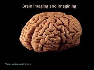
2. Brain
PET (Positron Emission Tomography) scans show localized glucose
uptake. A PET scan can discern metabolically active tumors
previously revealed in more detail with an MRI (Magnetic
Resonance Imaging). Photo: aboutcancer.com
3. FUNCTIONAL NEUROCOGNITIVE IMAGING
fMRI detects changes in neural activation while a patient is performing a
cognitive test, providing a three dimensional map of brain regions that are used
to perform the test. Photo: cognitivefxusa.com
4. Diffusion Tensor Imaging
White matter fiber tracts seen with syngo DTI Tractography. The technique
depends on water molecule interactions across membranes and other cell
structures. Photo: usa.healthcare.siemens.com
5. Brain
A 2.5-foot replica of a 3-D computer Diffusion Tensor image of the
brain’s white matter for an exhibition at the Franklin Institute..
Image: American Precision Prototyping
6. GLASS BRAIN
“….a state-of-the-art real-time brain visualization technology that is created on the Unity 3D
game-engine and powered by NVIDIA’s GPU computing. Its inputs include an individual’s brain
structure, both tissue and fiber tract architecture, obtained from high-resolution MRI-DTI brain
scans. Real-time brain activity and functional interactions among networks are superimposed
on the brain structure using high-density EEG (electroencephalography).
Photo: neuroscape.ucsf.edu
7. Another Glass Brain image from Neuroscape.ucsf.edu
Artist: Karen Norberg, Location: Boston Museum of Science