1. Assessment of Six1 Expression in Mouse Mammary Tumor Cells
Reina Hernandez; Mentor, Michael Cantrell; P.I. Dr. Carla Van Den Berg
College of Pharmacy, The University of Texas at Austin
Abstract
Six 1 is a homeoprotein overexpressed in numerous
cancers, where it plays a role in Epithelial-Mesenchymal
Transition (EMT) and metastasis. Six1 expression is
increased by JNK2 (c-Jun N-terminal kinase 2). Together;
JNK2 promotes EMT and Six1 expression in breast
cancer cell lines. To determine whether transforming
growth factor beta (TGFß) activates JNK2 to increase
Six1 expression, cell culture experiments were completed
throughout a 24, 48, and 72 hour period. P53: jnk2ko
GFP-JNK2 cancer cells were treated with serum free
media alone or with TGFß, fetal bovine serum, and
fibroblast growth factor-alpha added. Throughout the 24-
48 hour period, cells were harvested and Western blot
analysis was used to determine the protein expression of
Six1. Growth of mouse mammary tumor cells was seen in
a 24 to 72 hour period, and changes in morphology were
noted. PCR was also completed to establish a standard
curve that could be used in future qPCR experiments
using samples from the Six1 cell culture experiments.
Unexpectedly, multiple bands showed that there may be
changes in Six1 phosphorylation in addition to potential
expression differences. To better understand the
mechanisms behind Six1 expression and JNK2, further
experiments are required.
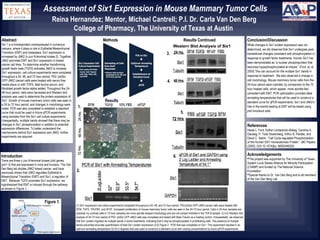
(1) Six1 expression cell culture experiments completed throughout a 24, 48, and 72 hour period. P53:jnk2ko GFP-JNK2 cancer cells were treated with
SFM, TGFß, 10%FBS, and ∂FGF. Increased proliferation of mouse mammary tumor cells are seen in the 24-72 hour period. Cells in 24-hour samples are
cuboidal; by contrast cells in 72-hour samples are more spindle shaped morphology and are not contact inhibited in the TGFβ sample. (3,4,5) Western blot
analysis of 24-72 hour period of P53: jnk2ko GFP-JNK2 cells was completed and tested with Beta-Tubulin as a loading control. Unexpectedly, we observed
that Six1 protein migrates as multiple bands in some treatments, indicating that it may be phosphorylated in proliferating cells. The presence of multiple
bands prevented accurate quantification of total Six1 protein expression (2,6) Figure 2. PCR that was completed on Six1. This experiment resulted in an
optimum annealing temperature 52-53.5 degrees that was used to construct a standard curve with varying concentrations by future qPCR experiments.
Results
Results ContinuedMethods
1.
2.
3.
6.
4.
Conclusion/Discussion
While changes in Six1 protein expression was not
determined, we did observed that Six1 undergoes post-
tranlastional changes consistent with phosphorylation in
response to growth factor treatments. Human Six1 has
been demonstrated as “a nuclear phosphoprotein that
becomes hyperphosphorylated at mitosis.” (Ford, et. al.
2000) This can account for the multiple Six1 bands in
response to treatment. We also observed a change in
cell morphology. Mouse mammary tumor cells from the
24-hour period were cuboidal, by comparison to the 72
hour treated cells, which appear more spindle-like,
consistent with EMT. PCR optimization provided ideal
annealing temperatures that will be used to construct a
standard curve for qPCR experiments. Six1 and JNK2’s
role in the events leading to EMT will be tested using
jnk2 knockout cells.
Introduction
There are three c-Jun N-terminal kinase (jnk) genes
(jnk1-3) that are expressed in mice and humans. The Van
Den Berg lab studies JNK2 breast cancer, and have
previously shown that JNK2 regulates Epithelial to
Mesenchymal Transition (EMT) and Six1, a regulator of
EMT. Because TGFß promotes Six1 expression, we
hypothesized that EMT is induced through the pathway
as shown in Figure 1.
Acknowledgements
This project was supported by The University of Texas
System Louis Stokes Alliance for Minority Participation
(LSAMP) and funded by The National Science
Foundation
Special thanks to Dr. Van Den Berg and to all members
of the Van Den Berg Lab
Figure 1.
5.
References
Heide L. Ford, Esther Landesman-Bollag, Caroline S.
Dacwag, P. Todd Stukenberg, Arthur B. Pardee, and
David C. Seldin. “Cell Cycle-regulated Phosphorylation
of the Human SIX1 Homeodomain Protein.” JBC Papers
(2000). DOI 10.1074/jbc. M002446200