FT-IR, NIR Microscopy and Imaging
•
0 likes•575 views

PerkinElmer Spotlight™ FT-IR, NIR Microscopy and Imaging Systems are built to the highest ISO-9001 manufacturing standards. This document presents technical information and typical performance specifications based on factory tests. The Spotlight systems take the proven and popular IR microscopy technique and add a new level of speed and applications capability. Spotlight systems incorporate high performance detectors, which deliver the ultimate in sensitivity, out-performing competitive top-of-the line IR microscopy systems. The revolutionary imaging capabilities enable previously time-consuming and difficult chemical composition studies to be performed without compromising data quality.

Recommended
Recommended
More Related Content
What's hot
What's hot (15)
Similar to FT-IR, NIR Microscopy and Imaging
Similar to FT-IR, NIR Microscopy and Imaging (20)
More from PerkinElmer, Inc.
More from PerkinElmer, Inc. (20)
Recently uploaded
Recently uploaded (20)
FT-IR, NIR Microscopy and Imaging
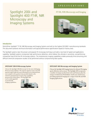
- 1. S P E C I F I C A T I O N S FT-IR, NIR Microscopy and ImagingSpotlight 200i and Spotlight 400 FT-IR, NIR Microscopy and Imaging Systems Introduction PerkinElmer Spotlight™ FT-IR, NIR Microscopy and Imaging Systems are built to the highest ISO-9001 manufacturing standards. This document presents technical information and typical performance specifications based on factory tests. The Spotlight systems take the proven and popular IR microscopy technique and add a new level of speed and applications capability. Spotlight systems incorporate high performance detectors, which deliver the ultimate in sensitivity, out-performing competitive top-of-the line IR microscopy systems. The revolutionary imaging capabilities enable previously time-consuming and difficult chemical composition studies to be performed without compromising data quality. SPOTLIGHT 200i IR Microscopy System Choose the Spotlight 200i Microscope for the most challenging IR microscopy problems. The superior signal-to-noise and wide spectral range enable the most information to be obtained from your samples in the quickest time possible thanks to the extensive automated microscope setup capabilities pioneered by Perkin Elmer. The Spotlight 200i can be easily upgraded to provide full imaging capabilities. SPOTLIGHT 400 Microscopy and Imaging System Choose the Spotlight 400 Imaging System for ultimate IR imaging speed, efficiency and accuracy. The Spotlight 400 incorporates a number of unique productivity tools and features plus the unique revolutionary PerkinElmer small array of detectors, providing optimized imaging for a range of mid-IR and NIR imaging applications.
- 2. 2 Optical System Microscope Platform All reflective, triple cassegrain optical system. One touch software switches between transmission and reflectance modes. Lower cassegrain automatically optimizes throughput for transmission measurements. All reflective, triple cassegrain optical system. One touch software switches between transmission and reflectance modes. Lower cassegrain automatically optimizes throughput for transmission measurements. Numerical Aperture (N.A.) 0.6 (IR mode). Automatic NA switching for visible illumination for improved depth of field in the visible range. 0.6 (IR mode). Automatic NA switching for visible illumination for improved depth of field in the visible range. Upgradability6 Fully compatible with PerkinElmer Frontier, Spectrum Two, Spectrum 65, 100, 400 FT-IR systems. Fully compatible with PerkinElmer Frontier, Spectrum 65, 100, 400 FT-IR systems. Spectrometer Improved Michelson interferometer, self-compensating for dynamic alignment changes due to tilt and shear, incorporating high reflectivity first-surface aluminum- coated optics. Improved Michelson interferometer, self-compensating for dynamic alignment changes due to tilt and shear, incorporating high reflectivity first-surface aluminum- coated optics. IR Source (MIR)1 Long-life source with proprietary hot-spot stabilization. User replaceable. Long-life source with proprietary hot-spot stabilization. User replaceable. IR Source (NIR)7 Tungsten halogen lamp Tungsten halogen lamp Micro-Attenuated Total Reflection (ATR)1 Optional integrated Micro-ATR with 100µ Silicon or Germanium ATR crystal. ATR objective does not require re-alignment before or after use. Optional integrated Micro-ATR with 100 µ Silicon or Germanium ATR crystal. ATR objective does not require re-alignment before or after use. Auto-Micro-Attenuated Reflection (ATR) Optional fully integrated automated micro-ATR with motorized drive and pressure control. Fully operated from software with automatic pressure sensing and adjustable force control. Optional fully integrated automated micro-ATR with motorized drive and pressure control. Fully operated from software with automatic pressure sensing and adjustable force control. Imaging-Attenuated Total Reflection (ATR)1 N/A Optional stage mounted Germanium ATR imaging crystal providing a maximum ATR imaging circular area of diameter of 500 µ or 1200 µ. ATR imaging software wizard automates alignment procedure and guides users through data collection ensuring the highest quality of data. Crystal can be inverted for easy cleaning. Purge Sample area purge available. Optional environmental enclosure available. Sample area purge available. Optional environmental enclosure available Sample Viewing Sample Illumination White light LED illumination for true color display. Auto-illumination function available as well as easy access to wide-range brightness and contrast controls. Focus software or joystick control of focus. One-touch autofocus mode available. White light LED illumination for true color display. Auto-illumination function available as well as easy access to wide-range brightness and contrast controls. Focus software or joystick control of focus. One-touch autofocus mode available. Mode Switching Dichroic mirrors provide mechanism-free switching between IR and visible viewing. IR and visible beam use identical beam paths for optimum geometric image accuracy. Micro-ATR accessories can be permanently mounted and lowered into measurement position. No manual re-alignment required. Dichroic mirrors provide mechanism-free switching between IR and visible viewing. IR and visible beam use identical beam paths for optimum geometric image accuracy. Micro-ATR accessories can be permanently mounted and lowered into measurement position. No manual re-alignment required. Drop-in stage mounted ATR imaging accessory enables quick and easy switching between ATR imaging and traditional transmission or reflectance measurements. Image On screen viewing of image with over 32,000 colors. Image size is scalable to full screen. On screen viewing of image with over 32,000 colors. Image size is scalable to full screen. Magnification Continuously variable magnification enables views of the samples at different magnifications to be viewed on-screen. Continuously variable magnification enables views of the samples at different magnifications to be viewed on-screen. Sample Area 75 x 50 mm (3 x 2 in.) 160 x 60 mm (6.3 x 2.4 in.) or 75 x 50 mm (3 x 2 in) Stage Accuracy Stage movement accuracy of 0.1u Stage movement accuracy of 0.1u Technical Description and Specifications Spotlight 200i Spotlight 400
- 3. 3 Software General A single software platform incorporates all of the functions required for infrared micro spectroscopy; instrument control, data-manipulation and analysis. Optional software packages provide advanced capabilities or functions designed for specific applications areas, Enhanced security (ES) version available for 21CFR11 technical compliance. A single software platform incorporates all of the functions required for infrared micro spectroscopy and imaging instrument control, data-manipulation and analysis. Optional software packages provide advanced capabilities or functions designed for specific applications areas. Data Display Real time data display. Full spectra are live during data collection. Real time image display. 10 updates per second. Show Structure Single button-click chemometrics-based function in SpectrumIMAGE viewer to automate contrast enhancement in collected images by identifying and revealing true spectral differences. Single button-click chemometrics-based function to automate contrast enhancement in collected images by identifying and revealing true spectral differences. Interactive Multimedia Integral video camera provides on-screen visible image. Sampling points for all data collection modes are defined by direct interaction with live visible image. Integral video camera provides on-screen visible image. Sampling points for all data collection modes are defined by direct interaction with live visible image. System Diagnostics Key microscope and instrument components are checked on startup. Status of all Spotlight components are displayed via PC. Key microscope and instrument components are checked on startup. Status of all Spotlight components are displayed via PC. Atmospheric Compensation Minimizes effect of atmospheric water and CO2 on the sample spectra without the need for reference or calibration spectra. Minimizes effect of atmospheric water and CO2 on the sample spectra without the need for reference or calibration spectra. Accessories Polarizers1 Visible and IR polarizers available. Visible and IR polarizers available. Hot and Cold Sample Stage Optional sample stage covering temperature range-196 ˚C-600 ˚C. Optional sample stage covering temperature range 196 ˚C-600 ˚C. Sampling Options Include compression cell, hot stage, diamond anvil cell and micro-sampling tools. Stage mounted tablet sample holders. Provided with 11, 15 and 22 mm diameter tablet Autosampler molds for accommodating irregular shaped samples. Compatible with Leica® EM Trim supports for depth profiling. Sample cup version for powder or blend analyses available. Include compression cell, hot stage, diamond anvi cell and micro-sampling tools. Stage mounted tablet sample holders. Provided with 11, 15 and 22 mm diameter tablet holders. Compatible with PerkinElmer Tablet Autosampler molds for accommodating irregular shaped samples. Compatible with Leica® EM Trim supports for depth profiling. Sample cup version for powder or blend analyses available. Large Detector Dewar2 Additional Liquid N2 dewar providing total detector cool time greater than 18 hours. Additional Liquid N2 dewar providing total detector cool time greater than 18 hours. IR Optics Description Objectives1 Ge, Si Micro-ATR Objective. Other crystal coating options available on request. Ge, Si Micro-ATR Objective. Other crystal coating options available on request. Ge ATR imaging Objective Z-fold magnification system provides automatic switching between wide area survey and high definition imaging modes. Detector Type Single element medium-band or wide-band MCT detector. Other point detector options, including DTGS and InGaAs, available on request. Single element medium-band MCT detector and dual mode detector (Duet) containing single element and array detector on common dewar. Requires no mechanical switchover. Wide- band MCT array option available for extended long wave- length coverage NIR InGaAs point and array detector option for high performance dedicated NIR microscopy and imaging. Detector Technology Single element: 100 µ wide-band or medium-band detector Single element: 100 µ medium-band MCT. Array: Photoconductive array exclusive for PerkinElmer with guaranteed 0 dead pixels. Wide band photoconductive MCT and NIR photovoltaic InGaAs options available. Detector Switching Upgrade required One-touch software switch between imaging and single element mode where applicable. Spotlight 200i Spotlight 400
- 4. For a complete listing of our global offices, visit www.perkinelmer.com/ContactUs Copyright ©2006-2013, PerkinElmer, Inc. All rights reserved. PerkinElmer® is a registered trademark of PerkinElmer, Inc. All other trademarks are the property of their respective owners. 007229G_02 PerkinElmer, Inc. 940 Winter Street Waltham, MA 02451 USA P: (800) 762-4000 or (+1) 203-925-4602 www.perkinelmer.com Single Element Specifications Wavelength Range with standard detector 7800-600 cm-1 7800-600 cm-1 N/A 10,000-4000 cm-13 Wavelength range with wide-band detector 7800-450 cm-1 N/A N/A N/A Signal-to-noise (2 min scan, 4 cm-1 resolution), >40,000:14 50,000:14 N/A 20000:15 standard detector IR Imaging Specifications7 Image Pixel Size Upgrade required 50 µ, 25 µ, 6.25 µ 50 µ, 25 µ, 6.25 µ 50 µ, 25 µ, 6.25 µ ATR Image Pixel Sizes Upgrade required 6.25 µ, 1.56 µ 6.25 µ, 1.56 µ N/A Number of dead pixels Upgrade required Zero Zero Zero Wavelength Range Upgrade required 7800-710 cm-1 7800-580 cm-1 7800-4000 cm-1 Sampling/detector fill-factor Upgrade required 100% 100% 100% Signal-to-noise (25 µ pixel size, 16 cm-1 spectral Upgrade required >800:1 >100:1 >1000:1 resolution, 4 scans) Signal-to-noise (6.25 µ pixel size, 16 cm-1 spectral Upgrade required >400:1 >50:1 >600:1 resolution, 4 scans) Image Size Upgrade required IR Imaging Collection Speeds 100 x 100 µ, 6.25 µ, full ramp Upgrade required 1.6 seconds 1.6 seconds 1.6 seconds 400 x 400 µ, 25 µ, full ramp Upgrade required 1.6 seconds 1.6 seconds 1.6 seconds 0.8 mm x 0.8 mm µ, 50 µ, full ramp Upgrade required 1.6 seconds 1.6 seconds 1.6 seconds Spectrum Acquisition Rate Upgrade required 170 full range spectra/second (16 cm-1 , 7,800-700 cm-1 ) Spotlight 200i Spotlight 400 MCT Duet W-B MCT Array InGaAs NIR Duet 1. Not generally applicable for NIR operation 2. Not applicable for InGaAs array detector option 3. Coverage of 10000-7800 cm-1 region may require filter reconfiguration 4. 2100-2000 cm-1 range 5. 4900-4700 cm-1 range 6. For further information, contact your local PerkinElmer representative. 7. Not applicable with Spectrum Two version Continuously variable image size and aspect ratio, limited by PC’s RAM memory.