2. Plastic and Reconstructive Surgery • January 2013
tal. We hypothesized that this was likely because
the majority of fingertip injuries can be treated
conservatively without operative intervention.
Based on this observation, we sought to prospec-tively
follow patients with fingertip injuries to as-sess
outcomes, including return to work, return of
protective sensation, and aesthetic result based on
type of injury and structures injured. In addition,
we hoped to obtain epidemiologic data on the
mechanism and severity of these injuries.
Bellevue Hospital, which is the oldest public
hospital in the United States, is a large metropol-itan
hospital that services many indigent patients
in New York City. Given the inherent socioeco-nomic
limitations and cultural values of this pa-tient
population, poor patient compliance, irreg-ular
follow-up, and delayed presentation are
common obstacles in our experience. As a result,
this study design proves ideal for assessing the
outcomes of conservative management for these
injuries. In addition, the high-volume emergency
room provides many patients sustaining fingertip
injuries. Therefore, we hypothesize that, despite
currently accepted algorithms, a large portion of
fingertip injuries can be treated with nonoperative
management and achieve optimal sensation, fine
motor control, and aesthetic results.
PATIENTS AND METHODS
After obtaining institutional review board ap-proval
(no. 09-0718) from the New York University
School of Medicine and Bellevue Hospital, a pro-spectively
collected chart review of all fingertip
injuries presenting to Bellevue Hospital between
January of 2011 and May of 2011 was conducted.
Patients were enrolled in an electronically col-lected
database on initial presentation to the
emergency room, and their follow-up care was
tracked through the electronic medical record.
Injuries were classified based on the patient’s age,
mechanism of injury, handedness, occupation, ge-ometry
of injury, size of defect, fracture, exposure
of bone, nail bed injury, emergency room proce-dure
performed, need for splinting or surgical
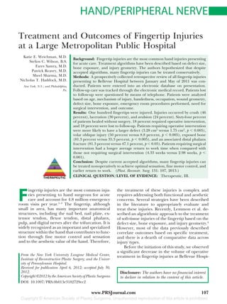
intervention, and overall outcome. Geometry of
injury was described using a schematic adapted
from the Fassler angles and levels of amputation
of the fingertip5 (Fig. 1). Patients who were lost to
follow-up were contacted by telephone and ques-tioned
about their outcomes. Statistical analysis
was performed using the t test and analysis of
variance using Minitab 16 (Minitab, Inc., State
College, Pa.).
RESULTS
During the 5-month period between January
and May of 2011, 100 fingertip injuries in 83 pa-tients
were prospectively registered by means of
the electronic medical record system at Bellevue
Hospital. There were 57 male patients (67.8 per-cent)
and 26 female patients (32.2 percent). Pa-tients
were students (27 percent), maintenance
workers (18 percent), employed in the food in-dustry
(cooks/butchers) (14 percent), teachers or
in the art industry (11 percent), clerical workers
(10 percent), construction workers (9 percent),
unemployed (8 percent), and health care workers
(3 percent). The majority of patients were right
hand dominant (75 percent).
Injuries were distributed between the domi-nant
and nondominant hands, 52 percent right
hand and 48 percent left hand. The most common
mechanism of injury was crush (45 percent), fol-lowed
by laceration (32 percent) and avulsion (23
percent). There was one digit injured in 86.7 per-cent,
two digits injured in 7.2 percent, three digits
Fig. 1. Fassler wound geometry. (Printed in Lemmon JA, Janis JE, Rohrich RJ. Soft-tissue
injuries of the fingertip: Methods of evaluation and treatment. An algorithmic ap-proach.
Plast Reconstr Surg. 2008;122:105e–117e. Reprinted with permission from
Fassler PR. Fingertip injuries: Evaluation and treatment. J Am Acad Orthop Surg.1996;
4:84 –92.) Copyright 1996 American Academy of Orthopaedic Surgeons.
108
3. Volume 131, Number 1 • Treatment of Fingertip Injuries
injured in 4.8 percent, and four digits in 1.2 per-cent.
There were 22 injured index fingers, 27 in-jured
long fingers, 23 injured ring fingers, 10 in-jured
small fingers, and 18 injured thumbs. Injury
patterns were type A in 34 digits, type B in 15 digits,
type C in 40 digits, and type D in 11 digits. The
average size of soft-tissue defect was 1.87 cm2.
Fifty digits required a nail bed repair in the
emergency room and 13 digits were treated with
a composite graft in the emergency room. Twelve
(92.3 percent) of these composite grafts healed
without requiring any further procedures, and
one was lost to follow-up. Sixty-eight digits healed
without surgery, 16 digits ultimately required sur-gical
intervention, and 13 digits required soft-tis-sue
surgery. Sixteen patients (16 digits) were lost
to follow-up after their initial presentation to the
emergency room. The average time from injury to
the operating room was 12.2 days. The surgical
procedures for soft-tissue management included
nail plate removal (n3), full-thickness skin graft
(n 3), cross-finger flap (n 2), completion
amputation (n 2), Atasoy flap (n 1), thenar
flap (n1), and first dorsal metacarpal artery flap
(n 1). Additional surgical procedures per-formed
included bony fixation (n 2) and ten-don
reconstruction (n 1).
Sensation was intact to two-point discrimina-tion
(7 mm) in 65 digits, impaired in eight, and
lost to follow-up or absent from notes in 27. Of
those eight digits with decreased two-point dis-crimination,
four (50 percent) were managed with
local wound care, three (37.5 percent) were
treated with nail plate removal, and one (12.5
percent) was treated with a cross-finger flap. Pa-tients
without documented examinations or who
were lost to follow-up were treated as follows: local
wound care, 24 patients (88.8 percent); cross-fin-ger
flap, one patient (3.7 percent); debridement,
one patient (3.7 percent); and full-thickness skin
graft, one patient (3.7 percent). Two patients with-out
documented examination treated with local
wound care were children younger than 3 years.
The average time until return to work was 3.26
weeks for all patients. Patients requiring surgical
intervention had a longer average return to work
time when compared with those not requiring
surgical intervention (4.33 weeks versus 2.98
weeks, respectively; p 0.0096). All patients not
lost to follow-up returned to work.
The 16 patients requiring surgical interven-tion
had a median age of 31 years. Nine were
manual laborers and six were nonmanual labor-ers.
Eight sustained a laceration, seven suffered an
avulsion injury, and one suffered a crush injury.
The majority of these injuries were volar oblique
with exposed bone (n 8), followed by transverse
(n 7) and then dorsal oblique (n 1). Thirteen
had fractures and 13 also had exposed bone. Four
injured their dominant hands and two injured
their nondominant hands.
When comparing patients requiring operative
intervention versus those healing with conserva-tive
measures in a univariate analysis, patients re-quiring
surgery were more likely to have suffered
a volar oblique injury [50 percent (n 8) versus
8.8 percent (n 6); p 0.001]. They were also
more likely to have exposed bone [81.3 percent
(n13) versus 35.3 percent (n24); p0.0009]
and an associated distal phalanx fracture [81.3
percent (n 13) versus 47.1 percent (n 32);
p 0.013]. Manual laborers were no more likely
to require surgical intervention [nine (56.3 per-cent)
versus 25 (36.7 percent; p 0.14] when
compared with nonsurgical intervention. Finally,
patients requiring operative intervention were
more likely to have a larger soft-tissue defect (3.28
cm2 versus 1.75 cm2; p 0.005) (Table 1). In the
multivariate analysis, mechanism, occupation, and
exposed bone were not found to be independent
predictors of need for surgical intervention.
DISCUSSION
Fingertip amputation is one of the most com-mon
injuries presenting to the emergency room.
The basic tenets of finger reconstruction are to
provide durable coverage, preserve sensation and
length, minimize discomfort, and expedite return
Table 1. Characteristics of Those Who Healed
without Surgery versus with Surgery
Healed with
Surgery (%)
Healed without
Surgery (%) p
No. of patients 16 68
Mean age, yr 31 10.8 32 18.2 0.86
Manual labor 9 (56.3) 25 (36.7) 0.14
Sex
Male 11 (68.8) 50 (73.5) 0.704
Female 5 (31.2) 18 (26.4) 0.698
Crush mechanism 1 (6.25) 15 (22.1) 0.146
Laceration
mechanism 8 (50) 22 (32.3) 0.183
Avulsion
mechanism 7 (43.7) 31 (45.5) 0.896
Orientation
A 0 (0) 29 (42.6) 0.0013
B 8 (50) 6 (8.8) 0.001
C 7 (43.7) 25 (36.7) 0.652
D 1 (6.25) 8 (11.7) 0.525
Exposed bone 13 (81.3) 24 (35.3) 0.0009
Fracture 13 (81.3) 32 (47.1) 0.013
Average soft-tissue
defect, cm2 3.28 1.75 0.001
109
4. Plastic and Reconstructive Surgery • January 2013
to work and normal activities.4 There are multiple
described techniques to treat fingertip amputa-tion.
To help navigate these options, treatment
algorithms have been developed.4 We follow a
standard algorithm in our center in an effort to treat
these patients in an expeditious manner. However,
secondary to our patient population, there is often
delay in presentation to the operating room despite
scheduled operative dates. On presentation, often
these wounds are healed and therefore no proce-dure
is performed. To better describe this, we per-formed
a prospectively enrolled retrospective review
of this patient population.
As previously mentioned, reconstructive strat-egies
will vary depending on the mechanism of
injury and severity of the injured digit(s). Other
factors include the patient’s preference, hand
dominance, occupation, age, sex, and reliability to
follow up. Standard procedures for fingertip re-construction
include revision amputation6 and
split-thickness7 or full-thickness skin grafts.8,9 Also,
various local flaps have been used, including the
V-Y volar advancement flap,10 the homodigital
neurovascular island flap,11 the first dorsal meta-carpal
artery flap,12 the Littler flap,13 the Moberg-
O’Brien flap,14 the Atasoy flap,15 the Hueston
flap,15 the Cutler flap,16 the modified volar ad-vancement
flap,17 the thenar flap,18,19 and the
cross-finger flap.20 In addition, free flaps have also
been shown to be effective when reconstructing
extensive fingertip defects secondary to trauma,
more specifically, the medial plantar venous flap,21
the glabrous flap,22 the free dorsoulnar artery per-forator
flap,23 the superficial palmar branch of the
radial artery flap,24 and various toe pulp flaps.
Of the 83 patients our group reviewed, 29
required nail bed repair on initial presentation to
the emergency room. Acute management of nail
bed injuries is well described.25–28 Nail bed repair
is often the first step in minimizing fingertip de-formities
and cosmetic and functional problems.28
The basic principles include sufficient cleaning,
minimal de´bridement of the nail bed (sterile and
germinal matrix), proper alignment of the injured
structures, preservation of marginal skin folds,
and an appropriate wound dressing.28 If the repair
is done properly, a new nail can grow that is in-distinguishable
from the patient’s original nail. If
the germinal matrix is not properly reapproxi-mated
or a wide scar is present, a permanent split
nail will result.28 Still, preservation of the nail bed
is not always attainable. Three of our patients ul-timately
underwent surgery for nail bed ablation.
Revision amputation is one of the mostcommon
operations of the hand.6 Regardless of wound ori-entation,
fingertip amputation injuries proximal to
the lunula often require revision amputation.4 The
reported advantage of revision amputation com-pared
with other reconstructive efforts is that it of-fers
the patient a relatively quick return to the
work force. The most common reported reason
for refusal of replantation is the inability to im-mediately
return to work.29 Only two digits in this
series were treated with revision amputation.[30]
Thirteen of the 100 digits in this review were
treated with a composite graft at the time of the
initial presentation. Composite grafts are typically
performed following a nonreplantable traumatic
distal fingertip amputation.11 This technique in-volves
excision of any bony segment and defatting
the pulp of the amputated digit, reapproximating
the prepared amputated segment to the remain-ing
digit, and using a bolster dressing. Some have
reported high success rates in terms of functional
and aesthetic outcomes with similar techniques.31
Specifically, Uysal et al. reported good retained
sensibility, acceptable aesthetic outcomes, and full
satisfaction from their patient population, who
were reported to have graft viability rates of almost
87 percent.32 Of the 13 digits treated with a com-posite
graft, 84.6 percent survived and 92 percent
of these had return of protective sensation.
Only 17 of the 100 digits reviewed ultimately
received surgical reconstruction. These interven-tions
included bone fixations, cross-finger flaps,
full-thickness skin grafts, local flaps, a thenar
flap, a dorsal metacarpal artery flap, and nail
bed ablation.
Furthermore, Lemmon et al. suggest that fin-gertip
amputation defect size less than or equal to
1.5 cm2 without exposed bone should be allowed
to heal by secondary intention. Our group found
an average size of soft-tissue defect to be 1.87 cm2
and, as one would expect, a significantly larger
average soft-tissue defect in fingertips that ulti-mately
required reconstruction compared with
those that did not require reconstruction. Of the
100 digits reviewed, 68 healed without surgery,
compared with just 13 requiring soft-tissue sur-gery.
The average defect size allowed to heal by
secondary intention was 1.75 cm2, compared with
the average defect size requiring surgery, which
was 3.28 cm2 (p 0.029) (Table 1). Interestingly,
of the six patients who ultimately reported hyper-sensitivity
on follow-up, five were treated with con-servative
wound management alone, which may
suggest inadequate soft-tissue volume in the af-fected
digit. There can be several explanations
that account for our relatively large average defect
size in patients who ultimately did not undergo
110
5. Volume 131, Number 1 • Treatment of Fingertip Injuries
reconstruction. First, 73 percent of our patients
were adults, with the majority employed as manual
laborers (27 percent) or in the food industry (13
percent) or other service industries (10 percent).
Presumably, these patients are compensated on an
hourly basis, with minimal or no paid sick leave.
Only 9.0 percent of our patient population was
employed as teachers, in the art industry, or cler-ical
workers. Our patient population often missed
operative appointments and presented later in the
healing process. This tendency also biased the
average size defect of our conservatively managed
patients despite our initial intention to treat in
these cases. Our treatment algorithm was not
based on a defect size cutoff but rather took into
account the type of injury, the necessity for our
patients to return to work, and our patient pop-ulation’s
generally poor reliability to return for
proper follow-up. Furthermore, 16 digits were lost
to follow-up.
There was a significant difference in average
return to work time when comparing the surgical
treatment arm to the nonsurgically treated pa-tients,
4.33 weeks compared with 2.98 weeks, re-spectively.
This may be influenced by our average
time from injury to the operation of 12.2 days. This
number may be elevated when compared with the
community because of the lack of appropriate fol-low-
up after initial injury in our patient popula-tion.
Accounting for these days, the average return
to work time would be similar in the nonoperative
group (2.98 weeks) and the operative group (2.68
weeks), arguing against surgical intervention pro-viding
quicker return to work for patients.
After evaluating the management of traumatic
injuries by prospectively assessing all fingertip in-juries
presenting to a large metropolitan public
hospital, it is clear that a large number of these
injuries can be treated by conservative manage-ment.
Despite this fact, suboptimal outcomes are
still being attained because of socioeconomic lim-itations,
poor patient compliance, poor follow-up
rates, and other factors. Although it is difficult to
mitigate the aforementioned factors, improve-ments
in patient education may help to improve
the patient’s understanding of the long-term se-quelae
of hand injuries. Also, patients should be
encouraged to speak with social workers to try to
gain workers’ compensation and other monetary
compensation to allow these patients to make de-cisions
based on their health and not on their job
status. Furthermore, increased surgical staffing
and operating room availability may decrease the
lag between the time of injury and the scheduled
operating room date to improve on intention-to-treat
outcomes in the face of a difficult-to-manage,
low-income, urban patient population.
Nicholas T. Haddock, M.D.
Department of Plastic Surgery
University of Texas Southwestern
1801 Inwood Road
Dallas, Texas 75390
haddockmd@gmail.com
REFERENCES
1. Chau N, Gauchard GC, Siegfried C, et al. Relationships of
job, age, and life conditions with the causes and severity of
occupational injuries in construction workers. Int Arch Occup
Environ Health 2004;77:60–66.
2. Sorock GS, Lombardi DA, Hauser RB, Eisen EA, Herrick RF,
Mittleman MA. Acute traumatic occupational hand injuries:
Type, location, and severity. J Occup Environ Med. 2002;44:
345–351.
3. Gavrilova N, Harijan A, Schiro S, Hultman CS, Lee C. Pat-terns
of finger amputation and replantation in the setting of
a rapidly growing immigrant population. Ann Plast Surg.
2010;64:534–536.
4. Lemmon JA, Janis JE, Rohrich RJ. Soft-tissue injuries of the
fingertip: Methods of evaluation and treatment. An algorith-mic
approach. Plast Reconstr Surg. 2008;122:105e–117e.
5. Fassler P. Fingertip injuries: Evaluation and treatment. J Am
Acad Orthop Surg. 1996;4:84–92.
6. Blair JW, Moskal MJ. Revision amputation achieving maxi-mum
function and minimizing problems. Hand Clin. 2001;
17:457–471, ix.
7. Moon SH, Lee SY, Jung SN, et al. Use of split thickness
plantar skin grafts in the treatment of hyperpigmented skin-grafted
fingers and palms in previously burned patients.
Burns 2011;37:714–720.
8. Wendt JR. Coverage of full-thickness volar hand skin defects
with lateral great toe skin grafts. Plast Reconstr Surg. 2001;
108:2069–2071.
9. Schenck RR, Cheema TA. Hypothenar skin grafts for finger-tip
reconstruction. J Hand Surg Am. 1984;9:750–753.
10. Mehling I, Hessmann MH, Hofmann A, Rommens PM. V-Y
flap for restoration of the fingertip (in German). Oper Orthop
Traumatol. 2008;20:103–110.
11. Chen SY, Wang CH, Fu JP, Chang SC, Chen SG. Composite
grafting for traumatic fingertip amputation in adults: Tech-nique
reinforcement and experience in 31 digits. J Trauma
2011;70:148–153.
12. Chen C, Zhang X, Shao X, Gao S, Wang B, Liu D. Treatment
of thumb tip degloving injury using the modified first dorsal
metacarpal artery flap. J Hand Surg Am. 2010;35:1663–1670.
13. Xarchas KC, Tilkeridis KE, Pelekas SI, Kazakos KJ, Kakagia
DD, Verettas DA. Littler’s flap revisited: An anatomic study,
literature review, and clinical experience in the reconstruc-tion
of large thumb-pulp defects. Med Sci Monit. 2008;14:
CR568–CR573.
14. Kapandji T, Bleton R, Alnot JY, Oberlin C. Digital flap au-tografts
for pulp coverage in distal amputations of the fin-gers:
68 flaps (in French). Ann Chir Main Memb Super. 1991;
10:406–416.
15. Vasseur C, Legre R, Leps P, et al. Qualitative retrospective
study comparing 43 advanced-rotated flaps to 19 island type
Venkataswami-Subramanian flaps (in French). Chir Main
2000;19:44–55.
16. Roberts AH. Kutler repair for amputated fingertip. Ann R
Coll Surg Engl. 1980;62:75–76.
111
6. Plastic and Reconstructive Surgery • January 2013
17. Souquet R. The asymmetric arterial advancement flap in
distal pulp loss (modified Hueston’s flap) (in French). Ann
Chir Main 1985;4:233–238.
18. Hugon S, Castus P, Schoofs M. Index reconstruction by
means of a fasciocutaneous thenar flap. Plast Reconstr Surg.
2010;126:43e–44e.
19. Melone CP Jr, Beasley RW, Carstens JH Jr. The thenar flap:
An analysis of its use in 150 cases. J Hand Surg Am. 1982;7:
291–297.
20. Mishra S, Manisundaram S. A reverse flow cross finger pedi-cle
skin flap from hemidorsum of finger. J Plast Reconstr
Aesthet Surg. 2010;63:686–692.
21. Yokoyama T, Tosa Y, Hashikawa M, Kadota S, Hosaka Y.
Medial plantar venous flap technique for volar oblique am-putation
with no defects in the nail matrix and nail bed.
J Plast Reconstr Aesthet Surg. 2010;63:1870–1874.
22. Orbay JL, Rosen JG, Khouri RK, Indriago I. The glabrous
palmar flap: The new free or reversed pedicled palmar fas-ciocutaneous
flap for volar hand reconstruction. Tech Hand
Up Extrem Surg. 2009;13:145–150.
23. Simsek T, Engin MS, Aslan O, Neimetzade T, Eroglu L.
Finger pulp reconstruction with free dorsoulnar artery per-forator
(DUAP) flap. J Reconstr Microsurg. 2011;27:543–549.
24. Lee TP, Liao CY, Wu IC, Yu CC, Chen SG. Free flap from the
superficial palmar branch of the radial artery (SPBRA flap)
for finger reconstruction. J Trauma 2009;66:1173–1179.
25. Van Beek AL, Kassan MA, Adson MH, Dale V. Management
of acute fingernail injuries. Hand Clin. 1990;6:23–35; discus-sion
37–38.
26. Shepard GH. Management of acute nail bed avulsions. Hand
Clin. 1990;6:39–56; discussion 57–58.
27. Shepard GH. Nail grafts for reconstruction. Hand Clin. 1990;
6:79–102; discussion 103.
28. Brown RE. Acute nail bed injuries. Hand Clin. 2002;18:561–575.
29. Ozer K, Kramer W, Gillani S, Williams A, Smith W. Replan-tation
versus revision of amputated fingers in patients air-transported
to a level 1 trauma center. J Hand Surg Am.
2010;35:936–940.
30. Heistein JB, Cook PA. Factors affecting composite graft sur-vival
in digital tip amputations. Ann Plast Surg. 2003;50:299–
303.
31. Chai Y, Kang Q, Yang Q, Zeng B. Replantation of amputated
finger composite tissues with microvascular anastomosis.
Microsurgery 2008;28:314–320.
32. Uysal A, Kankaya Y, Ulusoy MG, et al. An alternative tech-nique
for microsurgically unreplantable fingertip amputa-tions.
Ann Plast Surg. 2006;57:545–551.
Evidence-Based Medicine: Questions and Answers
Q: I’ll do my best to indicate the correct clinical question and Level of
Evidence (LOE) on my manuscript. How does the LOE grading process
work with PRS?
A: The authors’ own grading is the first step in the process toward
determining the “real” LOE of an article.
Once submitted, manuscripts are peer reviewed as part of the normal
review process. PRS is training its reviewer panels on how to determine
LOE clinical questions and grading. As part of the review process, we
will ask our reviewers to indicate their assessment of the LOE for the
papers they review. After manuscripts have been reviewed, revised, and
accepted for publication, they will be sent to independent evidence-based
medicine and LOE experts, who will rate the manuscripts for
clinical question and LOE grade. These experts will make the final
determination of the LOE of all accepted papers, and their decisions
will be reflected in the published LOE of the articles. For those papers
that are not gradable, we will leave the LOE grade off of the published
abstract.
112