
Recommended
More Related Content
What's hot
What's hot (20)
Similar to Application of laser in dermatology
Similar to Application of laser in dermatology (20)
Recently uploaded
Recently uploaded (20)
Application of laser in dermatology
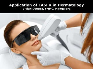
- 1. Application of LASER in Dermatology Vivian Dsouza, FMMC, Mangalore
- 2. Dr Leon Goldman Father of Laser Medicine and Surgery
- 5. TYPES OF LASER MEDIA GAS LIQUID SOLID Argon Carbon dioxide Copper vapour Helium-neon Krypton Xenon chloride Rhodamine dye dissolved in organic solvent Crystal •Alexandrite •Erbium doped yttrium aluminium garnet (YAG) •Holmium doped YAG •Neodymium doped YAG •KTP •Ruby Semiconductor diode •Aluminium •Gallium •Arsenide
- 6. EXCIMER LASER NON-LASER LIGHT SOURCES •Name is derived from the terms excited and dimers. •Use reactive gases, chlorine and fluorine mixed with inert gases such as Argon, Krypton or Xenon. •When electrically stimulated a pseudo-molecule (dimer) is produced. •Dimer produces light in the UV range. •Intense pulse light •Non coherent light within 500- 1200nm.
- 7. Chromophores • These are absorbing molecules. • Exhibit characteristic bands of absorption at certain wavelengths. • Three primary skin chromophores are : Water Haemoglobin Melanin
- 8. Chromophores in human skin – absorption spectra of Haemoglobin, melanin and water
- 9. Penetration of LASER depends upon : • Absorption and scattering • Depth of penetration increases with wavelength • Amount of scattering is inversely proportional to wavelength.
- 10. Other factors include • Thermal interactions such as photocoagulation and photo-vaporization. • Thermal relaxation time • Photochemical ablation • Selective photothermolysis • Skin cooling
- 12. Ablative (Vaporizing) Skin Resurfacing The ablative lasers are far-IR Carbon dioxide Erbium YAG Chromophore – water Very useful for treating 1. Photoaging 2. Scars 3. Epidermal nevi 4. Seborrheic keratoses Complications : scarring, hyper pigmentation, delayed onset permanent hypo pigmentation, prolonged erythema, bacterial, viral and fungal infections.
- 13. Treatment of vascular lesions A peak absorption of oxyhaemoglobin occurs at 577nm within the yellow spectrum. 1. Flashlamp-pumped pulse dye lasers (PDLs) with pulse durations ranging from 0.45-40 ms 2. Pulse neodymium YAG lasers 3. Alexandrite lasers Hypertrophic lesions PDL resistant vascular lesions 4. Copper vapour or copper bromide lasers
- 14. 5. Potassium titanyl phosphate (KTP) lasers Complications : • Purpura as a result of microvascular damage • Subsequent thrombosis • Delayed appearance of vasculitis
- 16. Interactions during treatment of pigmented lesion and tatoos • Red and near IR lasers are most selective • Mechanism : a) Treatment of tattoos with Q switched lasers, fragments the ink particles and selectively kills pigment containing cells b) This is associated with resultant ink particle release c) Subsequent removal of tattoo particles can occur via an epidermal crust and/or lymphatic transport, and some particles are re- phagocytosed by dermal cells
- 17. • LASER commonly used are 1. Q-switched ruby 2. Alexandrite lasers 3.Q-switched Nd:YAG laser Other conditions • Lentigines • Nevus of ota • Cafe-au-lait macules • Melanocytic nevi • Side effects : pigment and textural changes, allergic reactions, ink darkening, tissue aerosolization with possible infectious particles.
- 18. Interactions during Hair removal • Red to near IR region of the spectrum (deeply penetrating) • High energy • Mechanism : damage to follicular stem cells in the bulge region of the outer root sheath and/or the dermal papilla at the base of the hair follicle TEMPORARY HAIR LOSS PERMANENT HAIR LOSS Induction of catagen which can occur at very low fluences. Miniaturization to produce vellus like hairs. An 810nm diode laser source intended for home use Complete degeneration with local fibrosis.
- 19. Interactions during non- ablative skin rejuvination • Non-ablative facial facial rejuvination • Fine lines • Non-dynamic rhytides • Mid-IR lasers 1. 1320nm Nd:YAG 2. 1450nm diode 3. 1520nm erbium:glass lasers 4. IPL sources MECHANISM: These work by subtle thermal effects on the dermis, presumably stimulating a wound healing response.
- 21. Fractional Photothermolysis Thousand of nearly visible, microscopic zones of thermal injury are created Stimulating turnover Remodelling of both – epidermis and the dermis
- 22. NON ABLATIVE FP ABLATIVE FP Uses focused mid-IR laser microbeams to create a pixilated pattern of small columns of thermal damage called microthermal treatment zones uses carbon dioxide or erbium lasers to vaporize a similar pattern of vertical channels that can extend deeply into the skin USES : •Scars •Fine rhytides •Telengiectasias •Dermatoheliosis •Poikiloderma
- 23. LASER based Diagnostics 1. Optical coherence tomography - Near IR - low coherence light - used for high resolution cross sectional imaging of the body tissues
- 24. 2. LASER confocal microscopy - Captures light scattered or emitted from a thin plane “section” inside the skin. - Since histochemical stains are not used confocal microscopy of skin tumors reveal diagnostic features that are different from those of conventional histology. - Microvascular blood flow and trafficking of lymphocytes can be observed.
- 25. BIOPSY OF HUMAN SMALL INTESTINE VISUALIZED WITH A CONFOCAL LASER SCANNING MICROSCOPE
- 26. THANK YOU