
Recommended
More Related Content
Similar to October 2017 DISCOVER 27I’m lying on my back in the .docx
Similar to October 2017 DISCOVER 27I’m lying on my back in the .docx (15)
More from vannagoforth
More from vannagoforth (20)
Recently uploaded
Recently uploaded (20)
October 2017 DISCOVER 27I’m lying on my back in the .docx
- 1. October 2017 DISCOVER 27 I’m lying on my back in the tunnel of an MRI scanner, my skull immobilized in a head coil, which looks like a cage fighter’s mask. There’s a vitamin E capsule taped to the right side of my forehead. The head coil controls variations in the scanner’s magnetic field and the capsule has to do with scan orientation, in the same way that surgeons will write on your right leg so they don’t mistakenly operate on your left. A writer taking part in an aging study explores his senior moments. BY JEFF WHEELWRIGHT PHOTOS BY DAVID ZENTZ This Old Brain I’m sporting headphones and watching a projec- tion of a Tom and Jerry cartoon from the 1950s, and different parts of my brain are presumably paying attention. Meanwhile, the scanner is taking slices — noisy, virtual slices — of my gray and white matter. The purpose is to illuminate the fea- tures of my brain that are processing Jerry running from Tom. From that information, the scientists in charge can make a wiring diagram of my brain, flickering in time with the images.
- 2. I feel childlike and helpless. I may have seen this cartoon 60 years ago, but I can’t remember. Relax, I remind myself. Just stay still. Cognitive decline at my age is expected. “ H ow a r e yo u d o i n g , Je ff ? ” Tay l o r Kuhn asks through the headset. Kuhn, a post- doctoral research fellow in cognitive psychology at the University of California, Los Angeles, has a courtly Southern accent, like one of the aristo- cratic characters in Gone With the Wind. (What was his name? Ashley. Yes, Ashley something. “Oh, Ashley,” Scarlett gushes.) “Jeff,” Kuhn interrupts, “we’re going to do the name-association task.” I notice that the scanner is silent, waiting. “OK,” I say. We practiced this exercise beforehand. The MRI cranks up again, sounding like a cross between a jackhammer and a dentist’s drill. A face flashes on the screen directly above me, along with a name. I try to memorize the pairing, after which I press a button on a console resting on my lap. Another face and name appear, and my working memory, such as it is, gamely records the association. And so on, perhaps a dozen in all. I set hasty mnemonic cues, like bread crumbs. Peter has dark hair. Mary’s hair is long and lank — lank hair equals Mary. Stuart is the guy who looks blah. Here’s another blah guy. Wait, was he Allen? My recall is supposed to be tested later, outside the scanner, but Kuhn and Susan Bookheimer, the clinical neuropsychologist who is the principal investigator of my brain, have
- 3. assured me that my recall doesn’t matter as much as the neurological tracks of my memorization. 28 DISCOVERMAGAZINE.COM They are interested in the brain’s connectivity. This is all part of a UCLA study of the human con- nectome, senior division. I think of it as neuroscience 2.0. Connectome, connectomics and connectivity are newish terms stemming from the technical capacity to capture and visualize neural networks. Research has moved beyond the exploration of parts, i.e., the ana- tomical and functional description of the brain’s com- ponents. For human studies, neuroscience 1.0 depended largely on effects of injuries. When a brain-damaged person lost function in a particular region, scientists learned what that area does in the normal state. But the brain’s parts don’t run in isolation. With imaging tools like positron emission tomography (PET) and especially functional magnetic resonance imaging (fMRI), the connections among brain areas are literally becoming clearer. Where X-rays and first-generation MRI depict structure, the newer scans show activity. For example, scientists have known for decades that the hippocampus, a structure in the middle of the brain, has the lead role in the formation and preservation of memories, and is one of the regions that shape a person’s identity. The hippocampus is a few inches from the pre- frontal cortex, about as far, relatively, as the alternator and carburetor are in an automobile engine. The fMRI can show the two parts working together under the hood.
- 4. Connectomics not only maps the circuitry of the links and nodes, the so-called structural connectivity of the brain, but also the dynamic correlations that arise among regions when circuits are active: the functional connec- tivity. Even when it’s not thinking or doing work at all, just fleeting for no reason over a scene in Gone With the Wind, the brain is fully powered. Systems are humming and active, as in an idling car before the light turns green. MIND AND MATTER The Human Connectome Project (HCP), a joint ven- ture of a dozen universities that launched in 2012 with support from the National Institutes of Health, aims to create a database of connectivity patterns. The HCP has sketched an engine manual, so to speak, of normal and healthy brain function, drawn from hundreds of fMRI scans. The subjects of these scans were young adult siblings. In the latest phase of the project, chil- dren and older people are being enrolled so that scien- tists can learn how connectivity changes as the brain waxes in youth and wanes in old age. Bookheimer’s lab at UCLA is one of the research sites processing the latter group. The overall HCP-A (A for aging) cohort, when complete, will have involved 1,208 people ranging from 36 to over 100 years old. Autopsy and scanning studies indicate that a healthy 69-year-old like me has been shedding brain matter at a rate of 0.5 percent per year for a decade and probably longer. My hippocampus, if I’m typical, has begun to lose neurons even faster, perhaps 2 percent annually. Senior moments have become laughably common, as the bonds between the names of things and the things themselves loosen and in a few cases vanish altogether. But I can still write a pretty good sentence, heartened
- 5. that the cognitive decline that might be expected to accompany the brain’s atrophy is neither linear nor predictable, partly because older brains compensate by forging workarounds in connectivity. For familiar and basic tasks, the aging brain opens new pathways, enlisting untapped regions and neurons, installing patches where needed. By contrast, the loss of neurons in Alzheimer’s disease is pronounced and rapid, and so too the October 2017 DISCOVER 29 mental failing. MRI scans suggest that structural and functional connectivity has gone haywire. Orderly networks give way to disorderly new webs, as if the brain were a drowning person flailing and grabbing onto whatever it can for support. About 10 percent of Americans older than 65 have Alzheimer’s, roughly 5 million people, a number that can only grow as the nation’s population ages. Research has centered on detecting and countering amyloid plaques and neurofibrillary tangles — the protein complexes that clog synapses and destroy neurons. Unfortunately, normal aging entails plaques and tangles, too. MRI and X-rays cannot see them accumulate in the hippocampus and other structures. By the time the build-up is visible on a PET scan, it’s too late to stop the effects of disease. So as they ana- lyze connectivity patterns, the scientists of the HCP-A project hope to establish points where healthy aging and unhealthy degeneration diverge. “Where do you go off that trajectory to a disease state?” asks Beau Ances
- 6. of Washington University in St. Louis, who is HCP-A’s lead investigator. If the connectome project can set the baseline parameters of normal aging, Ances notes, “other investigators can take the scans and ask, ‘Where do my patients fit?’ ” In effect, we are the control group for studies yet to come. To the extent that the HCP-A inventory contains dementias in waiting, scientists may explore hypotheses about the nascent stages of disease. Nearly half of us will be scanned again in two years, and our health checked thoroughly. Beyond that, the plan is uncertain. “We will probably request funds to continue follow-up when this [five-year] grant expires,” says Bookheimer. “It is definitely important.” PINWHEEL FOR THE BRAIN The MRIs comprise only part of the data. Each HCP-A participant is put through a battery of cognitive, psy- chological and even physical testing. Working with Bookheimer’s team — Kuhn and Mirella Diaz-Santos, who are postdocs, and Kevin Japardi, a staff research associate — I answer scores of questions while they take notes. I rate my feelings about the situations in my life in terms of “a lot,” “a little” or “not at all.” Several times I am asked, in different ways, if I have ever suffered a head injury. I provide measures of blood pressure, eye- sight and grip strength. I give a blood sample for genetic analysis. Somewhat to my discomfort, the question- naires pinpoint my psychological flaws. As a gauge of stamina, I walk rapidly for two minutes around traffic cones on the carpet in the hallway. Now it’s time for cognitive tests. Seated in front of a screen, I am instructed to pronounce a series of very obscure, polysyllabic words. While being timed, I draw
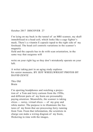
- 7. lines between ascending numbers and alphabetically ordered letters randomly spread about a page, moving from a number to a letter to the next number and so forth. Next, my short-term memory is examined — “From the series of words I just read to you, tell me as many as you can remember.” (Dismally few, it turns out.) I react to a stream of geometric shapes, pushing a button for a circle or a square but not for any other shapes. Mistakes are OK, the young researchers chirp. Easy for them to say. I’m older than any two of them put together. The scientific discipline that incorporates these tasks, tests and surveys is called neuropsychology. The neuro- psychologists for the connectome project will collect a large number of metrics from a large group of healthy (more or less) seniors, and set statistical boundaries As part of the study, the author had a capsule taped to his forehead (far left) and went through a battery of tests that included (from left) grip strength, walking around
- 8. cones, timed cognitive tests and reading an eye chart. 30 DISCOVERMAGAZINE.COM SC IE N C E SO U RC E reflecting the high, low and middle responses. The spec- trum will show all of us aging “normally” except where a few might fall out of range. What’s new is the correlation of our group’s neu- ropsychological variation with images of our neural mechanisms at work. Thus Bookheimer’s team has me repeat a few of the simpler tasks inside the scanner. In addition to the name-association task, I do what they call the checkerboard game, which is not much of a game but rather a test to see how quickly I push a button when
- 9. squares are illuminated on a pinwheeling circle. The wheel has a black-and-white checkerboard pattern. As it spins and the scanner rhythmically pounds, one or two squares on the left or right side of the pattern light up in red. The task comes near the end of my second session in the machine. I get out feeling like I’ve been at a bad heavy metal concert. Again, my score doesn’t matter as much as the nodes and links that the activity uncovered. Since individual results won’t be released, none of us who sign up for HCP-A will know where we fit into the overarching data. But the investigators, though blind to our identities, will know. “We will be able to use your data points,” Kuhn explains, “to compare vasculature, corti- cal thickness, functional networks, etc., across the entire group as a function of age.” Earlier I’d squeezed a dynamometer, a device to mea- sure grip strength, and I ask the team why they didn’t put me to the same test inside the scanner. According to recent literature, older people employ a dif- ferent brain network than younger people when performing this task, especially when squeezing with their less-dominant hand, which loses strength faster than the other. It’s because the time in an MRI scanner is so expensive, I am told. Another reason
- 10. is that the lab’s dynamometers have metal grips, which would mess with the scanner’s magnets. Still, the dyna- mometer results and other external measures can be assessed indirectly, by comparing them with the fMRI images of my default mode network, or DMN. The DMN represents the idling state I mentioned above. When the brain transitions from performing a task to a resting state, it engages a unique network, linking a half-dozen regions in the cerebral cortex and the hippocampus, just below the cortex. The DMN has become one of the most-studied networks of the human connectome. The scanner probed my DMN twice; I was advised in so many words to simply daydream for the eight minutes the scan required. But the DMN is more than a daydream factory. Scientists believe it orchestrates the rehearsal of focused activity, in the microseconds before you decide to squeeze a dyna- mometer, say. The network tidies up the circuits before memories are retrieved — just before you reach for the name of that person who’s just come over and said hello. JUICY HIPPOCAMPUS Considering my role as both participant and journal- ist, the UCLA researchers agree to show me my brain structures; other seniors in the project won’t get that opportunity. At the end of the day, I meet with Susan Bookheimer for her quick take on my brain’s nuts and bolts. (The computer processing of my functional connectivity scans, such as the DMN and the chain of regions that lit up during the checkerboard task, will require much more time.) Previously, Bookheimer had cautioned me by email: “There is little to report in an individual brain scan unless there is an abnormality. We have all scans read by a radiologist for these, and if there
- 11. is an abnormality that requires some action, we would tell you about it.” She calls up the black-and-white images on her desk- top computer and riffles through the slices, zooming from the left side of my skull to the right. She sees no sign of cerebral vascular disease or tumors, benign or otherwise. Just “normal age-related change.” “May I say,” she adds, “you have a very nice brain.” I hope she means it’s healthier-looking than others — but actually she means that the quality of the image is clear and pleasing. I must have held quite still. Bookheimer points out my corpus callosum, the band of fibers that join the two hemispheres, and just below it the dark linings of the ventricles. “The ventricles hold fluid, and in abnormal aging the fluid expands into the spaces made as tissue atrophies,” she says. It’s not hap- pening much here. So far, so good. Then to the major features of my brain. On a tablet, Bookheimer accesses a scan configured to capture my hippocampus, one in each hemisphere. The left hippo- campus tends to be more involved in verbal memory and the right more involved in nonverbal and visual memory. She mentions the name-face association task, which is “to take two arbitrary things and bind them together, just like we do in real life when we meet new people. Seniors have a harder and harder time doing this.” The hippocampus in profile is a thin, elongated, curl- ing structure, which is said to resemble a sea horse (the Latin translation of hippocampus). Bookheimer zooms from front to back, and stops at a cross section. “You have a nice, fat, juicy hippocampus,” she says, calling
- 12. to my mind a sirloin steak, yet she’s just commenting once more on the visual reproduction. “This scan is very pretty. You can see it’s beautiful. We will be able to make fine measurements of the subregions. Look here. It’s like a Cinnabon.” As she zooms in further, faintly swirling lines like tree rings come into focus. “It’s gorgeous,” she says before catching herself. “I’m such a geek!” The hippocampus contains “reverberating circuits,” Bookheimer says. The cells are communicating across PET scans reveal a slight decrease in brain activity (shown in bright colors) between a 20-year-old (left) and an 80-year-old person. October 2017 DISCOVER 31 TO P: C O U RT ES Y JE FF W H
- 14. N C E SO U RC E the inner curvature and their synapses are forming mem- ories. Their specific tasks? “Some respond to anything new, some are repeaters,” she says. The hippocampus reviews and consolidates experiences and eventually sends them out for storage in other regions. “Some of the cells are discriminating between similar items, some are able to reconstruct a memory from partial cues. . . . This hippocampus is getting information from the left side of the brain, and it is well developed.” I see a well- aged steak again. “The hippocampus can grow new neurons — one of the few areas that can. Brain stimulation, learning new information, does seem to help.” She mentions the brain’s plasticity, its ability to compensate and find ways around damage and decline. I’m starting to appreciate this wispy curling tissue, floating at the heart of multiple networks. When the brain is at rest or in sleep mode, the hippocampus works with the DMN in memory forma- tion. The region may also be a place to look for early signs of cognitive decline or of Alzheimer’s. “The hippocampus is a target,”
- 15. Bookheimer says with relish, “because it can shed light on the rate at which the brain is aging.” Our business complete, B o o k h e i m e r ’s t e a m escorts me down to Westwood Avenue on the UCLA campus. It has been a tiring but stimulat- ing day and a half on the frontiers of research. My data right now are being crunched, I hope to good purpose. I take my bearings on the Westwood sidewalk. “Can you find your way back to the garage?” they ask. D Jeff Wheelwright is a contributing editor at Discover. “May I say,” she adds, “you have a very nice brain.” A cross section of the author’s brain, showing (from top) the cerebrum, the corpus callosum and the feathery-looking cerebellum. Comparison of brain scans of a 20-year- old (left) and an Alzheimer’s patient. Bright areas represent neural activity. © 2017 Discover Magazine
- 16. Sheet1schoolid23school name24school address221dfa000 kjhd2ddf98 suifh3daf777dkjh Sheet1schoolid1school name1school address11dfa000 kjhd2ddf98 suifh3daf777dkjh Sheet1schoolid2school name2school address21dfa000 kjhd2ddf98 suifh3daf777dkjh