Extended field Radiotherapy with chemotherapy in treatment of cervical carcinoma with retroperitoneal lymphadenopathy.
•Download as PPTX, PDF•
2 likes•157 views

Extended field Radiotherapy with chemotherapy in treatment of cervical carcinoma with retroperitoneal lymphadenopathy.

Recommended
Recommended
More Related Content
What's hot
What's hot (20)
Similar to Extended field Radiotherapy with chemotherapy in treatment of cervical carcinoma with retroperitoneal lymphadenopathy.
Similar to Extended field Radiotherapy with chemotherapy in treatment of cervical carcinoma with retroperitoneal lymphadenopathy. (20)
Recently uploaded
Recently uploaded (20)
Extended field Radiotherapy with chemotherapy in treatment of cervical carcinoma with retroperitoneal lymphadenopathy.
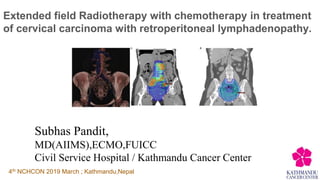
- 1. Extended field Radiotherapy with chemotherapy in treatment of cervical carcinoma with retroperitoneal lymphadenopathy. Subhas Pandit, MD(AIIMS),ECMO,FUICC Civil Service Hospital / Kathmandu Cancer Center 4th NCHCON 2019 March ; Kathmandu,Nepal
- 2. Lymph node status in cervical cancer ● Previously FIGO Staging was essentially clinical. ● Lymph node status did not contribute to FIGO stage BUT impact survival Stage 5 year survival IB/IIA 95 vs 78% (Kim SM IJGC 2000). ● Surgical para-aortic nodal involvement increases with stage from about 5% in patients with stage I disease to 25% in those with stage III disease
- 3. Adding notation of r (imaging) and p (pathology) to indicate the findings that are used to allocate the case to Stage IIIC. IIIC Involvement of pelvic and/or para‐aortic lymph nodes, irrespective of tumor size and extent (with r and p notations)c IIIC1 Pelvic lymph node metastasis only IIIC2 Para‐aortic lymph node metastasis
- 4. How to detect PA nodes? Sensitivity Specificity Ultra sonogram Variable CT-scan Pelvis >8mm PA >10mm 70-80% 85-92% MRI Size, rounded, irregular margin, clustering, necrosis 60-80% 80-90% PET CT 85-91% 95% *Surgerical – gold standard
- 5. So, should all patients undergo surgical LN sampling? ● Requires advanced surgical skills. ● Since 85% of cases presently occur in low‐resource settings-skill and infrastructure not widely available ● Cochrane Review (2013) - Pre‐treatment surgical para‐aortic lymph node assessment in locally advanced cervical cancer lacks sufficient data ● Pathological confirmation is the gold standard but imaging can be used to interpret disease extent. NO
- 6. How to treat PA nodes? ● Extended field Radiotherapy (EFRT) ○ 2D ○ 3D ○ IMRT / VMAT
- 7. 2D Technique ● Based on X-ray simulation ● Dose distribution not seen ● Toxicity was concern ○ No dose escalation ○ CT+RT difficult
- 8. Conformal techniques: 3D Vs. IMRT/VMAT
- 9. Why IMRT / VMAT ? ● Reduce dose to normal structure translate to reduced toxicity ● Simultaneous Integrated boost – Dose escalation without prolonging overall treatment time.
- 10. What IMRT can save? ● Bowel ● Bone Marrow
- 11. IMRT plan – conformal dose with sparing of normal structures
- 12. Bone marrow Bowel Bag Dose Volume Histogram- Sparing of Bowel and Bone marrow
- 13. EFRT using Simultaneous Integrated Boost 54Gy 45Gy
- 14. • A biologic response, as measured by the change in SUV for metastatic lymph nodes, was observed at a dose threshold of 54 Gy. We recommend that involved lymph nodes be treated to this minimum dose What Dose ? >54Gy
- 15. Anatomic distribution of FDG -avid lymph nodes in patients with cervical cancer. (190 LN+ in 50 pt.) Practical Radiation Oncology: January-March 2013 Location of PA node ?
- 16. Anatomic distribution of FDG -avid lymph nodes in patients with cervical cancer. (190 LN+ in 50 pt.) Practical Radiation Oncology: January-March 2013
- 17. Location of PA Nodes
- 18. Location in relation to IVC and Aorta
- 19. Location of PA LN in Left para-aortic area.
- 20. Location of PA LN in intra-aortocaval and Rt paracaval position
- 21. 3 5 2 Intra Aortocaval Lt lateral PA Rt Paracaval Location of PA Ln in KCC series
- 22. 2 5 Location of PA Node in relation to renal vessel. Superior to Renal Vessel Inferior to Renal Vessel
- 23. Concurrent chemotherapy ● RT Dose: 45 - 60Gy ● 4 patients required hospital admission during their treatment course. 5 2 Chemotherapy (wkly CDDP) Yes No 1 3 1 5 cycles 3 cycles 2 cycles No of chemo cycles
- 25. 1. Contour the inferior vena cava and aorta. 2. The superior extent should be the left renal vein. 3. The inferior extent should be the level of the bifurcation of the aorta.
- 26. 4. Expand the aorta by a margin of 10 mm anteriorly, posteriorly and medially, and 15 mm laterally. 5. Expand the inferior vena cava by 8 mm anteriorly and medially, and 6 mm posteriorly and laterally.
- 27. 1 cm 1.5 cm 0.6 cm 0.8 cm
- 29. Conclusions ● If PAN+ (FIGO IIIC) consider EFRT with concurrent cisplatin. ● No role of prophylactic EFRT at present but is area of active investigation ● IMRT/VMAT reduces toxicity and improves compliance to concurrent chemo-radiotherapy ● Consensus Contouring Guideline is available for uniformity.
- 30. Snowfall in Kathmandu cancer center after 12 years.
Editor's Notes
- Surgical series suggest that the incidence of para-aortic nodal involvement increases with stage from about 5% in patients with stage I disease to 25% in those with stage III disease
- . The type of imaging modality or pathology technique used should always be documented.
- PA nodal inv olvement is difficult. 8% of PET-CT –ve pt have positive PALNSurgicopathological assessment of lymph node involvement requires advanced surgical skills, whether performed by conventional or minimally invasive surgery. Since 85% of cases presently occur in low‐resource settings, the required professional skills and infrastructure facilities are presently not widely available. Pathological confirmation is the gold standard but imaging can be used to interpret disease extent. The choice of imaging modality for nodal evaluation has not been fixed by FIGO. It depends upon the availability of the imaging modality and patients’ affordability. Non‐availability of an imaging modality should not be a reason for undue delay in initiation of treatment. FIGO does not define criteria to discriminate between malignancy and inflammation/infection on imaging, which is left to the discretion of the clinician. The clinician must opine on whether these look suspicious enough to upstage the case or not. The best available technology should be used for assessment, and the lowest appropriate stage should be assigned—i.e., when in doubt assign the lower stage. At the present time, lack of facilities universally is recognized and clinical assessment of staging with the use of other facilities as available is permissible. The method of assigning the stage is to be recorded and reported.
- Requires advanced surgical skills, whether performed by conventional or minimally invasive surgery. Since 85% of cases presently occur in low‐resource settings, the required professional skills and infrastructure facilities are presently not widely available. Cochrane Review (2013) - Pre‐treatment surgical para‐aortic lymph node assessment in locally advanced cervical cancer lacks sufficient data Pathological confirmation is the gold standard but imaging can be used to interpret disease extent.
- GI toxicity and Bone marrow
- Location