2. been studied and compared extensively.3,11,12
After 12
training sessions, improvements in the classic strength
training group, that trained with sustained contractions,
were higher for maximal force compared to the group that
trained explosive contractions, but improvements in explo-
sive force were higher in the group that trained explosively
compared to the group that trained with sustained contrac-
tions. Based on surface EMG analysis, Tillin & Folland
(2014) suggested that the training-specific performance
improvements might be explained by specific neural adap-
tations. Due to methodological restrictions, it is not possi-
ble to extract the location (eg, spinal or supraspinal) of
such changes within the central nervous system from the
EMG signal alone. As a consequence, many studies in the
past years used electrical stimulation of peripheral nerves
or transcranial magnetic stimulation (TMS) to get a deeper
insight into the underlying mechanisms. However, these
studies often showed contradictory results. The H-reflex
size, which can be used as an indicator of neural changes
at spinal level, remained identical,5,13-16
or increased6,8,17
after strength training when measured during an ongoing
contraction. Similarly, motor evoked potential (MEP) size
elicited by TMS, which can be seen as an indicator of neu-
ral changes at corticospinal level, remained the same,18
increased14,19,20
or decreased.4,21
A likely reason for these
contradictory results is, that depending on the type and
duration of the strength training intervention, peripheral
adaptations occur7
which hinder the proper interpretation of
surface EMG data, especially during maximal or near-max-
imal muscle contractions.22
Thus, we designed this study to answer mainly two
questions. First, does short-term strength training induce
task-specific neuroplasticity and second, does neuroplastic-
ity happen more at supraspinal or spinal level? For this,
we had two groups of subjects that trained only one of
two different knee extensor isometric strength tasks, that
is explosive vs ramped-up then sustained contractions. We
hypothesized that the training would lead to task-specific
changes in the CNS. We expected to find neural differ-
ences between pre- and post-training, but only when sub-
jects perform the trained task, and not when they perform
the untrained task. To compare directly the neurophysio-
logical measurements before and after the training, we
used a very short training duration in order to limit
peripheral adaptations as much as possible. We timed the
H-reflex and TMS measurements right at the onset of
muscle contraction to detect alterations in the descending
motor command which would allow us to attribute such
changes to supraspinal sites.23
Moreover, we assessed
corticospinal excitability with MEPs and controlled for
spinal changes with H-reflexes, which we both elicited at
the onset of the trained and untrained strength task
respectively.
2 | RESULTS
For all ANOVAs, the degree of freedom was 17. For all
within-group paired t tests, the degree of freedom was 9
for the SUS group and 8 for the EXPL group. Groups were
matched by age (P = .19), height (P = .71) and weight
(P = .95). Before the training, as displayed in Table 1,
there was no difference between SUS and EXPL group in
MVCa (P = .35), RFD50 (P = .95), RFD100 (P = .73),
RFD150 (P = .82), EMG50 (P = .57), EMG100 (P = .92),
EMG150 (P = .97), EMGMVC (P = .87), VA (P = .19),
Ptw (P = .53), RFDPtw (P = .27) and AMT (P = .39).
2.1 | H-reflexes and MEPs
There were no group, task or group 9 task interaction
effects on the pre-training values of M-wave amplitude
(Figure 1B). However, a task effect was detected for H-
reflex amplitude and MEP area (P = 3.23E-8 and 1.07E-7,
respectively; Figures 1C and 2B). Post hoc comparisons
revealed smaller H-reflex amplitudes and MEP areas during
the sustained contractions compared with the explosive
contractions for both the SUS group and the EXPL group
(H-reflex amplitude: P = 6.12E-5 and 2E-4 and MEP area:
P = 1.13E-4 and 3.1E-4 respectively).
A three-way ANOVA showed no difference in M-wave
amplitude between the pre- and post-training measurements
(Figure 1B). However, a task effect was detected for the
H-reflexes (P = 5.96E-13), but no effect of time (P = .58),
group (.26) or any interaction effects (Figure 1C). Two-
way ANOVAs performed on each group showed a task
effect (P = 2.3E-7 for SUS and 9.28E-7 for EXPL). Post
hoc t tests performed between tasks at pre- and post-train-
ing time point were significant (for pre-training values, see
Key points
• Improvements in performance after physical
exercise seems to follow the specificity principle:
“You get what you train for!”.
• After only 4 training sessions, the corticospinal
but not spinal excitability was lower during the
execution of the trained task, but not while per-
forming the untrained task.
• Strength training elicits task-specific neuroplas-
ticity, possibly at supraspinal level, that can
underlie the task-specific performance improve-
ments seen after prolonged training.
• Neural adaptations after the first few sessions of
strength training are similar to those reported
after skill learning.
2 of 11
| GIBOIN ET AL.
3. above; P = 1.12E-6 for SUS post and P = 1.4E-4 for
EXPL post), demonstrating higher H-reflex amplitude dur-
ing the explosive task than during the sustained task.
For MEP area pre- and post-training (Figure 2B), the
three-way ANOVA revealed a time (P = .009), task
(P = 6.71E-8) and group 9 time 9 task interaction effect
(P = .021). Two-way ANOVA performed for each task
revealed a time effect (P = .003), but no group 9 time
interaction (P = .10) for the sustained contraction and a
time (P = .041) and a group 9 time interaction (P = .042)
for the explosive contraction. Post hoc paired t tests
revealed a time effect during the sustained contraction only
for the SUS group, explained by a smaller MEP area dur-
ing the post-training measurements (SUS: P = .034,
gav = 0.52; EXPL: P = .08, gav = 0.004), and a time effect
during the explosive contraction only for the EXPL group,
explained by a smaller MEP area during the post-training
measurements (SUS: P = .98, gav = 0.75; EXPL: P = .021,
gav = 0.89).
2.2 | Force
All results are displayed in Table 1. There was a time
effect (P = 4.88E-4) for MVCa, explained by an increase
in MVCa for both groups (SUS: P = .014, gav = 0.53;
EXPL: P = .016, gav = 0.83). There was no effect of train-
ing on RFD50 and RFD100. However, a time effect was
seen for RFD150 (P = .005), which can be explained by
an increase in RFD150, which turned out to be significant
for EXPL (P = .038, gav = 0.75) but not for SUS
(P = .069, gav = 0.53). No significant effects were detected
for Ptw and RFDPtw.
2.3 | EMG and VA
All results are displayed in Table 1. No effects were
detected for EMG50, EMG100, EMG150 and EMGMVC.
However, a two-way ANOVA revealed a group
(P = .036) and a time (P = .002) effect for VA, which
was explained by a difference pre- and post-training for
both groups (SUS: P = .029, gav = 0.91; EXPL: P = .038,
gav = 0.82) and a group difference after the training due
to a higher VA in SUS (P = .001). No effect was detected
for AMT.
3 | DISCUSSION
The present study demonstrated a significantly decreased
MEP area in the EXPL group after training at the onset of
the trained task (explosive contraction), but not at the onset
of the untrained task (ramped-up then sustained contrac-
tion) indicating that the short-term strength training induced
task-specific neuroplasticity.
3.1 | Observing the excitability of neural
networks at the onset of movement
We triggered the neurophysiological measurements at EMG
onset of a knee extensor muscle for three reasons. First,
observation of task-specific neuroplasticity at the onset of
movement supports the hypothesis of neural changes hap-
pening at supraspinal level. Second, despite an already
ongoing background EMG, forces at the onset of a muscle
contraction are still low, which enables us to elicit MEPs
TABLE 1 Force and neuromuscular variables pre- and post-training
Dependent variable
SUS group EXPL group 2-way ANOVA
Pre Post Pre Post Group Time Group 3 time
EMG50 6.5 3.7 6.2 2.9 7.4 2.4 6.92 2 0.51 0.62 0.91
EMG100 6.5 4.3 6.1 2.6 6.7 1.8 6.7 1.7 0.76 0.72 0.77
EMG150 6.1 3.9 6.1 2.6 6.2 1.5 6.9 2.3 0.7 0.59 0.59
EMGMVC 5.8 2 5.7 1.6 6.2 1.5 6.35 2.1 0.98 0.39 0.78
RFD50 3542 1185 3620 1187 3572 1008 3613 907 0.98 0.76 0.92
RFD100 2959 647 3276 812 3065 668 3216 437 0.93 0.1 0.55
RFD150 2549 514 2848 574 2603 484 2943 381* 0.72 0.005 0.84
MVCa 615 104 671 94* 576 66 636 71* 0.33 4.88E-04 0.86
VA 92.1 7.1 96.6 2.4* 88.1 5.4 91.8 3.1* 0.035 0.002 0.7
Ptw 143.1 35.1 133.7 2 134.4 19.6 130.7 13.6 0.59 0.14 0.52
RFDPtw 2772 727 2540 420 2456 408 2514 406 0.38 0.51 0.31
AMT 41.7 6.1 40.8 6.2 38.8 5.3 39.4 3.6 0.43 0.92 0.61
RMS EMG values are expressed in % Mmax amplitude. RFD values are expressed in [N/s]. MVCa and Ptw values are given in [N]. VA and AMT values are expressed
in % maximal stimulator output. A star corresponds to a significant difference between the pre- and post-measurements in the training group (SUS or EXPL).
GIBOIN ET AL. | 3 of 11
4. and H-reflexes with similar biomechanical conditions
before and after the training intervention.24
Third, the onset
of an explosive or ramped-up contraction seems to be
under the control of many neuronal systems,25,26
which
may be sensitive to training.26
3.2 | Task-specific motor command
The significantly different pre-training H-reflex peak-to-
peak amplitudes and MEP areas between the two tasks
clearly demonstrated a different motor command at the
onset of muscle contraction. H-reflexes and MEPs were lar-
ger at the onset of the explosive task compared with those
measured at the onset of the ramped-up then sustained task
suggesting a higher excitability at both the cortical and
spinal level. More than one mechanism could explain such
a difference. First, the higher contraction speed during
(A)
(B)
(C)
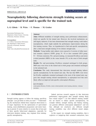
FIGURE 1 H-reflex pre- and post-training. A, Averaged EMG
traces from one representative subject of the SUS group during the
explosive (solid line) and ramped-up then sustained (dashed line) task.
EMG is plotted in % of Mmax for each task and in function of time (in
ms). B and C, Group average of M-wave amplitude (B) and H-reflex
amplitude (C) in % Mmax pre- (white circles) and post-training (blue
circles) for the SUS and EXPL group when performing the ramped-up
then sustained and explosive task. The thin black vertical lines represent
SD. The thin horizontal grey lines represent variation in the potential
amplitude pre- and post-training for each subject
(A)
(B)
FIGURE 2 MEP pre- and post-training. A, Averaged then
rectified EMG traces from one representative subject of the EXPL
group pre- (solid line) and post-training (dashed line) during the
explosive task. EMG is plotted in % of the amplitude of Mmax (pre-
and post-training) and in ms. B, Group average of MEP area
expressed in % Mmax area pre- (white circles) and post-training (blue
circles) for the SUS and EXPL group when performing the ramped-
up then sustained or explosive task. The thin black vertical lines
represent SD. The thin horizontal grey lines represent variation in the
MEP area pre- and post-training for each subject. A star represents a
significant time difference (P-value .05). The bracket with a star
represents a time 9 group interaction for the explosive task (P-
value = .042)
4 of 11
| GIBOIN ET AL.
5. explosive, compared to sustained contractions, could have
increased the excitability of cortico-motoneuronal cells at
the onset of the movement for the explosive task.27
Sec-
ond, the higher force during explosive contractions, com-
pared to the force level reached during sustained
contractions, could have also increased motoneuronal
excitability due to suppression of presynaptic inhibition of
Ia fibres at the onset of muscle contraction.25
Nevertheless, the observed differences in excitability at
different levels of the CNS in the present study highlight
the existence of different motor commands for both tasks.
Thus, it seems intuitive to assume that the training of only
one task and, therefore one motor command, should result
in specific neural adaptations.28
3.3 | Task-specific neuroplasticity
After training, we observed in the EXPL group a reduction
in MEP area in the trained (explosive task), but not in the
untrained task (ramped-up and sustained task), which
demonstrate clearly task-specific neuroplasticity. For the
SUS group, the MEP area was also significantly smaller after
training during the trained, but not during the untrained task.
Nevertheless, the absence of a significant group 9 time
interaction effect indicates a weaker effect compared to the
EXPL group in the explosive task. It could be argued that the
timing of our measurements at the onset of muscle contrac-
tion was better suited to detect neuroplasticity for the explo-
sive task and that having performed our measurements closer
to the end of the ramp would have been more relevant for the
ramp-up then sustained contractions. However, this seems
not to be a likely explanation, as it has already been sug-
gested that the measurement of neuronal mechanisms at the
onset of a ramp (eg, presynaptic inhibition) is in conformity
with measurements during later segments of the task.25
Another explanation could be that the target force level was
different for the two tasks (75% in the sustained task vs 90%
in the explosive task), which may account for the weaker
effect in MEP size changes with training, as the magnitude
of neuroplasticity following strength training may depend
more on contraction strength than contraction speed.29
A
third explanation could be related to the size of the MEP
obtained during the sustained task. As MEPs were consider-
ably smaller during the ramp-up then sustained contractions,
compared to MEPs elicited during the explosive contrac-
tions, they were possibly not in the linear part of the input-
output relationship within the motor neurone pool any more.
Thus, the smaller MEPs could have been less sensitive to a
change in excitatory and inhibitory inputs, which might have
partly masked neuroplasticity during the sustained task.30
A decrease in corticospinal excitability after strength
training has already been reported in previous studies4,21
and has been consistently interpreted to reflect a higher
efficiency in corticospinal transmission.21,31,32
Several
explanations on a mechanistic level have been proposed,
the most probable being: (i) a task-specific modulation of
the interneuronal circuitry sensitive to TMS. (ii) An
increase in firing rate and afterhyperpolarization of motor
neurones which reduces the response probability of motor
neurones to the descending volleys elicited by TMS. (iii) A
task-specific modulation of the intrinsic properties of motor
neurones.4
However, in the present experiment, it does not seem
very likely that neuroplasticity observed at the onset of
movement took place solely on the motoneuronal level. It
has been shown that during voluntary contractions, corti-
cospinal and Ia inputs recruit the motor neurones in similar
manner.33
Thus, it can be assumed that if the decreased
MEPs were related to a task-specific difference in the firing
rate, afterhyperpolarization or a change in intrinsic proper-
ties of motor neurones, we would have expected to mea-
sure a similar effect for the H-reflex peak-to-peak
amplitudes. This clearly was not the case, as H-reflex
amplitudes remained unaltered after both training interven-
tions, suggesting that the task-specific neuroplasticity likely
occurred at the cortical or subcortical level, rather than the
spinal level.34
However, we cannot preclude the existence of task-spe-
cific neuroplasticity at the spinal level in this study. Indeed,
as the H-reflex is not a direct measure of the motor neu-
rone pool excitability,35
it must be acknowledged that a
modulated Ia monosynaptic excitatory efficiency (eg, presy-
naptic inhibition) and a modulation of polysynaptic spinal
networks may affect the MEP and the H-reflex differ-
ently.36,37
Moreover, the H-reflex cannot directly assess
any changes occurring at cortico-motoneuronal synapses, a
site which seems to be modifiable by strength training.
Several studies have shown that one acute session of
strength training could increase cervicomedullary evoked
potential (cMEP), which estimates the cortico-motoneuronal
synapse efficiency and motoneuronal excitability.29,38
Moreover, cMEPs are larger during strong contractions in
chronically strength-trained subjects than in untrained sub-
jects.39
On the other hand, recent results demonstrated that
after 4 weeks of strength training, no changes could be
observed in cMEP, suggesting that the improvement in
force was induced by neural changes “upstream” of the
cortico-motoneuronal synapse.40
Moreover, even if we pre-
sume that neuroplasticity occurred also at the corticospinal-
motoneuronal synapse in our study, a control from suprasp-
inal structures would still be required to modulate it
accordingly to the planned task at least at the onset of con-
traction. We suggest that the location of the neuroplasticity
induced by strength training may vary according to the
duration of the training. During short duration training, like
in the present study, adaptations may happen mostly at the
GIBOIN ET AL. | 5 of 11
6. corticospinal level. With a longer training duration (eg,
4 weeks and more), adaptations may occur at spinal
level.41,42
We propose that the task-specific MEP reduction
observed in this study, and the MEP reduction seen in pre-
vious studies,4,21
could be explained by a task-specific
decreased primary motor cortex activity, as observed during
a fast skill learning phase.43
This decrease in excitability
could be interpreted as a reduction in the neuronal
resources used to perform the task.44
It has been shown in
the rat that, during a week of skilled forelimb reaching
training, a subpopulation of M1 movement-encoding neu-
rones displayed less variable and more correlated firing pat-
tern as the trained task was performed more and more
accurately, suggesting a more temporally synchronous or
amplified signal output from M1 leading to a better task
execution.45
This task-specific remodelling of M1 neurone
populations throughout learning may explain why in our
study the decreased MEP, possibly induced by a more syn-
chronous M1 output, is observable only during the trained
task.
To conclude, our results question the proposition that
strength training, contrarily to skill training, does not
induce significant cortical reorganization but mostly adapta-
tions at spinal level.38,46,47
Moreover, in the light of our
present results, we suggest that strength training should be
seen, at least partly, as skill training.
3.4 | Limitations
Due to the high amount of muscle contractions our subjects
had to perform, we measured RFD and MVC during the
same contraction in order to avoid fatigue. This might not
be optimal for obtaining a maximal RFD value. It has been
advised recently, to perform around 10 brief contractions
with the instruction to contract “as fast as possible,” in
order to reach the highest RFD possible.48
However, we
gave the instructions to contract “as fast as possible and
then as hard as possible,” and thereby ensured reliable con-
ditions to measure both RFD and MVC during one con-
traction.
Another limitation comes from possible errors by the
visual determination of the MEP areas (onsets and offsets)
during muscle contractions. We used the following proce-
dure to ensure that even small MEPs could be reliably dis-
criminated from the ongoing background EMG. To avoid
systematic errors, always the same investigator determined
the onsets with the help of the onset latencies which could
easily be determined during the stimulations performed at
rest. In order to reduce random errors as much as possible,
we averaged MEP traces and thereby reduced the stochastic
part of the ongoing EMG, which allowed to discriminate the
MEP and H-reflex onsets and offsets much clearer.
We have analysed MEP area and H-reflex amplitude, as
we suggest that VL MEPs are better characterized with
their area, contrarily to H-reflexes, which are better charac-
terized with their amplitude. We explain this by the lesser
synchronicity of volleys reaching the muscle during a MEP
than during a H-reflex in the VL. Indeed, desynchroniza-
tion of volleys increases phase cancellation, which proba-
bly affects more MEP amplitude than area.49
3.5 | Conclusions
Four strength training sessions decreased MEP area for the
trained, but not for the untrained task, clearly demonstrat-
ing task-specific neuroplasticity. Decreased MEP areas
without any changes in H-reflex amplitudes at the onset of
the movement indicate that neural adaptations are located
rather supraspinal, than spinal and might reflect an
improved task-specific corticospinal efficiency after short-
term strength training. Our findings provide evidence for a
high similarity between the neural mechanism underlying
the beginning of a strength training programme and the
learning of a new skill. Task-specific neuroplasticity proba-
bly explains the task-specific improvement that can be
observed after short-term strength training.
4 | METHODS
Twenty-two young healthy male participants, without any
lower limb injury during the previous year, were recruited.
The participants were asked to continue their regular sport-
ing activities throughout the duration of the study. Three
participants had to be excluded from the study; one
dropped out due to schedule and in the two others we were
not able to elicit a distinct H-reflex. The participants were
randomly divided into two groups after the first experimen-
tal session, in order to match both groups by age, weight,
height and pre-training MVC and RFD50 values (see
Table 1). The EXPL group (N = 9, mean standard devi-
ation, age: 24 3 years, weight: 80 10 kg, height:
182 8 cm) trained with explosive contractions and the
SUS group (N = 10, age: 26 3 years, weight:
80 9 kg, height: 181 5 cm) trained with sustained
contractions. All participants signed a written informed
consent, and experiments were approved by the ethics com-
mittee of the University of Konstanz and in accordance
with the declaration of Helsinki.
4.1 | Experimental procedure
Subjects participated in two experimental sessions; the pre-
testing was performed 48 hours before the first training
session and the post-testing 48 hours after the last training
6 of 11
| GIBOIN ET AL.
7. session. The procedure within the pre- and post-experimen-
tal sessions remained identical (see Figure 3 for a full over-
view of the experimental procedure).
At the beginning of each experimental session, the par-
ticipants performed a general warm-up routine consisting
of 3 sets of 10 body weight squats and 2 sets of 3 counter-
movement jumps.
A mark was then drawn with waterproof pen on the
right Achilles tendon, 2 cm higher than the line parallel to
the floor passing below the lateral malleolus in sitting posi-
tion, to ensure identical placement of the force transducer
in the pre- and post-measurements.
Surface EMG sensors (Bagnoli Desktop EMG, Delsys,
Natick, MA, USA) were fixed on the muscle belly of the
M. vastus lateralis (VL) and M. biceps femoris (BF) after
shaving, abrading with sandpaper and cleaning the respec-
tive sites with alcohol. We followed the SENIAM guideli-
nes for EMG sensor locations. EMG signals were
amplified (91000 or 9100 for Mmax in some subjects),
high-pass- and low-pass-filtered (20 Hz 10% and
450 Hz 10%, respectively), sampled with a Power 1401
interface (Cambridge Electronic Design, Cambridge, UK)
at 4000 Hz and stored on a computer with the Signal soft-
ware (Cambridge Electronic Design).
Participants were then seated and strapped tightly at
chest and hip level to a custom-made chair, with hip and
knee angles positioned at around 90°. Thereafter, the right
ankle was fixed to a force transducer (Model 9321A, Kis-
tler, Winterthur, Switzerland) exactly at the line that had
been drawn before.
Then, to stimulate the femoral nerve (Stimulator DSH7,
Digitimer), a cathode (copper, circular, 2 cm diameter,
wrapped in a water soaked sponge) was fixed with tape at
the femoral triangle and an anode (copper, 7 9 5 cm,
wrapped in a soaked sponge) at the lower level of the glu-
teus maximus. A small sandbag was placed directly above
the cathode and firmly pressed down on the femoral trian-
gle by a laboratory assistant throughout the duration of the
experiment. We stimulated with square pulse duration of
1 ms and gradually increased the intensity of the stimula-
tion until reaching Mmax in the VL at rest and maximum
unpotentiated twitch at rest of the knee extensors. The
intensity required for reaching Mmax was multiplied by
1.5 and used for all following Mmax measurements and
interpolated twitch methods.
Throughout the experiments, the participants were fac-
ing a computer screen where we were able to display the
exerted forces during muscle contractions as well as cursors
that we used to indicate the respective target forces. Right
before the start of the experiment, the participants per-
formed an additional specific warm-up procedure that con-
sisted of around 15 knee extensions with gradually
increasing intensity and duration of rest periods, until
reaching a force close to what could be perceived as 90%
their MVC (step 1 in Figure 3).
4.1.1 | MVC and VA measurements
Participants then performed 3 MVC trials, with 90 seconds
of rest between trials. For each MVC, subjects were
instructed to contract first “as fast as possible and then as
hard as possible (step 2 in Figure 3).” Strong verbal
encouragement was given by the experimenter throughout
the MVC. We determined voluntary activation (VA) of the
FIGURE 3 Experimental procedure performed pre- and post-training. Blue surfaces represent knee extensor contractions (performed at
100%, 90%, 75% or 30% MVC). Vertical arrows represent supramaximal peripheral nerve stimulations used to measure VA or Mmax. (1) The
participants performed a warm-up with incremental isometric contractions up to 90% perceived MVC. (2) The participants performed 3 MVCs
“as fast then as hard as possible” (1 min of rest in between), during which VA is measured. (3) Mmax is measured during the explosive and
ramped-up and sustained contraction (Figure 4B). (4) TMS and peripheral nerve stimulation intensity are calibrated to elicit MEP and H-reflex
while the participant performed the calibration task (Figure 4A). (5) Main measurements: MEP and H-reflexes are elicited during explosive and
ramped-up then sustained contractions (10 sets of 4 explosive or ramped-up then sustained contractions). (6) Mmax is measured again during the
explosive and ramped-up and sustained contraction. In point (2), the investigator triggered stimulations. In point (3, 4, 5 and 6), participant self-
initiated contractions and stimulations were triggered automatically on the rise in EMG at the onset of VL contraction
GIBOIN ET AL. | 7 of 11
8. knee extensor muscles according to the interpolated twitch
method.50
Stimulations were triggered manually by the
investigator during the MVC trials after reaching a stable
force plateau which was detected visually.
4.1.2 | Main experiment
The main measurements consisted of 10 sets of 4 contrac-
tions (step 5 in Figure 3). The 4 contractions were sepa-
rated by a rest period of 20 seconds and the 10 sets by a
rest period of 1 minutes. The 4 contractions of one set
were always of the same mode, that is either explosive or
ramped-up then sustained contractions. To limit the occur-
rence of fatigue during the experiment, the duration of the
sustained contraction at 75% MVC was reduced to 2 sec-
onds instead of 3 seconds (Figure 4B). The order of the
contraction mode was always alternated between sets, with
half of the subjects starting with explosive and the other
half with sustained contractions. The order was counterbal-
anced for the two groups and identical for pre- and post-
measurements. Each contraction was self-initiated by the
subject after receiving oral approvement by the investiga-
tor. During each set, 2 H-reflexes and 2 MEPs were eli-
cited in random order. Thus, during a total of 40
contractions, we elicited 10 H-reflexes and 10 MEPs for
the sustained and the same amount for the explosive task.
4.2 | Stimulations
For the H-reflex, we first started to stimulate the femoral
nerve at rest. We gradually increased the intensity from
subthreshold intensities (no burst in VL EMG visible) up
to an intensity at which the amplitude of the H-reflex
started to decline again. Then, we reduced the intensity of
the stimulation to obtain a stable M-wave and H-reflex,
which could be clearly discriminated from the M-wave.
With this intensity, H-reflexes were elicited while the par-
ticipant performed the calibration task. The calibration task
was chosen as being a hybrid task between the explosive
and the sustained tasks, and the intensity of contraction
remained low to prevent the development of fatigue (30%
MVC with a ramp of 0.5 seconds and sustained contraction
during 2 seconds, see Figure 4A). The stimulation intensity
was then adjusted accordingly (see the H-reflex and TMS
intensity set-up section).
For TMS, we used a figure-of-eight coil designed to
stimulate the lower limb cortical areas (MC-B70, MagVen-
ture), which was connected to a MagPro R30 Stimulator
(MagVenture) and delivered biphasic pulses (current flow
in the coil in AP/PA direction). During stimulation, the coil
was oriented with the figure of “8” perpendicular to the
interhemispheric fissure. We searched for the optimal coil
position by slightly moving the coil while delivering pulses
that induced MEPs of around 1 mV during a 10% MVC
isometric contraction of the knee extensors. The optimal
coil position was defined as the position eliciting the high-
est MEP in VL. The optimal coil position was drawn on a
swim cap to allow identical placement throughout the
whole experiment.
4.2.1 | H-reflex and TMS intensity set-up
Stimulations were triggered by a rise in the VL EMG. To
do so, one investigator visually inspected the unrectified
VL EMG in the 200 ms preceding a calibration contraction
and set an upper and a lower threshold cursor at the maxi-
mal and minimal peaks in the EMG signal. The aim was to
trigger the stimulation at the very first burst in the EMG,
so the H-reflex or the MEP occurred before the force onset.
However, the setting of threshold cursors at the minimum
and maximum peaks of just one rest EMG signal increased
too much the risk of false-positive stimulation triggered by
(A)
(B)
FIGURE 4 Strength tasks. Force is displayed as % MVC and
plotted against time in (s). A, The task used to calibrate the intensity
of stimulations eliciting H-reflexes and MEPs. B, Explosive (thin
line) and ramped-up then sustained tasks (thick and dashed lines).
The ramped-up then sustained task with a plateau at 75% MVC
lasting 3 s is the task used during training (thick line). The ramped-
up then sustained task with a 2-s plateau is the task used during pre-
and post-training experiments to avoid fatigue throughout the
experiment (thick dashed line)
8 of 11
| GIBOIN ET AL.
9. the random activation of a motor unit at rest. Therefore,
the threshold cursors were adjusted during at least 5 con-
tractions to reach the best compromise between the need of
eliciting a potential before the force onset and the absence
of false-positive stimulations. This resulted in average
amplitude of the upper and lower thresh-
olds = 0.035 0.018 mV; average standard deviation of
the EMG signal of the VL muscle in the 50 ms epoch
before the trigger = 0.0048 0.002 mV; average ampli-
tude of the upper and lower thresholds in number of stan-
dard deviation of the EMG = 7.6 3.7 standard deviation,
that is around 3.8 standard deviations per side; as a relative
comparison, the average of Mmax amplitude was
6.7 2.9 mV. Subjects were asked to stay as still as pos-
sible before the contraction.
With this method, we were able to elicit reliably the
potentials induced by the stimulations near the onset of
force. Latencies of the potentials (H-
reflex = 21.4 1.7 ms, MEP = 25.8 1.6 ms) were on
average smaller than the latency of the onset of force
(28.8 20 ms). The onset of force for every contraction
was determined at the time the force was equal or higher
than 1% of the highest force amplitude measured during
the 3 initial MVC trials.
To calibrate stimulation intensities, we first measured
Mmax with supramaximal femoral nerve stimulation dur-
ing the explosive and the ramped-up then sustained con-
traction (step 3 of Figure 3). This procedure was repeated
at the end of the main measurements (step 6 of Figure 3),
to examine whether neuromuscular changes induced by
the contractions during the experiment changed muscle
membrane properties and, thus, the EMG signal. There
was no difference of Mmax amplitude between tasks or
between the beginning and the end of the main measure-
ments. Then (step 4 of Figure 3), we calibrated the inten-
sity of femoral nerve stimulation to elicit H-reflex in the
VL, as well as the intensity of TMS to elicit a MEP in
the VL according to the following standardized procedure.
Subjects were asked to perform the calibration task to
trigger stimulations. Stimulation intensity for the H-reflex
was adjusted so that the H-reflex amplitude was always
in the ascending part of the H-reflex amplitude/current
intensity curve with a M-wave of around 10% of Mmax.
In post-measurements, we adjusted the stimulation inten-
sity so that the H-reflex amplitude always was in the
ascending part of the H-reflex amplitude/current intensity
curve and the M-wave size was identical to the M-wave
size of the pre-training experiment (both normalized to
their respective Mmax amplitude). With regard to TMS,
the intensity was set at the intensity required for the
active motor threshold (AMT) and then multiplied by 1.3.
AMT corresponded to the intensity where at least 3 of 6
stimuli produced any discernable potential (or silent
period) in the VL EMG. The average AMT corresponded
to 40% of maximum stimulator output.
4.3 | Training
The training consisted of 4 sessions of isometric knee
extensions separated by 48 hours. The protocol used was
similar to the protocol used by Tillin and Folland (2014).
The EXPL group performed 4 sets of 10 contractions, last-
ing around 1 seconds, performed as fast as possible with
the aim of reaching 90% MVC (see Figure 4B). EXPL
contractions were separated by a rest period of 5 seconds.
The SUS group performed 4 sets of 10 contractions, con-
sisting of a ramped-up contraction lasting 1 seconds, which
was then sustained for another 3 seconds at 75% MVC
(see Figure 4B). SUS contractions were separated by a rest
period of 2 seconds. For both groups, there was 2 minutes
of rest between each set.
4.4 | Data analysis
For MVCa (maximal amplitude of force measured during
MVC), RFD, EMG, VA, Ptw and RFDPtw values, we aver-
aged the measurements over the 3 initial MVCs. MVCa was
determined as the maximum force value before or after the
interpolated twitch occurred. RFD was calculated as the dif-
ference between forces measured at 50, 100 or 150 ms and
the force at onset, divided by time [N/s]. We then averaged
the 3 best values. EMG in VL was analysed as root mean
square in the corresponding time intervals (0-50, 0-100 and
0-150 ms, zero corresponding to the onset of force). This
was normalized to Mmax obtained from the explosive task.
EMGMVC was calculated as the root mean square in the
time interval of 250 ms prior to the potentiated twitch. VA
was determined as the ratio of the size of the interpolated
twitch and the potentiated twitch at rest.50
We further deter-
mined Ptw as the highest force value of the twitch [N] and
RFDPtw as the highest force value of the twitch divided by
the time between the onset of force and the highest force
value of the twitch [N/s].
H-reflex and M-wave peak-to-peak amplitudes were
normalized to Mmax, which corresponded to the mean of
the two Mmax values obtained at the beginning and end of
the main experiment. If an M-wave amplitude was over
twice the mean M-wave amplitude of the subject, or below
the mean divided by 2, we did not take into account the
stimulation in the analysis (9% total H-reflex stimulations).
M-wave and H-reflex elicited by these stimulations were
excluded a posteriori from our analysis. M-wave and H-
reflex amplitudes were normalized to Mmax amplitude.
The 10 MEP traces for the sustained as well as the
explosive tasks were separately averaged and rectified.
MEP onset and offset were visually determined and
GIBOIN ET AL. | 9 of 11
10. thereafter the area was calculated. This procedure allowed
us to extract even small MEPs from the background EMG
activity. All MEP areas were normalized to Mmax area.
4.5 | Statistics
All statistics were conducted with R (version 3.3.0, The R
foundation for Statistical Computing). Values following
a correspond to SD. Unpaired t tests were used to assess
whether a group difference existed for weight, height, age,
and whether there were any group differences in the pre-
training values of MVCa, RFD, EMG, VA, Ptw, RFDPtw
and AMT. The effect of time (training) and of group was
tested on each dependent variable with a two-way
ANOVA, with time being the within-subject variable and
group the between-subject variable.
A two-way ANOVA was performed on the pre-training
values of H-reflex latency, MEP latency, M-waves ampli-
tudes, H-reflexes amplitudes and MEPs areas to search for
any group or task baseline difference.
The effect of time, group and task (explosive vs sus-
tained contraction) was tested on M-waves amplitudes, H-
reflexes amplitudes and MEPs areas with a three-way
ANOVA, with group being the between-subject variable
and time and task within-subjects variables. In case of a
significant main or interaction effect, post hoc two-way
ANOVA and paired or unpaired t test were calculated to
clarify the effect of training. Moreover, within-subject
effect sizes (Cohen’s dav) with a Hedge’s g correction (gav)
were calculated.51
ACKNOWLEDGEMENTS
The authors would like to thank Tamara Poppendieker,
Eric Jung, Kristijan Milekic and Tyler Breedlove for their
help during data collection.
CONFLICT OF INTEREST
The authors declare having no conflict of interest.
AUTHORS’ CONTRIBUTIONS
LSG and MG conceptualized and designed experiments.
LSG, BW and FT collected data. LSG analysed data. LSG
and MG interpreted data. LSG drafted the manuscript, and
all authors contributed to the manuscript revision.
REFERENCES
1. Baker D, Wilson G, Carlyon B. Generality versus specificity: a com-
parison of dynamic and isometric measures of strength and speed-
strength. Eur J Appl Physiol Occup Physiol. 1994;68:350-355.
2. Blazevich A. Are training velocity and movement pattern impor-
tant determinants of muscular rate of force development enhance-
ment? Eur J Appl Physiol. 2012;112:3689-3691.
3. Balshaw TG, Massey GJ, Maden-Wilkinson TM, Tillin NA, Fol-
land JP. Training-specific functional, neural, and hypertrophic
adaptations to explosive- vs. sustained-contraction strength train-
ing. J Appl Physiol (1985). 2016;120:1364-1373.
4. Carroll TJ, Riek S, Carson RG. The sites of neural adaptation
induced by resistance training in humans. J Physiol.
2002;544:641-652.
5. Del Balso C, Cafarelli E. Adaptations in the activation of human
skeletal muscle induced by short-term isometric resistance train-
ing. J Appl Physiol (1985). 2007;103:402-411.
6. Duclay J, Martin A, Robbe A, Pousson M. Spinal reflex plasticity
during maximal dynamic contractions after eccentric training.
Med Sci Sports Exerc. 2008;40:722-734.
7. Moritani T, deVries HA. Neural factors versus hypertrophy in the
time course of muscle strength gain. Am J Phys Med.
1979;58:115-130.
8. Aagaard P, Simonsen EB, Andersen JL, Magnusson P, Dyhre-
Poulsen P. Increased rate of force development and neural drive
of human skeletal muscle following resistance training. J Appl
Physiol (1985). 2002;93:1318-1326.
9. Schubert M, Beck S, Taube W, Amtage F, Faist M, Gruber M.
Balance training and ballistic strength training are associated with
task-specific corticospinal adaptations. Eur J Neurosci.
2008;27:2007-2018.
10. Selvanayagam VS, Riek S, Carroll TJ. Early neural responses to
strength training. J Appl Physiol (1985). 2011;111:367-375.
11. Tillin NA, Folland JP. Maximal and explosive strength training
elicit distinct neuromuscular adaptations, specific to the training
stimulus. Eur J Appl Physiol. 2014;114:365-374.
12. Tillin NA, Pain MT, Folland JP. Short-term training for explosive
strength causes neural and mechanical adaptations. Exp Physiol.
2012;97:630-641.
13. Vila-Cha C, Falla D, Correia MV, Farina D. Changes in H reflex
and V wave following short-term endurance and strength training.
J Appl Physiol (1985). 2012;112:54-63.
14. Beck S, Taube W, Gruber M, Amtage F, Gollhofer A, Schubert
M. Task-specific changes in motor evoked potentials of lower
limb muscles after different training interventions. Brain Res.
2007;1179:51-60.
15. Ekblom MM. Improvements in dynamic plantar flexor strength
after resistance training are associated with increased voluntary
activation and V-to-M ratio. J Appl Physiol (1985). 2010;109:19-
26.
16. Fimland MS, Helgerud J, Gruber M, Leivseth G, Hoff J. Func-
tional maximal strength training induces neural transfer to single-
joint tasks. Eur J Appl Physiol. 2009;107:21-29.
17. Holtermann A, Roeleveld K, Engstrom M, Sand T. Enhanced H-
reflex with resistance training is related to increased rate of force
development. Eur J Appl Physiol. 2007;101:301-312.
18. Jensen JL, Marstrand PC, Nielsen JB. Motor skill training and
strength training are associated with different plastic changes in
the central nervous system. J Appl Physiol (1985). 2005;99:1558-
1568.
19. Griffin L, Cafarelli E. Transcranial magnetic stimulation during
resistance training of the tibialis anterior muscle. J Electromyogr
Kinesiol. 2007;17:446-452.
10 of 11
| GIBOIN ET AL.
11. 20. Kidgell DJ, Stokes MA, Castricum TJ, Pearce AJ. Neurophysio-
logical responses after short-term strength training of the biceps
brachii muscle. J Strength Cond Res. 2010;24:3123-3132.
21. Carroll TJ, Barton J, Hsu M, Lee M. The effect of strength train-
ing on the force of twitches evoked by corticospinal stimulation
in humans. Acta Physiol (Oxf). 2009;197:161-173.
22. Farina D, Merletti R, Enoka RM. The extraction of neural strate-
gies from the surface EMG: an update. J Appl Physiol (1985).
2014;117:1215-1230.
23. Seidler RD, Noll DC, Thiers G. Feedforward and feedback pro-
cesses in motor control. NeuroImage. 2004;22:1775-1783.
24. Zehr EP. Considerations for use of the Hoffmann reflex in exer-
cise studies. Eur J Appl Physiol. 2002;86:455-468.
25. Meunier S, Pierrot-Deseilligny E. Gating of the afferent volley of
the monosynaptic stretch reflex during movement in man. J Phys-
iol. 1989;419:753-763.
26. Geertsen SS, Lundbye-Jensen J, Nielsen JB. Increased central
facilitation of antagonist reciprocal inhibition at the onset of dor-
siflexion following explosive strength training. J Appl Physiol
(1985). 2008;105:915-922.
27. Nielsen J, Petersen N. Changes in the effect of magnetic brain
stimulation accompanying voluntary dynamic contraction in man.
J Physiol. 1995;484(Pt 3):777-789.
28. Thompson AK, Chen XY, Wolpaw JR. Acquisition of a simple
motor skill: task-dependent adaptation plus long-term change in
the human soleus H-reflex. J Neurosci. 2009;29:5784-5792.
29. Nuzzo JL, Barry BK, Gandevia SC, Taylor JL. Acute strength
training increases responses to stimulation of corticospinal axons.
Med Sci Sports Exerc. 2016;48:139-150.
30. Capaday C. Neurophysiological methods for studies of the motor
system in freely moving human subjects. J Neurosci Methods.
1997;74:201-218.
31. Falvo MJ, Sirevaag EJ, Rohrbaugh JW, Earhart GM. Resistance
training induces supraspinal adaptations: evidence from move-
ment-related cortical potentials. Eur J Appl Physiol.
2010;109:923-933.
32. Lee M, Gandevia SC, Carroll TJ. Short-term strength training
does not change cortical voluntary activation. Med Sci Sports
Exerc. 2009;41:1452-1460.
33. Bawa P, Lemon RN. Recruitment of motor units in response to
transcranial magnetic stimulation in man. J Physiol.
1993;471:445-464.
34. Carroll TJ, Selvanayagam VS, Riek S, Semmler JG. Neural adap-
tations to strength training: moving beyond transcranial magnetic
stimulation and reflex studies. Acta Physiol (Oxf). 2011;202:119-
140.
35. Burke D, Gandevia SC. Properties of human peripheral nerves:
implications for studies of human motor control. Prog Brain Res.
1999;123:427-435.
36. Burke D, Gandevia SC, McKeon B. Monosynaptic and oligosy-
naptic contributions to human ankle jerk and H-reflex. J Neuro-
physiol. 1984;52:435-448.
37. Nielsen J, Petersen N. Is presynaptic inhibition distributed to cor-
ticospinal fibres in man? J Physiol. 1994;477:47-58.
38. Giesebrecht S, van Duinen H, Todd G, Gandevia SC, Taylor JL.
Training in a ballistic task but not a visuomotor task increases
responses to stimulation of human corticospinal axons. J Neuro-
physiol. 2012;107:2485-2492.
39. Philpott DT, Pearcey GE, Forman D, Power KE, Button DC.
Chronic resistance training enhances the spinal excitability of the
biceps brachii in the non-dominant arm at moderate contraction
intensities. Neurosci Lett. 2015;585:12-16.
40. Nuzzo JL, Barry BK, Jones MD, Gandevia SC, Taylor JL.
Effects of four weeks of strength training on the corticomotoneu-
ronal pathway. Med Sci Sports Exerc. 2017;49:2286-2296.
41. Gruber M, Gruber SB, Taube W, Schubert M, Beck SC, Goll-
hofer A. Differential effects of ballistic versus sensorimotor train-
ing on rate of force development and neural activation in
humans. J Strength Cond Res. 2007;21:274-282.
42. Gruber M, Taube W, Gollhofer A, Beck S, Amtage F, Schubert
M. Training-specific adaptations of H- and stretch reflexes in
human soleus muscle. J Mot Behav. 2007;39:68-78.
43. Dayan E, Cohen LG. Neuroplasticity subserving motor skill
learning. Neuron. 2011;72:443-454.
44. Poldrack RA. Imaging brain plasticity: conceptual and method-
ological issues–a theoretical review. NeuroImage. 2000;12:1-13.
45. Li Q, Ko H, Qian ZM, et al. Refinement of learned skilled move-
ment representation in motor cortex deep output layer. Nat Com-
mun. 2017;8:15834.
46. Adkins DL, Boychuk J, Remple MS, Kleim JA. Motor training
induces experience-specific patterns of plasticity across motor
cortex and spinal cord. J Appl Physiol (1985). 2006;101:1776-
1782.
47. Remple MS, Bruneau RM, VandenBerg PM, Goertzen C, Kleim
JA. Sensitivity of cortical movement representations to motor
experience: evidence that skill learning but not strength training
induces cortical reorganization. Behav Brain Res. 2001;123:133-
141.
48. Maffiuletti NA, Aagaard P, Blazevich AJ, Folland J, Tillin N,
Duchateau J. Rate of force development: physiological and
methodological considerations. Eur J Appl Physiol.
2016;116:1091-1116.
49. Rosler KM, Petrow E, Mathis J, Aranyi Z, Hess CW, Magistris
MR. Effect of discharge desynchronization on the size of motor
evoked potentials: an analysis. Clin Neurophysiol.
2002;113:1680-1687.
50. Gandevia SC. Spinal and supraspinal factors in human muscle
fatigue. Physiol Rev. 2001;81:1725-1789.
51. Lakens D. Calculating and reporting effect sizes to facilitate
cumulative science: a practical primer for t-tests and ANOVAs.
Front Psychol. 2013;4:863.
How to cite this article: Giboin L-S, Weiss B,
Thomas F, Gruber M. Neuroplasticity following
short-term strength training occurs at supraspinal
level and is specific for the trained task. Acta
Physiol. 2018;222:e12998.
https://doi.org/10.1111/apha.12998
GIBOIN ET AL. | 11 of 11