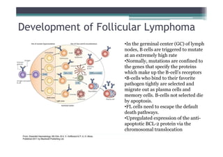
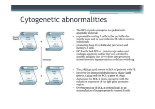
Follicular Lymphoma
Yuan Yao
Follicular Lymphoma (FL) Overview
The second most common B-cell lymphoma
• FL accounts about 20% of all lymphomas
• FL affects predominantly adults with a median age
of 60 at diagnosis
• FL slightly affects more females (M:F ratio of 1:1.7)
• FL involves mostly lymph nodes
• Caused by accumulation of malignant B cells in the
germinal centres of lymph nodes
• Neoplastic B cells show a follicular growth pattern
Normal reactive
lymph node
Follicular
Lymphoma
Morphology
Warnke et al.,1995
Discrete follicles, well sparated Nodularity throughout the node
Cytological grading of FL
•The neoplastic follicles are composed of cells that resemble the normal cells
of the germinal center, including centrocytes and centroblasts
•Centrocytes are small to medium sized cells with cleaved nuclei,
inconspicuous nucleoli, irregular nucleus, and scanty pale blue cytoplasm.
•Centroblasts are larger cells with round to oval nuclei, one to three nucleoli
and a narrow border of pale blue cytoplasm
•FL with a predominance of small cleaved lymphocytes tends to be a relatively
low-grade or indolent neoplasm, whereas FL with a higher proportion of large
lymphoid cells tends to be clinically aggressive
Development of Follicular Lymphoma
•In the germinal center (GC) of lymph
nodes, B cells are triggered to mutate
at an extremely high rate
•Normally, mutations are confined to
the genes that specify the proteins
which make up the B-cell’s receptors
•B-cells who bind to their favorite
pathogen tightly are selected and
migrate out as plasma cells and
memory cells. B-cells not selected die
by apoptosis.
•FL cells need to escape the default
death pathways.
•Upregulated expression of the anti-
apoptotic BCL-2 protein via the
chromosomal translocation
Cytogenetic abnormalities
• The BCL-2 proto-oncogene is a potent anti-
apoptotic molecule
• expressed in resting B cells in the perifollicular
mantle zone and in post-follicular B cells in normal
individuals
• promoting long-lived follicular precursor and
memory B cells
• GC B-cells lack BCL-2 protein expression and
undergo apoptosis unless they are selected by
specific antigens that drive them into processes
termed somatic hypermutation and class switching
• T(14;18)(q32;q21) occurs in 85% of patients with FL
• Involves the immunoglobulin heavy chain (IgH)
gene at 14q32 and the BCL-2 gene at 18q21
• Juxtapose the BCL-2 proto-oncogene with the
enhancer sequences of the IgH gene promoter
region
• Overexpression of BCL-2 protein leads to an
accumulation of inappropriately rescued B cells
• multiple genetic events are required for the development
of FL
x secondary cytogenetic alterations are generally observed in
most FL cells, which include gains in 1q, 2p, 3, 7, 8, 12q, 18q
and X as well as deletions in 1p, 6q, 10q, 13q, and 17p
• Approximately 10% of patients with FL lack
t(14;18)(q32;q21) and do not show increased BCL-2
expression by immunostaining
x It has been postulated that other genetic defects along a similar
antiapoptotic pathway may occur in these patients, which give
a similar disease pattern
Cytogenetic abnormalities
Diagnosis of the FL
• Lymph node biopsy
-- Histological staining: determine the grade
-- Immunostaining: high levels of BCL-2 protein
expression correlate with a worse outcome
--PCR/FISH: demonstrate the t(14;18), BCL-2 gene
expression
• Blood tests
• Chest X-ray
• CT and PET scanning
• Bone marrow biopsies
follicular lymphoma.pdf

follicular lymphoma.pdf

  • 1.
  • 2.
    Follicular Lymphoma (FL)Overview The second most common B-cell lymphoma • FL accounts about 20% of all lymphomas • FL affects predominantly adults with a median age of 60 at diagnosis • FL slightly affects more females (M:F ratio of 1:1.7) • FL involves mostly lymph nodes • Caused by accumulation of malignant B cells in the germinal centres of lymph nodes • Neoplastic B cells show a follicular growth pattern
  • 3.
    Normal reactive lymph node Follicular Lymphoma Morphology Warnkeet al.,1995 Discrete follicles, well sparated Nodularity throughout the node
  • 4.
    Cytological grading ofFL •The neoplastic follicles are composed of cells that resemble the normal cells of the germinal center, including centrocytes and centroblasts •Centrocytes are small to medium sized cells with cleaved nuclei, inconspicuous nucleoli, irregular nucleus, and scanty pale blue cytoplasm. •Centroblasts are larger cells with round to oval nuclei, one to three nucleoli and a narrow border of pale blue cytoplasm •FL with a predominance of small cleaved lymphocytes tends to be a relatively low-grade or indolent neoplasm, whereas FL with a higher proportion of large lymphoid cells tends to be clinically aggressive
  • 5.
    Development of FollicularLymphoma •In the germinal center (GC) of lymph nodes, B cells are triggered to mutate at an extremely high rate •Normally, mutations are confined to the genes that specify the proteins which make up the B-cell’s receptors •B-cells who bind to their favorite pathogen tightly are selected and migrate out as plasma cells and memory cells. B-cells not selected die by apoptosis. •FL cells need to escape the default death pathways. •Upregulated expression of the anti- apoptotic BCL-2 protein via the chromosomal translocation
  • 6.
    Cytogenetic abnormalities • TheBCL-2 proto-oncogene is a potent anti- apoptotic molecule • expressed in resting B cells in the perifollicular mantle zone and in post-follicular B cells in normal individuals • promoting long-lived follicular precursor and memory B cells • GC B-cells lack BCL-2 protein expression and undergo apoptosis unless they are selected by specific antigens that drive them into processes termed somatic hypermutation and class switching • T(14;18)(q32;q21) occurs in 85% of patients with FL • Involves the immunoglobulin heavy chain (IgH) gene at 14q32 and the BCL-2 gene at 18q21 • Juxtapose the BCL-2 proto-oncogene with the enhancer sequences of the IgH gene promoter region • Overexpression of BCL-2 protein leads to an accumulation of inappropriately rescued B cells
  • 7.
    • multiple geneticevents are required for the development of FL x secondary cytogenetic alterations are generally observed in most FL cells, which include gains in 1q, 2p, 3, 7, 8, 12q, 18q and X as well as deletions in 1p, 6q, 10q, 13q, and 17p • Approximately 10% of patients with FL lack t(14;18)(q32;q21) and do not show increased BCL-2 expression by immunostaining x It has been postulated that other genetic defects along a similar antiapoptotic pathway may occur in these patients, which give a similar disease pattern Cytogenetic abnormalities
  • 8.
    Diagnosis of theFL • Lymph node biopsy -- Histological staining: determine the grade -- Immunostaining: high levels of BCL-2 protein expression correlate with a worse outcome --PCR/FISH: demonstrate the t(14;18), BCL-2 gene expression • Blood tests • Chest X-ray • CT and PET scanning • Bone marrow biopsies