5. 5
Eye
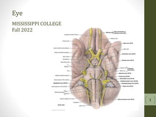
CommonTendinousRing
• 4 recti muscles arise from the
common tendinous ring
• Structures that pass through the ring
lie within the cone formed by the
muscles
• Lacrimal, trochlear and frontal nerves
enter the orbit above the ring
• Optical canal contains the optic nerve
and the ophthalmic artery
11. 11
Innervation
SomaticMotor
• Oculomotor (CN III)
o Superior division
Levator palpebrae superioris
Superior rectus
o Inferior division
Inferior oblique
Medial rectus
Inferior rectus
• Abducens (CN VI)
o Lateral rectus
• Trochlear (CN IV)
o Superior oblique
12. 12
Extraocular Muscles
Innervation
• Trochlear nerve
o CN IV
o Only cranial nerve to emerge from the
brain dorsally
o Emerges medially near the mid-brain-
pontine junction
o Passes laterally around the cerebral
peduncle
o Continues anteriorly through the
lateral wall of the cavernous sinus
o Enters the orbit through the superior
orbital fissure above the common
tendinous ring
o General somatic motor input to the
superior oblique (GSE)
14. 14
Extraocular Muscles
Innervation
• Abducent
o CN VI
o General somatic motor input to the
lateral rectus (GSE)
o Emerges from the groove between the
pons and the medulla
o Courses anteriorly
o Pierces the dura at the clivus in the
posterior cranial fossa
o Runs anteriorly through the cavernous
sinus
o Enters the orbit through the superior
orbital fissure through the common
tendinous ring
o Abducts the eye
15. 15
Extraocular Muscles
Innervation
• Oculomotor
o CN III
o Provides somatic motor (GSE)
innervation to 5 of the 7 extraocular
muscles
Levator palpebrae superioris
Superior rectus
Medial rectus
Inferior rectus
Inferior oblique
o Also provides parasympathetic
innervation to the constrictor pupillae
and ciliary muscles
16. 16
Innervation
SomaticMotor
• Oculomotor (CN III)
o Superior division
Levator palpebrae superioris
Superior rectus
o Inferior division
Inferior oblique
Medial rectus
Inferior rectus
• Abducens (CN VI)
o Lateral rectus
• Trochlear (CN IV)
o Superior oblique
18. 18
Innervation
Sympathetic
• Sympathetic
o Preganglionic sympathetic fibers originate in the
thoracic intermediolateral cell column
o Sympathetic nerves travel in the sympathetic
trunk to synapse in the superior cervical ganglion
o Postganglionic fibers gain access to the head by
way of the carotid nerves; a fine meshwork of
fibers which invest the carotid artery
o Postganglionic fibers branch off the internal
carotid artery at the opthalmic artery
o Enter the orbit through the common tendinous
ring
o Run along with the nasociliary nerve (primary
sensory nerve of the eye)
o These long ciliary nerves enter the eye and
innervate the dilator pupillae.
o Some sympathetic fibers may pass through the
cililary ganglion and emerge as short ciliary
nerves
19. 19
Innervation
SympatheticandParasympathetic
• Sympathetic
o Preganglionic sympathetic fibers originate in the
thoracic intermediolateral cell column
o Sympathetic nerves travel in the sympathetic
trunk to synapse in the superior cervical ganglion
o Postganglionic fibers gain access to the head by
way of the carotid nerves; a fine meshwork of
fibers which invest the carotid artery
o Postganglionic fibers branch off the internal
carotid artery at the opthalmic artery
o Enter the orbit through the common tendinous
ring
o Run along with the nasociliary nerve (primary
sensory nerve of the eye)
o These long ciliary nerves enter the eye and
innervate the dilator pupillae.
o Some sympathetic fibers may pass through the
cililary ganglion and emerge as short ciliary
nerves
20. 20
Eye
TunicsoftheEye
• Middle vascular tunic
o Choroid – posterior
o Ciliary body
o Iris
Pigments
Projects outward from the ciliary body
Central opening – pupil
Muscles
Sphincter pupillae – parasympathetic - decrease the pupillary opening
Dilator – sympathetic – increase the pupillary opening
21. 21
• Parasympathetic
o Originate in the accessory oculomotor nucleus
o Travel with CN III (oculomotor)
o Enter the head with the inferior division of CN III
o Synapse in the ciliary ganglion
o Postganglionic fibers reach the eye by way of the
short ciliary nerves
o Contraction of the sphincter pupillae
o Contraction of ciliary muscles
o GVE
Innervation
Parasympathetic
22. 22
Eye
TunicsoftheEye
• Middle vascular tunic
o Choroid – posterior
o Ciliary body
Forms a complete ring around the
eyeball
Extends from the anterior border of
the choroid
Components include ciliary muscle
Smooth muscle
Parasympathetic innervation by
CN III
Upon contraction decreases the
size of the ring formed by the
ciliary body leading to
constriction of the pupil
Components include ciliary process
Longitudinal ridges projecting
from the inner surface of the
ciliary body which attach to the
lens to create the suspensory
ligament
o Iris
23. 23
Carr BJ, Stell WK. The Science Behind Myopia. 2017 Nov 7. In: Kolb H, Fernandez E,
Nelson R, editors. Webvision: The Organization of the Retina and Visual System
[Internet]. Salt Lake City (UT): University of Utah Health Sciences Center; 1995-.
Figure 7. [The effect of ciliary muscle...]. Available from:
https://www.ncbi.nlm.nih.gov/books/NBK470669/figure/myopia.F7/
Accommodation
for near vision
27. 27
Innervation
Horner’sSyndrome
• Absence of cervical sympathetic trunk
• Defect in sympathetic innervation
• Caused by stroke, tumor or spinal cord injury or
idiopathic
• Affects function on the ipsilateral side of the face
• Signs
o Miosis – constriction of the pupil
Ciliary nerves innervate (sympathetic)
the dilator pupillae muscles
Unopposed parasympathetic
innervation (constriction)
Symptom may be subtle and may
require a dark room (Stanford Med)
o Ptosis - drooping of the superior eyelid
Inactivation of the superior tarsal
muscle
o Enophthalmos – recession of the eyeball
Paralysis of the orbitalis muscle
(Muller’s muscle)
o Vasodilation – redness and increased
temperature of the skin
o Anhydrosis – absence of sweating
28. TABLE 7.8. EXTRAOCULAR MUSCLES OF ORBIT
Muscle Origin Insertion Innervation Main Actiona
Levator
palpebrae
superioris
Lesser wing of
spheroid bone,
superior and
anterior to optic
canal
Superior tarsus and skin of
superior eyelid
Oculomotor nerve (CN III);
deep layer (superior tarsal
muscle) is supplied by
sympathetic fibers
Elevates
superior
eyelid
Superior
oblique (SO)
Body of spheroid
bone
Its tendon passes through a
fibrous ring or trochlea, changes
its direction, and inserts into
sclera deep to superior rectus
muscle
Trochlear nerve (CN IV) Abducts, depresses,
and medially rotates
eyeball
Inferior
oblique
(IO)
Anterior part of floor
of orbit
Sclera deep to lateral rectus muscle Abducts, elevates,
and laterally rotates
eyeball
Superior rectus (SR)
Inferior rectus(IR)
Common
tendinous ring
Sclera just posterior to
corneoscleral junction
Oculomotor nerve (CN III)
Elevates, adducts,
androtates eyeball
medially
Depresses, adducts,
androtates eyeball
laterally 28