

Recommended
More Related Content
What's hot
What's hot (20)
Similar to classification RPD.pptx
Similar to classification RPD.pptx (20)
Recently uploaded
Recently uploaded (20)
classification RPD.pptx
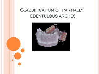
- 1. CLASSIFICATION OF PARTIALLY EDENTULOUS ARCHES
- 2. Prime purpose : To enable dentist to clearly communicate to a listener An aid in learning of the fundamentals of design
- 3. To formulate a good treatment plan To anticipate the difficulties common to occur for that particular design To communicate with a professional about a case Standardization of work principle
- 4. Allow visualization of type of partially edentulous arches that is being considered Allow differentiation between tooth supported and tooth- tissue supported partial dentures Serve as a guide to the type of design to be used Be universally accepted
- 5. Cummer Kennedy’s Applegate-Kennedy Bailyn Neurohr Mauk Friedman Godfrey Skinner Austin and Lidge Craddock Wild Watt et al Costa Osborne and Lammie Beckett Swenson ACP classification ICK classification
- 6. KENNEDY’S CLASSIFICATION Most widely used method of classification Proposed in 1923 by Dr. Edward Kennedy of New York It is based on the relationship of the edentulous spaces to the abutment teeth
- 7. Class I - Bilateral edentulous areas located posterior to the remaining natural teeth Class II - Unilateral edentulous area located posterior to the remaining natural teeth Class III - Unilateral edentulous area with natural teeth both anterior and posterior to it Class IV - Single, bilateral edentulous area located anterior to the remaining natural teeth
- 8. APPLEGATE KENNEDY’S SYSTEM It is a modification of the Kennedy’s system It takes into consideration the capabilities of the teeth, which bound the spaces to serve as abutments for the prosthesis
- 9. Dr. O.C Applegate (1960) later attempted to expand the Kennedy system by adding class V and VI Class V : Edentulous area bounded anteriorly and posteriorly by the natural teeth but in which the anterior abutment (the lateral incisor) is not suitable for the support Class VI : An edentulous situation in which the teeth adjacent to the space are capable of total support of the required prosthesis
- 10. Applegate also provided the following 8 rules to govern the application of the Kennedy system: Rule 1- Classification should follow rather than precede extractions that might alter the original classification Rule 2- If the third molar is missing and not to be replaced it is not considered in the classification Rule 3- If the third molar is present and is to be used as an abutment, it is considered in the classification APPLEGATE’S RULES
- 11. Rule 4 - If the missing second molar is not to be replaced that is the opposing second molar is also missing, it not considered in the classification Rule 5 – The most posterior edentulous area or areas always determines the classification Rule 6 – Edentulous areas other than those determining the classification are referred to as the modification spaces and are designated by their number
- 12. Rule 7 – The extent of the modification is not considered, only the number of additional edentulous areas are considered Rule 8 – There can be no modification areas in class IV arches
- 13. CUMMER’S CLASSIFICATION SYSTEM Proposed By Cummer in 1920 Cummer stated “ for working purposes all the cases may be made to fall into 4 simple classes, which have as their basis the choice of number and position of the direct retainer”
- 14. Class I – Diagonal : 2 diagonally opposite teeth are chosen as abutment teeth for the attachment of direct retainer Class II – Diametric: 2 diametrically opposite teeth are chosen as abutment teeth for the attachment of the direct retainers
- 15. Class III – Unilateral : one or more teeth on the same side are chosen as abutment teeth for the attachment of the direct retainers Class IV – Multilateral : Three or more teeth are chosen as abutment teeth for the attachment of the direct retainers
- 16. BAILYN’S SYSTEM Based on whether the prosthesis is tooth borne, tissue borne or a combination of the two: Bailyn divided all R.P.Ds into- A : Anterior restorations : Saddle area anterior to the 1st bicuspids P : Posterior restorations : Saddle area posterior to the cuspids
- 17. Subdivided as: Class I : Bounded saddle (not more than 3 teeth missing) Eg: P.I Class II : Free end saddle (no distal abutment) Eg: P.II Class III : Bounded saddle (more than 3 teeth missing) Eg P.III
- 18. Class A.III : •Edentulous space anterior to the 1st bicuspid •Bounded saddle (more than three teeth missing) Class A.I. P.II – •Edentulous area anterior to the first bicuspid and not more than 3 teeth missing •other edentulous space being posterior to the cuspid with only one tooth available as an abutment
- 19. Class P.I P.II – Both the edentulous spaces are posterior to the cuspids • One with only one tooth for anchorage • Other with two available teeth separated by a distance of less than three teeth
- 20. NEUROHR’S CLASSIFICATION Proposed in 1939, this classification is based on the support derived It is not commonly used due to its complexity Many of his denture designs did not match his principles of classification
- 21. Class I : Tooth bearing Teeth posterior to all spans, and when there are not more than four teeth missing in any space
- 22. There are two possible variation in this class - Variation 1 : Missing posteriors predominate a. Posteriors missing, anteriors in place b. Posteriors missing, some anteriors missing
- 23. Variation 2 : Missing anteriors predominate a) Anteriors missing, posteriors present b) Anteriors missing, some posteriors missing
- 24. Class II : Tooth-and-tissue bearing No teeth posterior to one or more spans • More than 4 teeth (which include a canine) in one or more spans
- 25. Class II is further subdivided into divisions with variation under each Division 1: When there are no teeth posterior to 1 or more span Variation 1 : Missing posteriors predominate a. Posteriors missing, anteriors in place b. Posteriors missing, some anteriors missing Variation 2 : Missing anteriors predominate a. None b. Anteriors missing, some posteriors missing
- 26. Division 2: when there are teeth posterior in all spans, but when there are more than 4 teeth in any one or more spans. Variation 1 : Missing posteriors predominate a. None b. Posteriors missing, some anteriors missing Variation 2 : Missing anteriors predominate • Anteriors missing, posteriors in place • Anteriors missing, some posteriors missing CLASS III : Tissue bearing complete dentures
- 27. MAUK’S SYSTEM Class I – Bilateral posterior spaces and teeth remaining in a segment in the anterior region Class II – Bilateral posterior spaces and one or more teeth at the posterior end of one space Based on number, length and position of the spaces and number and position of the remaining teeth
- 28. Class III - Bilateral posterior spaces and one or more teeth at the posterior end of both spaces Class IV – Unilateral posterior space with or without teeth at the posterior end of the space. Opposing arch is unbroken
- 29. Class V – It has an anterior space only. Posterior part of the arch is unbroken on either side Class VI – Has irregular spaces around the arch. The missing teeth are single or in groups
- 30. WILD’S CLASSIFICATION Proposed simple and self explanatory classification Class I : Interruption of the dental arch (i.e bounded) Class II : Shortening of the dental arch (i.e free end) Class III : combination of I & II
- 31. GODFREY’S SYSTEM (1951) Based on the location and the extent of the edentulous spaces Class A – Tooth borne denture bases in the anterior part of the mouth It may be an unbroken 5 - tooth space; a broken 5-tooth space; or an unbroken 4-tooth space
- 32. Class B – Mucosa borne denture base area in anterior of the mouth Unbroken six tooth space; an unbroken 5-tooth space; a broken 5- tooth space Class C – Tooth borne denture base in the posterior part of the mouth Unbroken 3-tooth space; a broken 3-tooth space; an unbroken 2-tooth space; a broken 2-tooth space
- 33. Class D – Class D has mucosa borne denture bases in the posterior part of the mouth It may be an unbroken 4 – tooth space or a 3 tooth; 2 tooth or single tooth space
- 34. FRIEDMAN’S CLASSIFICATION Introduced ‘ABC’ classification in 1953. According to this classification - A: Anterior B: Bounded posterior C: Cantilever
- 35. BECKETT AND WILSON CLASSIFICATION Beckett and Wilson based their ideas on Bailyn’s classification(1928) Based on proportionate amount of support provided by the teeth and tissues Beckett S L, The influence of saddle classification on the design of partial removable restorartions, J Prosthet Dent, 1953;3(4): 506-16
- 36. Class I : Bounded saddle. Abutment teeth qualified to support the denture. Mucosa is not used for support Class II : Free end a. Tooth and tissue borne b. Tissue borne
- 37. Class III : Bounded saddle. Abutment teeth not so qualified to support the denture as described in class I. Wilson in 1957 elaborated the classification as follows: • Mandibular Kennedy’s class III should be treated as class I • Maxillary Kennedy’s class III should be treated as class I or III
- 38. CRADDOCK’S CLASSIFICATION In 1954 Class I – Saddle supported on both sides by substantial abutment teeth Class II – Vertical biting forces applied to denture resisted entirely by soft tissue Class III – Tooth supported at only 1 end of the saddle
- 39. AUSTIN AND LIDGE CLASSIFICATION In 1957 Describes the position of teeth Class A: Missing anteriors A1 : Missing anteriors on one side A2 : Missing anteriors on both sides A B1: Missing anteriors with bilateral construction
- 40. Class P : Missing posteriors P1 – Missing posteriors on one side P2 – Missing posteriors on both sides P B1 : Posterior missing on both sides (distal extension)
- 41. Class AP : Missing anteriors and posteriors AP1 – Missing anteriors and posteriors on one side AP2 – Missing anteriors and posteriors on both sides
- 42. SKINNER’S SYSTEM This system was based on the relationship of the abutment teeth to the supporting residual alveolar ridge He said that the value of RPD is directly related to quantity and the degree of support, which it receives, from the abutment teeth and residual ridge Skinner C N, A Classification of removable partial dentures based upon the principles of anatomy and physiology; J Prosthet Dent,1959;9(2):240-46
- 43. Class I – Abutment teeth are located both anterior and posterior to the denture bases, spaces may be unilateral or bilateral Class II – All teeth are posterior to the denture base. It may be unilateral or bilateral
- 44. Class III – All the abutment teeth are anterior to denture base and may occur unilaterally or bilaterally Class IV – Denture bases are located both anterior and posterior to the remaining teeth. They may be unilateral or bilateral Class V – Abutment teeth are unilateral in relation to denture base
- 45. SWENSON’S CLASSIFICATION The 4 primary classes represent only slight modification of the Kennedy’s system Class I – Its an arch with one free end denture base
- 46. Class II – It is an arch with 2 free end denture base Class III – It is an arch with edentulous space posteriorly on one or both the sides but with teeth present anteriorly or posteriorly to each space Class IV – It is an arch with anteriorly edentulous space and with 5 or more anterior teeth missing
- 47. Subdivision – • A : Anterior • P : Posterior • AP : Anterior and posterior Class II A – It is basic class II with an anterior space Class IV P – Basic class IV with posterior space
- 48. TERKLA AND LANEY MODIFICATION (1963) Combined Kennedy’s and Swenson’s classification Kennedy’s class II = Swenson’s class I Kennedy’s Class I = Swenson’s Class II
- 49. WATT ET AL CLASSIFICATION Proposed the classification in 1958. It was based on the type of support derived Entirely tooth borne : Entire denture rests on the abutment teeth
- 50. Entirely tissue borne : Entire denture rests on soft tissue Partially tooth borne and partially tissue borne : These dentures rest on both teeth and the tissues
- 51. OSBORNE AND LAMMIE’S CLASSIFICATION Proposed in 1974. It is similar to Watt et al’s classification Class I : Mucosa borne Class II : Tooth borne Class III : combination of Mucosa – borne and tooth borne
- 52. COSTA’S CLASSIFICATION This system developed in 1974 was based on describing rather than classifying partially dentulous arches Costa E, A simplified system for identifying partially edentulous dental arches, J Prosthet Dent 1974;32(6):639-45
- 53. • Anterior : Edentulous space located in the anterior part of dental arch • Lateral : Edentulous space bounded both mesially and distally by remaining teeth • Terminal : Edentulous space not bounded distally by remaining teeth
- 54. PROSTHODONTIC DIAGNOSTIC INDEX The American College of Prosthodontists (ACP) has developed a classification system for partial edentulism based on diagnostic findings McGarry T H, Classsification system for partial edentulism, J prosthodont, 2002;11:181-193
- 55. THIS PARTIALLY EDENTULOUS CLASSIFICATION SYSTEM OFFERS THE FOLLOWING POTENTIAL BENEFITS Improved intra-operator consistency Improved professional communication An objective method for patient screening in dental education Standardized criteria for outcome assessment and research Improved diagnostic consistency Simplified, organized aid in the decision to refer a patient McGarry T H, Classsification system for partial edentulism, J prosthodont, 2002;11:181-193
- 56. CLASSIFICATION SYSTEM FOR THE PARTIALLY EDENTULOUS PATIENT DIAGNOSTIC CRITERIA 1. Location and extent of the edentulous area(s) 2. Condition of the abutment teeth 3. Occlusal scheme 4. Residual ridge
- 57. CRITERIA 1 LOCATION AND EXTENT OF EDENTULOUS AREA(S)
- 58. IDEAL OR MINIMALLY COMPROMISED EDENTULOUS AREA The edentulous span is confined to a single arch and one of the following: Any anterior maxillary span that does not exceed 2 missing incisors Any anterior mandibular span that does not exceed 4 missing incisors Any posterior maxillary or mandibular span that does not exceed 2 premolars or 1 premolar and 1 molar
- 59. MODERATELY COMPROMISED EDENTULOUS AREA The edentulous span is in both arches and one of the following: Any anterior maxillary span that does not exceed 2 missing incisors Any anterior mandibular span that does not exceed 4 missing incisors Any posterior maxillary or mandibular span that does not exceed 2 premolars or 1 premolar and 1 molar The maxillary or mandibular canine is missing
- 60. SUBSTANTIALLY COMPROMISED EDENTULOUS AREA Any posterior maxillary or mandibular span that is greater than 3 missing teeth or 2 molars Any edentulous span including anterior and posterior areas of 3 or more missing teeth
- 61. SEVERELY COMPROMISED EDENTULOUS AREA Any edentulous area or combination of edentulous areas requiring a high level of patient compliance
- 62. CRITERIA 2 ABUTMENT TEETH CONDITION
- 63. IDEAL OR MINIMALLY COMPROMISED ABUTMENT TEETH CONDITION No pre-prosthetic therapy is indicated
- 64. MODERATELY COMPROMISED ABUTMENT TEETH CONDITION Abutment Condition: Insufficient tooth structure to retain or support intra-coronal restorations – in one or two sextants Abutments require localized adjunctive therapy, i.e., periodontal, endodontic or orthodontic procedures in one or two sextants
- 65. Abutment condition Insufficient tooth structure to retain or support intracoronal or extracoronal restorations - three or more sextants Abutments require extensive adjunctive therapy, i.e., periodontal, endodontic or orthodontic procedures - in three or more sextants SUBSTANTIALLY COMPROMISED ABUTMENT TEETH CONDITION
- 66. SEVERELY COMPROMISED ABUTMENT TEETH CONDITION Abutments have a guarded prognosis
- 68. IDEAL OR MINIMALLY COMPROMISED OCCLUSAL SCHEME No preprosthetic therapy required Class I molar and jaw relationships
- 69. MODERATELY COMPROMISED OCCLUSAL SCHEME Occlusal scheme requires localized adjunctive therapy (e.g. enameloplasty on premature occlusal contacts) Class I molar and jaw relationships
- 70. SUBSTANTIALLY COMPROMISED OCCLUSAL SCHEME Entire occlusal scheme requires reestablishment but without any change in the vertical dimension of occlusion Class II molar and jaw relationships
- 71. SEVERELY COMPROMISED OCCLUSAL SCHEME Entire occlusal scheme requires reestablishment with changes in the vertical dimension of occlusion Class II Division 2 and Class III molar and jaw relationships
- 73. The criteria published for the Classification System for complete edentulism are used to categorize any edentulous span present in the partially edentulous patient
- 74. CLASSIFICATION SYSTEM FOR PARTIAL EDENTULISM
- 75. Class I Location and extent of edentulous area Condition of the abutments Occlusion Residual ridge characteristics Ideal or minimal compromise Ideal or minimally compromised no need for pre prosthetic treatment Ideal or minimally compromised class I molar and jaw relation Class I ridge
- 76. Class II Location and extent of edentulous area Condition of the abutments Occlusion Residual ridge characteristics One or both arches edentulous Extent anterior maxillary not exceeding 2 I, anterior mandibular not exceeding 4 I and any posterior span not exceeding 2 PM or 1 PM and 1 M or any missing canine Abutment moderately compromised Insufficient tooth structure for support Localized adjunctive therapy required Localized adjunctive therapy required Class I molar relationship Residual ridge: class II
- 77. Class III Location and extent of edentulous area Condition of the abutments Occlusion Residual ridge characteristics Present in one or both arches Posterior are greater than 3 teeth or 2 molars anterior and posterior areas of 3 or more teeth If 3 sextants have insufficient tooth structure Localized adjunctive therapy Abutments have a fair prognosis Requires reestablishment of the entire occlusal scheme without accompanying change in the OVD. Class II molar Class III residual ridge
- 78. Class IV Location and extent of edentulous area Condition of the abutments Occlusion Residual ridge characteristics Extensive and in both arches Acquired or congenital maxillofacial defects included In 4 or more sextants, have insufficient tooth structure and require extensive localized adjunctive therapy Re-establishment of entire occlusal scheme including changes in OVD required. Class II div 2 or class III molar Class IV ridge
- 79. GUIDELINES FOR THE USE OF THE CLASSIFICATION SYSTEM FOR PARTIAL EDENTULISM
- 80. Consideration of future treatment procedures must not influence the decision as to which diagnostic level to place the patient in Initial pre-prosthetic treatment and/or adjunctive therapy can change the initial classification level. The classification may need to be reassessed after the removal of existing prostheses
- 81. Periodontal health is intimately related to the diagnosis and prognosis for partially edentulous patients For the purpose of this system, it is assumed that patients will receive periodontal therapy to achieve and maintain periodontal health so that prosthodontic care can be accomplished
- 82. In the situation where the patient presents with an edentulous maxilla opposing a partially edentulous mandible, each arch is diagnosed with the appropriate classification system In this situation, the maxilla would be classified according to the complete edentulism classification system and the mandible according to the partial edentulism classification system
- 83. A single exception to this rule is when the patient presents with an edentulous mandible opposed by a partially edentulous or dentate maxilla This clinical situation presents significant complexity and long-term morbidity and as such, should be diagnosed as a Class IV in either system
- 84. ICK CLASSIFICATION SYSTEM ICK classification system will follow the Kennedy method with the following guidelines: 1. No edentulous space will be included in the classification if it will be restored with an implant supported fixed prosthesis Al-Johany et al, ICK Classification System for Partially Edentulous Arches, J Prosthodont 2008;17: 502–507
- 85. To avoid confusion, maxillary arch is drawn as half circle facing up and mandibular arch as as half circle facing down. The drawing will appear as if looking directly at the patient; right and left quadrants are reversed
- 86. The implant classification will always begin with the phrase “Implant Corrected Kennedy (class)” followed by the description of the classification. It can be abbreviated as follows: ICK I, for Kennedy class I situations ICK II, for Kennedy class II situations ICK III, for Kennedy class III situations and ICK IV, for Kennedy class IV situations
- 87. •The abbreviation “max” for maxillary and “man” for mandibular can precede the classification. The word modification can be abbreviated as “mod”. • man ICK I mod 2(#18, 22, 26, 31) •Roman numerals will be used for the classification, and Arabic numerals will be used for the number of modification spaces and implants • The tooth number using the American dental Association (ADA) system is used to give the number and exact position of the implant in the arch
- 88. The classification of any situation will be according to the following order: main classification first, then followed by the number of modification spaces, followed by the number of implants in parentheses according to their position in the arch preceded by the number sign(#).eg; man ICK I mod 2(#18, 22, 26, 31) The classification can be used either after implant placement to describe any situation of RPD with implants, or before implant placement to indicate the number and position of future implants with an RPD
- 89. The arrangement of implants will be from right to left in the maxillary arch and from left to right in the mandibular arch following the tooth numbering system ICK I (#2,15 ) ICK I (# 18, 22, 31)
- 90. Class I (Div A-D) Class II (Div A-D) Class III (Div A-D) Class IV (Div A-D) CLASSIFICATION OF PARTIALLY EDENTULOUS ARCHES IN IMPLANT DENTISTRY Misch CE, Judy WMK, classification of partially edentulous arches for implant dentistry, Int J Oral Implantal 1987;4:7-12
- 95. CONCLUSION
- 96. Stewart, Rudd, Kuebker, Clinical removable partial prosthodontics, 2nd edition, India, IEA Publishers, 1997, pp 1-18 McCracken, Removable partial denture prosthodontics,11th edition, India, Elsevier Publisher’s, 2005, pp 19-24 Avant E W, A Universal classification for removable partial denture situation, J Prosthet Dent,1996;533-39 References
- 97. Nallaswamy D, Textbook of Prosthodontics, 1st edition, India, Jaypee Publications, 2003, pp 270-287 McGarry T H, Classsification system for partial edentulism, J prosthodont, 2002;11:181-193 Friedman J, The ABC classification of partial denture segments, J Prosthet Dent 1953;3:517-23 Costa E, A simplified system for identifying partially edentulous dental arches, J Prosthet Dent 1974;32(6):639-45
- 98. Miller L E, System for classifying partially dentulous arches, J Prosthet Dent 1970;24(1):25-40 Beckett S L, The influence of saddle classification on the design of partial removable restorartions, J Prosthet Dent, 1953;3(4):506-16 Skinner C N, A Classification of removable partial dentures based upon the principles of anatomy and physiology, J Prosthet Dent,1959;9(2):240-46 Al-Johany et al, ICK Classification System for Partially Edentulous Arches, J Prosthodont, 2008;17:502–507