
Recommended
More Related Content
Similar to elbowdislocations-180623080147 (1).pptx
Similar to elbowdislocations-180623080147 (1).pptx (20)
More from sonalidas935894
More from sonalidas935894 (16)
Recently uploaded
Recently uploaded (20)
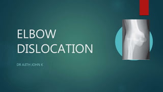
elbowdislocations-180623080147 (1).pptx
- 2. ▶ Simple elbow dislocation is one in which there are no associated fractures ▶ The elbow joint is the second most commonly dislocated joint in the adult population ▶ Adolescent males are the highest-risk group
- 3. GROSS ANATOMY ▶ Articulations The elbow joint is made up of three articulations ▶ Radiohumeral: capitellum of the humerus with the radial head ▶ Ulnohumeral: trochlea of the humerus with the trochlear notch (with separate olecranon and coronoid process articular facets) of the ulna ▶ Radioulnar: radial head with the radial notch of the ulna (proximal radioulnar joint)
- 4. MOVEMENTS ▶ The elbow is a trochoginglymoid (combination hinge and pivot) joint ▶ the hinge component (allowing flexion-extension) is formed by the ulnohumeral articulation ▶ the pivot component (allowing pronation-supination) is formed by the radiohumeral articulation and the proximal radioulnar joint
- 5. LIGAMENTS ▶ medial (ulnar) collateral ligament complex ▶ lateral (radial) collateral ligament complex ▶ oblique cord ▶ inconstant thickening of supinator muscle fascia and functionally insignificant ▶ runs from tuberosity of the ulna to just distal to radial tuberosity ▶ quadrate ligament (of Denuce) ▶ thickening of the inferior aspect of the joint capsule ▶ runs from just inferior to the radial notch of the ulna to insert to the medial surface of the radial neck
- 6. PATHOANATOMY AND APPLIED ANATOMY ▶ soft tissue restraints can be divided into both static and dynamic stabilizers ▶ static stabilizers joint capsule and the LCLs and MCLs
- 7. LCL ▶ Primary varus and posterolateral rotational stabilizer ▶ LCL has three components- The radial collateral ligament annular ligament The lateral ulnar collateral ligament
- 8. ▶ The radial head is surrounded by the annular ligament which attaches to the anterior and posterior margins of the radial notch of the proximal ulna ▶ The radial collateral ligament arises from the lateral epicondyle and blends with the annular ligament ▶ The lateral ulnar collateral ligament is posterior to the radial collateral ligament
- 9. ▶ MCL consists of the anterior and posterior bundles ▶ The anterior bundle is the key valgus stabilizer of the elbow, arising from the anteriorinferior aspect of the medial epicondyle to insert on the sublime tubercle of the proximal ulna ▶ The posterior bundle provides a secondary restraint to valgus load and also resists ulnar rotation
- 11. Dynamic restraints ▶ Biceps, brachialis, and triceps
- 12. ▶ Patients with simple elbow dislocations routinely have disruption of both the MCL and LCL and the elbow capsule ▶ The muscular origins may be disrupted as well; typically the injury to the lateral common extensor origin is more extensive than the medial common flexor origin ▶ Most activities of daily living exert a varus force on the elbow than a valgus force, residual instability is usually due to incompetence of the LCL in the majority of patients
- 13. ▶ The radial head causes an impression fracture of the posterior capitellum which can contribute to recurrent instability
- 14. Mechanisms of Injury ▶ Fall on an outstretched hand ▶ The soft tissue injury is thought to begin on the lateral side of the elbow with disruption of the lateral collateral ligament (LCL) and then proceeds through the capsule to the medial side with the medial collateral ligament (MCL) being injured last
- 16. Associated Injuries ▶ Simple elbow dislocations are not associated with fractures ▶ Disruption of the collateral ligaments, elbow capsule, and forearm flexor and extensor muscle origins ▶ Injury to the brachial artery has been described in closed simple dislocations and nerve palsies are possible ▶ The ulnar nerve is the most commonly injured nerve following elbow dislocation
- 17. Signs and Symptoms ▶ Obvious deformity and pain about the affected elbow ▶ Elbow flexed to 90 degrees, the medial and lateral epicondyles and the olecranon process should form an isosceles triangle ▶ Complete peripheral neurologic examination should be performed
- 18. Imaging and Other Diagnostic Studies ▶ Anteroposterior, lateral, and oblique radiographs are used to diagnose elbow dislocation and help to rule out associated fractures ▶ Although rarely required in practice, a line drawn along the anterior margin of the humerus (anterior humeral line) and one along the long axis of the radius should intersect near the centre of the capitellum
- 19. ▶ Computed tomography (CT) scanning is rarely needed but can be useful if there is a questionable associated fracture
- 20. ▶ MRI is not needed unless there is concern for ulnar nerve entrapment in the joint since the pathology of the soft tissue injury associated with elbow dislocations has been well established
- 21. CLASSIFICATION ▶ Based on the direction of dislocation ▶ Simple versus complex displacement of the ulna relative to the humerus Posterior ■ Posterolateral ■ Posteromedial ■ Lateral ■ Medial ■ Anterior
- 23. Post
- 24. Medial
- 25. Outcome Measures ▶ DASH and the Oxford Elbow Questionnaire
- 26. Fracture-Dislocations ▶ Associated radial head fracture ▶ Medial or lateral epicondyle fracture ▶ Coronoid process fracture
- 27. INJURY PATTERNS ▶ Posterior dislocation with a fracture of the radial head ▶ Posterior dislocation with fractures of the radial head and coronoid process—the so-called “terrible triad” injury ▶ Varus posteromedial rotational instability pattern injuries associated with anteromedial facet of the coronoid fractures ▶ Anterior olecranon fracture-dislocations ▶ Posterior olecranon fracture-dislocations
- 28. TYPES OF ELBOW INSTABILITY Posterolateral rotatory instability (elbow dislocations with or without associated fractures) Varus posteromedial rotational instability (anteromedial coronoid facet fractures) Olecranon fracture-dislocations
- 29. TREATMENT
- 30. NONOPERATIVE TREATMENT ▶ The majority of simple elbow dislocations can be treated nonoperatively with closed manipulative reduction evaluation of stability and an early rehabilitation program
- 31. TECHNIQUES-PARVINS METHOD ▶ The medial and lateral epicondyles are palpated and their relationship to the olecranon is determined in order to first correct and medial/lateral displacement in the coronal plane ▶ The elbow is typically flexed to approximately 30 degrees, and traction is placed through the forearm while stabilizing the humerus ▶ Direct pressure over the olecranon may help to guide it over the distal humerus and into joint ▶ Supination of the forearm may be helpful to gain the reduction
- 33. ▶ After reduction-the elbow is taken through an arc of flexion– extension in pronation, neutral, and supination in order to evaluate for residual instability ▶ The elbow redislocates when flexed to less than 30 degrees, operative treatment should be considered ▶ Most patients will have varus–valgus instability
- 34. ▶ The elbow is then immobilized in a light plaster splint with the forearm in st pronation, neutral, or supination (depending on the position of maximal ability) and the elbow at 90 degrees of flexion Radiographs are performed to ensure a congruous reduction has been achieved and to evaluate for the presence of fractures not visualized on the prereduction radiographs
- 35. ▶ Immobilization greater than 3 weeks should be avoided as this has been demonstrated to cause an increased incidence of stiffness and poorer functional outcomes
- 36. OUTCOMES ▶ Several studies have reported good to excellent outcomes in the majority of patients after simple elbow dislocation ▶ Prolonged immobilization after injury was associated with a worse result with increasing duration of immobilization leading to increased flexion contracture and more severe residual pain: In general, prolonged immobilization is to be avoided in this setting
- 37. OPERATIVE TREATMENT ▶ The main indication for operative management of simple elbow dislocations is an inability to maintain a concentric elbow joint after closed reduction or a recurrent dislocation ▶ irreducible dislocations are also indications for operative treatment but these are rare injuries
- 38. SURGICAL PROCEDURE ▶ Patient is placed supine on the operating table with a radiolucent arm table on the affected side ▶ Preoperative examination of the shoulder should be performed to be sure that there is adequate external rotation of the shoulder in order to approach the medial side of the elbow
- 39. ▶ Surgical Approach-posterior midline incision is employed and a full thickness lateral flap is elevated on the deep fascia ▶ If the medial structures require repair, full thickness elevation of the medial flap is performed
- 40. Soft Tissue Repair ▶ Vast majority of cases, a medial ligament repair is not required and the surgery is complete ▶ The lcl can be repaired using transosseous bone tunnels or suture anchors ▶ Locking krackow stitches are placed in the lcl while a second suture is placed in the extensor fascia
- 42. ▶ In the unusual setting that the elbow remains unstable in spite of repair of the lateral structures, the medial side of the elbow is approached with care taken to protect the ulnar nerve ▶ The flexor–pronator muscles are also repaired if they have been avulsed ▶ elbow is still unstable, then a static or hinged external fixator should be placed or, as a last resort, the elbow should be transfixed with a screw or robust Steinman pin
- 43. EXTERNAL FIXATION ▶ A hinged fixator will allow for range of motion exercises to be performed while the external fixator is in place and should be considered if the surgeon has access to this and the experience to apply it ▶ Static fixators are easier to apply and are more widely available ▶ The key to all hinged devices is an understanding of the axis of elbow rotation ▶ Two pins are placed in the humeral shaft laterally and two pins are placed in the ulnar shaft laterally in a position that allows for forearm rotation
- 44. ▶ Open pin placement is recommended to avoid injury to the radial nerve ▶ A static frame is assembled with the elbow joint reduced ▶ The external fixator is left in place for approximately 4 weeks and then a range of motion protocol is initiated as outlined above for closed treatment
- 46. ▶ Bridge Plate Indications are conditions where maintenance of reduction is challenging such as morbid obesity and patients with neurologic injuries such as spasticity or flaccid paralysis triceps-splitting approach Three to four locking screws are placed in the ulna and the distal humerus avoiding the articulation and fossae
- 47. ▶ The plate is removed at 4 weeks, and a posterior capsulectomy and an elbow manipulation can be considered at the time of plate removal to increase the recovery of motion
- 48. ADVERSE OUTCOMES
- 50. COMPLICATIONS ▶ Loss of motion (stiffness): Stiffness following complicated or uncomplicated elbow dislocation is usually the rule. Immobilization of the elbow should generally not go beyond 2 weeks
- 51. ▶ Compartment syndrome (Volkmann contracture): This may result from massive swelling due to soft tissue injury
- 52. ▶ Persistent instability/redislocation: This is rare after isolated, traumatic posterior elbow dislocation; the incidence is increased in the presence of an associated coronoid process and radial head fracture (terrible triad of the elbow)
- 53. THANK U