
Recommended
More Related Content
Similar to HERPES SIMPLEX VIRUS 12032019 TUESDAY pptx
Similar to HERPES SIMPLEX VIRUS 12032019 TUESDAY pptx (20)
More from PulkitMittal54
More from PulkitMittal54 (8)
Recently uploaded
Recently uploaded (20)
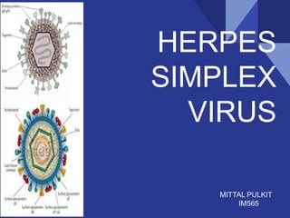
HERPES SIMPLEX VIRUS 12032019 TUESDAY pptx
- 2. INTRODUCTION ● Herpes (Greek: creep or crawl) ● Herpes simplex viruses belong to the ubiquitous Herpesviridae family ● Human herpes simplex virus (HSV) causes contagious infection with a large reservoir in the general population ● Herpesviruses are able to establish lifelong persistent infections in their hosts and undergo periodic reactivation ; incurable ● HSV has a potential for significant complications in the immunocompromised host
- 3. INTRODUCTION ● HSV-1 is normally associated with orofacial infections and encephalitis ● HSV-2 usually causes genital infections and can be transmitted from infected mothers to neonates ● Both viruses establish latent infections in sensory neurons and, upon reactivation, cause lesions at or near point of entry into the body
- 4. CLASSIFICATION OF HUMAN HERPESVIRUSES Biologic properties Examples Subfamily( “herpesviri nae) Growth cycle and cytopathology Latent infections Genus (“virus) Official name (“Human herpesvirus”) Common name Alpha Short, cytolytic Neurons Simplex 1 Herpes simplex virus type 1 2 Herpes simplex virus type 2 Varicello 3 Varicella-zoster virus Beta Long, cytomegalic Glands, kidneys Cytomegalo 5 Cytomegalovirus Long, lymphoproliferative Lymphoid tissue Roselo 6 Human herpesvirus 6 7 Human herpesvirus 7 Gamma Variable, lymphoproliferative Lymphoid tissue Lymphocrypto 4 Epstein-Barr virus Rhadino 8 Kaposi's sarcoma- associated herpesvirus
- 5. TRANSMISSION ● Transmission of both HSV types is by direct contact with virus-containing secretions or with lesions on mucosal or cutaneous surfaces ● HSV-1 is spread by contact, usually by infected saliva ● HSV-1 primarily infects skin above the waist ● HSV-2 is transmitted sexually or from a maternal genital infection to a newborn ● HSV-2 primarily infects skin below the waist
- 6. REPLICATION i. Virus adsorption and penetration i. Viral DNA replication and nucleocapsid assembly i. Acquisition of the viral envelope i. Latency
- 7. PATHOGENESIS ● Ballooning of infected cells ● Production of Cowdry type A intranuclear (Lipschutz) inclusion bodies ● Margination of chromatin ● Formation of multinucleated giant cells
- 8. CLINICAL SIGNIFICANCE ● HSV-1 ▪ Acute gingivostomatitis ▪ Recurrent herpes labialis (cold sores) ▪ Herpetic whitlow ▪ Keratoconjunctivitis ▪ Encephalitis ● HSV-2 ▪ Genital herpes ▪ Neonatal herpes (may be by HSV-1 as well)
- 9. CLINICAL SIGNIFICANCE ● Primary infections of the upper body Fig. Herpes simplex gingivostomatitis Fig. Herpetic whitlow Fig. Recurrent herpes labialis (cold sores) Fig. Keratoconjunctivitis
- 10. CLINICAL SIGNIFICANCE ● Primary infections of the genital tract Fig. Genital herpes simplex infections
- 11. CLINICAL SIGNIFICANCE ● Latency ▪ HSV-1: Trigeminal ganglia ▪ HSV-2: Sacral ganglia Fig. Primary and recurrent herpes simplex infections
- 12. LABORATORY DIAGNOSIS A. Cytopathology: ▪ A rapid cytologic method ▪ Scrapings obtained from the base of a vesicle is stained with 1% aq. solution of toluidine blue ‘0’ for 15 seconds ▪ Presence of multinucleated giant cells or ‘Tzanck cells’ = + HSV ▪ Intranuclear inclusion bodies with Giemsa-stained smears
- 13. LABORATORY DIAGNOSIS B. Isolation and identification: ▪ Inoculation of tissue cultures in human diploid fibroblasts is preferred for viral isolation ▪ Typical cytopathic changes may be seen in 24-48 hrs C. Polymerase chain reaction: D. Serology: ▪ Antibodies appear in 4–7 days after infection; reach a peak in 2–4 weeks ▪ Rise in Ab titre may be demonstrated by ELISA or complement fixation tests
- 14. TREATMENT
- 15. PREVENTION currently there are no licensed vaccines against either HSV type. ● Transmission can be reduced by: ▪ avoidance of contact with potential virus-shedding lesions ▪ safe sexual practice ▪ antiviral therapy
- 16. THANK YOU