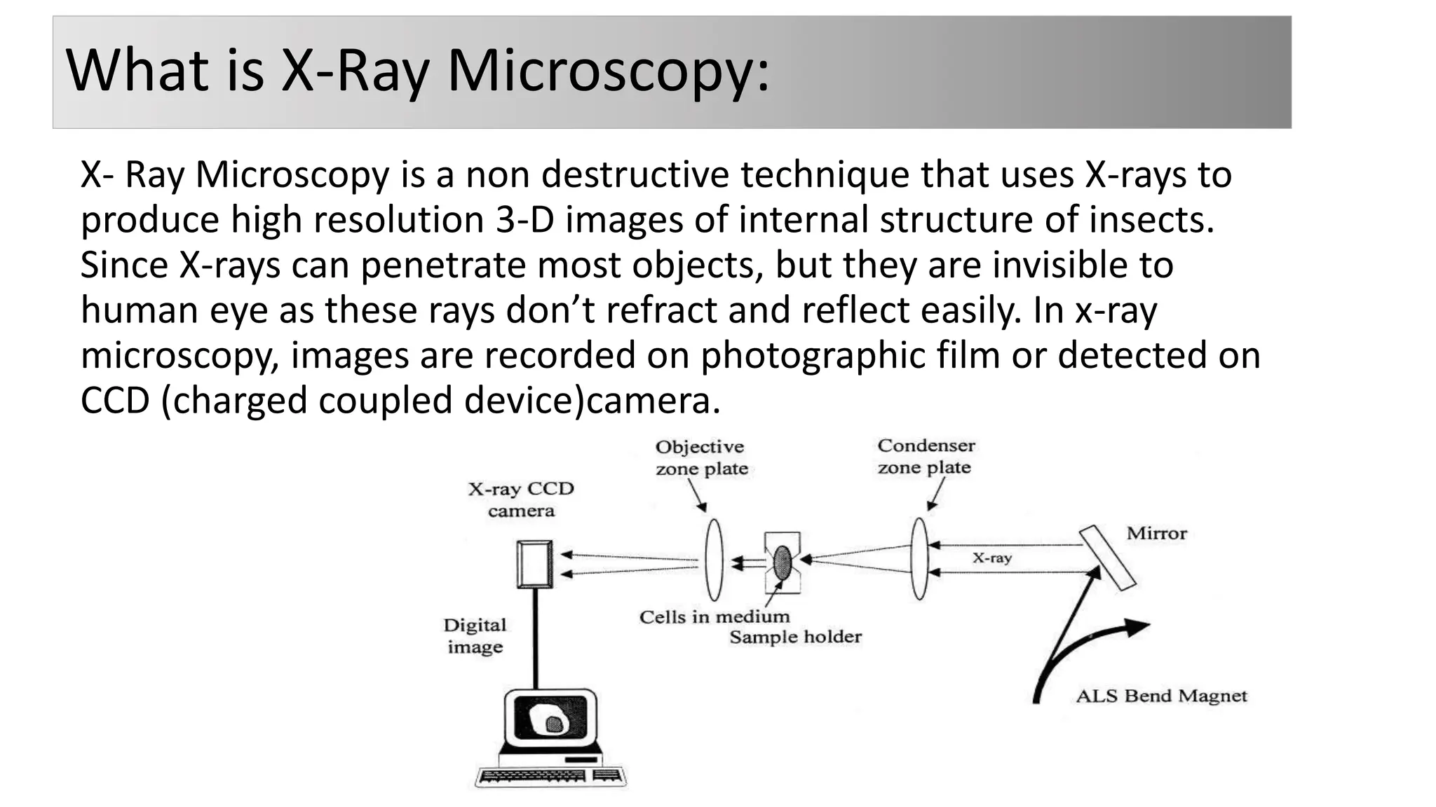
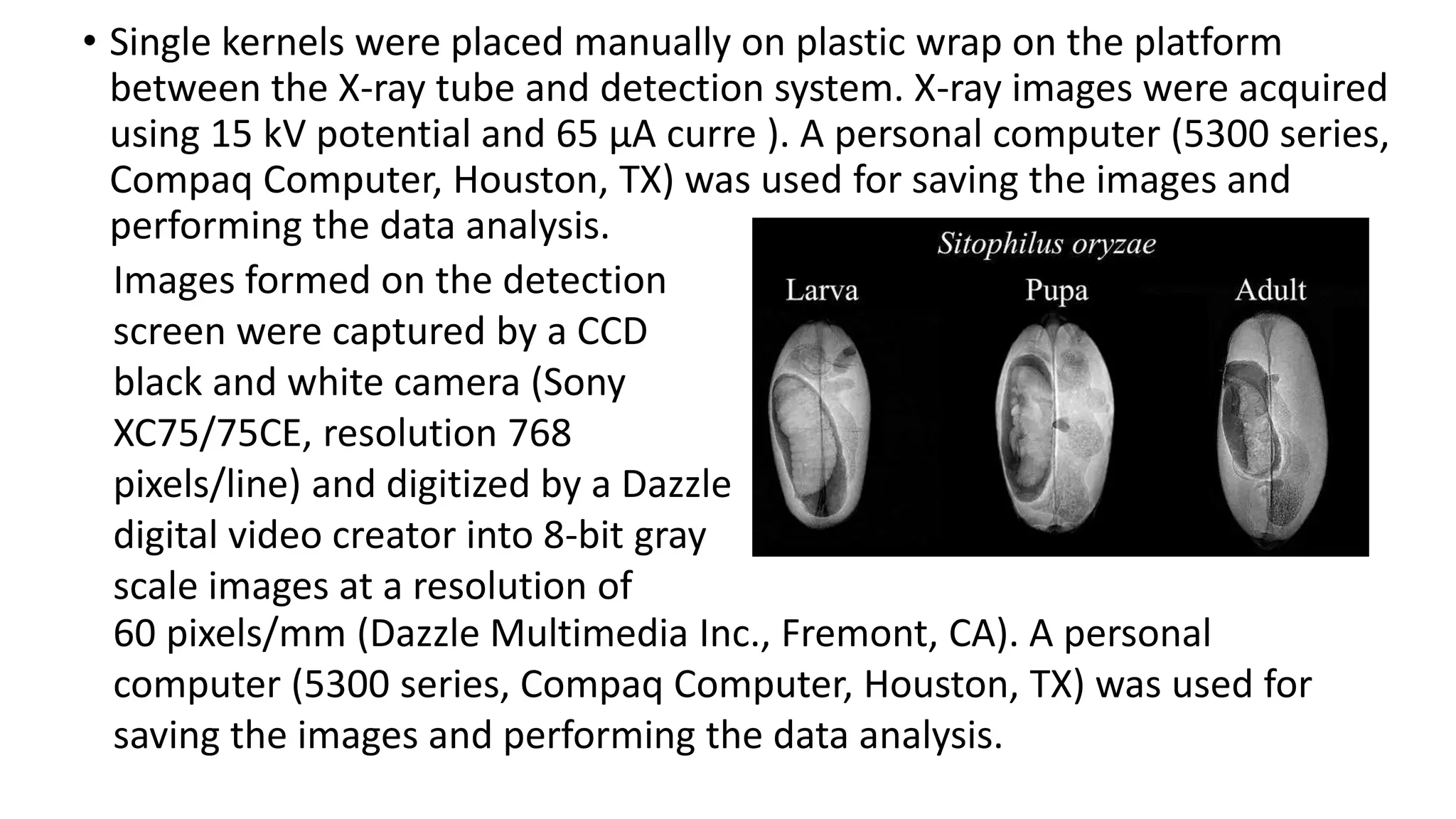
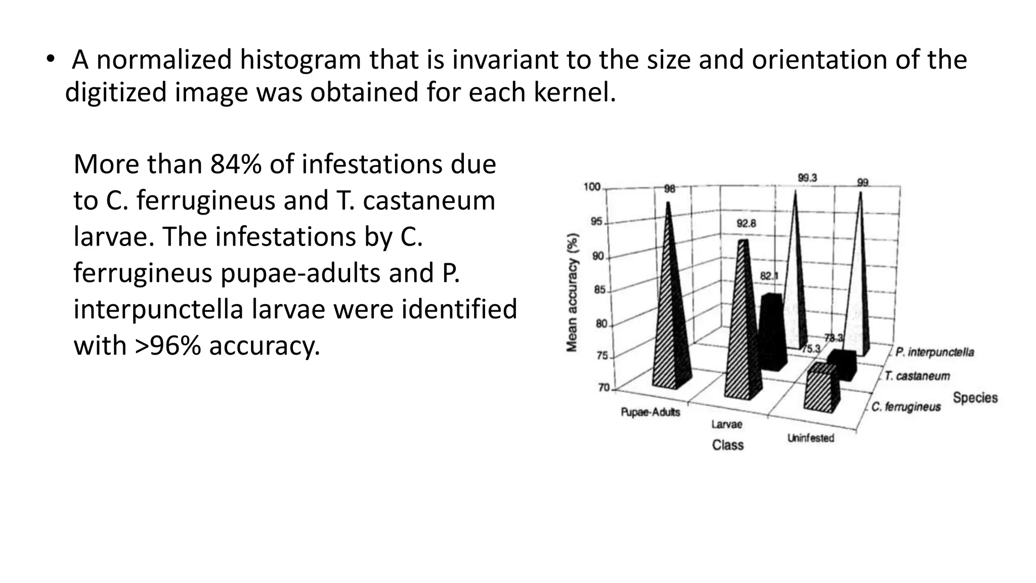
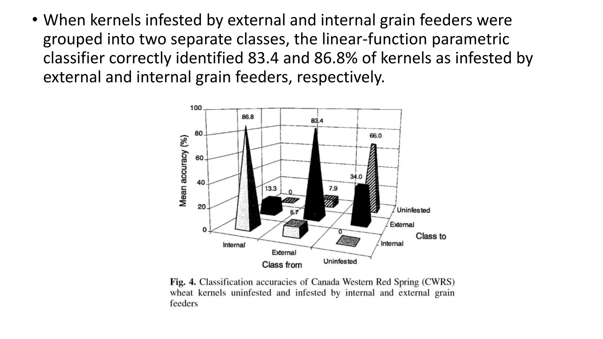
X-Ray Microscopy
Presented by: Mahnoor Baig
Submitted to : Dr. Bashir Ahmad
Department of zoology
Program: MPhil (1st semester)
What is X-Ray Microscopy:
X- Ray Microscopy is a non destructive technique that uses X-rays to
produce high resolution 3-D images of internal structure of insects.
Since X-rays can penetrate most objects, but they are invisible to
human eye as these rays don’t refract and reflect easily. In x-ray
microscopy, images are recorded on photographic film or detected on
CCD (charged coupled device)camera.
History of X-Ray Microscopy:
• Rontgen discovered X-rays in 1895 & in 1948, the first microscope was
developed by Kirkpatrick & Alfred Baez.
An X-ray microscopic image of living 10-days
old Canola Plant.
Advances in X-ray microscopy
X ray imaging can be performed using both soft and hard X rays. Hard
X rays have wavelength shorter than 0.2 nm, while soft x rays have
wavelength longer than that. Hard X rays have greater penetrating
power and greater energy but can induce more damage on the sample
during imaging.
X-ray imaging results in a two-dimensional radiograph, showing the
transmission of X-rays through an object. Low-density volumes of the
object allow high transmission, while high density volumes allow low
transmission.
• With a point X-ray source, magnification and illuminated volume can be
adjusted. Larger coverage is achieved by moving the object closer to the
detector, while higher magnification is achieved by moving the object
closer to the source. The source to detector distance also plays a pivotal
role in balancing magnification and coverage. Moving the detector closer
to the object results in higher coverage while moving the detector further
away yields higher resolution.
• Several projection images are
collected as the object is rotated.
The object is digitally
reconstructed, revealing the
position of features in the object.
C. Karunakaran,1 D. S. Jayas,1,2 and N. D. G. White3
Introduction:
• Insects consume grain and also contaminate it with their metabolic by-
products and body parts. If insect-infested grain is fumigated and cleaned,
the excreta and fragments remain in the grain. Insect-infested flours are
unacceptable in the baking industry for aesthetic reasons and consequent
health concerns to the consumers. Insect infestations adversely affect the
chemical characteristics of wheat flour and its baking qualities.
• Different methods have been used to detect insect infestations in grain.
The soft X-ray method is efficient in detecting infestations caused by
internal and external grain feeders and can distinguish between live and
dead insects inside the grain kernels. Movements by live insect stages were
evident in the consecutive X-ray images of the same kernels and using an
image subtraction algorithm, the live and dead insects inside the grain
kernels were distinguished.
Study Objective:
• To determine the efficiency of the soft X-ray method to detect infestations caused
by Cryptolestes ferrugineus (Stephens), Tribolium castaneum (Herbst), Plodia
interpunctella (Hübner), Sitophilus oryzae (L.), and Rhyzopertha dominica (F.) in
wheat kernels.
• 500 Wheat kernels were prepared by artificial implantation of insect eggs in the
germ of wheat kernels.
• Kernels infested by different stages of the insects were X-rayed until the adults
emerged from the kernels.
MATERIALS AND METHODS :
• Single kernels were placed manually on plastic wrap on the platform
between the X-ray tube and detection system. X-ray images were acquired
using 15 kV potential and 65 µA curre ). A personal computer (5300 series,
Compaq Computer, Houston, TX) was used for saving the images and
performing the data analysis.
Images formed on the detection
screen were captured by a CCD
black and white camera (Sony
XC75/75CE, resolution 768
pixels/line) and digitized by a Dazzle
digital video creator into 8-bit gray
scale images at a resolution of
60 pixels/mm (Dazzle Multimedia Inc., Fremont, CA). A personal
computer (5300 series, Compaq Computer, Houston, TX) was used for
saving the images and performing the data analysis.
• A normalized histogram that is invariant to the size and orientation of the
digitized image was obtained for each kernel.
More than 84% of infestations due
to C. ferrugineus and T. castaneum
larvae. The infestations by C.
ferrugineus pupae-adults and P.
interpunctella larvae were identified
with >96% accuracy.
Berlese funnel test:
• Wheat kernels (dyed with food coloring for identification) infested by
C. ferrugineus were X-rayed first and mixed with 1 kg of wheat at 15%
moisture content.
The number of extracted insects in the collecting jars
containing water was recorded every 2 hr for the first 6
hr and after 18 hr. After the end of experiments, the
infested kernels were dissected and the number of
trapped insects was recorded.
Some insects were in the pupal stage and some
developed adults were unable to exit the kernels and
hence were trapped and killed by the lamp heat.
RESULTS:
• wheat kernels infested by C. ferrugineus adults and P. interpunctella
larvae were identified with higher accuracies than the identification
of kernels infested by T. castaneum larvae.
• The internal grain feeders, S. oryzae and R. dominica damage germ
and starchy endosperm and cause considerable loss to the wheat
kernel mass. Grain kernels infested by internal grain feeders can be
detected by visual examination only after the third or fourth instar
larvae (Nicholson et al 1953; Russell 1988). The Xray method detected
more infested kernels than the visual examination and cracking
flotation methods.
• In this study, wheat kernels infested by S. oryzae and R. dominica
larvae were identified with >98% accuracy
• When kernels infested by external and internal grain feeders were
grouped into two separate classes, the linear-function parametric
classifier correctly identified 83.4 and 86.8% of kernels as infested by
external and internal grain feeders, respectively.
CONCLUSIONS
• The soft X-ray method has the potential to detect infestations caused
by one or more insect species in grain samples rapidly. Wheat kernels
infested by external grain feeders were identified with >84 and 96%
accuracies at the larval and pupal-adult stages of the insects.
Infestations caused by different life stages of internal grain feeders
were identified with >96% accuracy. The soft X-ray method identified
all kernels infested by C. ferrugineus larvae,
x ray microscopic image analysis to detect infestations caused by insects in grain. Look at description below

x ray microscopic image analysis to detect infestations caused by insects in grain. Look at description below

  • 1.
    X-Ray Microscopy Presented by:Mahnoor Baig Submitted to : Dr. Bashir Ahmad Department of zoology Program: MPhil (1st semester)
  • 2.
    What is X-RayMicroscopy: X- Ray Microscopy is a non destructive technique that uses X-rays to produce high resolution 3-D images of internal structure of insects. Since X-rays can penetrate most objects, but they are invisible to human eye as these rays don’t refract and reflect easily. In x-ray microscopy, images are recorded on photographic film or detected on CCD (charged coupled device)camera.
  • 3.
    History of X-RayMicroscopy: • Rontgen discovered X-rays in 1895 & in 1948, the first microscope was developed by Kirkpatrick & Alfred Baez. An X-ray microscopic image of living 10-days old Canola Plant.
  • 4.
    Advances in X-raymicroscopy X ray imaging can be performed using both soft and hard X rays. Hard X rays have wavelength shorter than 0.2 nm, while soft x rays have wavelength longer than that. Hard X rays have greater penetrating power and greater energy but can induce more damage on the sample during imaging. X-ray imaging results in a two-dimensional radiograph, showing the transmission of X-rays through an object. Low-density volumes of the object allow high transmission, while high density volumes allow low transmission.
  • 5.
    • With apoint X-ray source, magnification and illuminated volume can be adjusted. Larger coverage is achieved by moving the object closer to the detector, while higher magnification is achieved by moving the object closer to the source. The source to detector distance also plays a pivotal role in balancing magnification and coverage. Moving the detector closer to the object results in higher coverage while moving the detector further away yields higher resolution. • Several projection images are collected as the object is rotated. The object is digitally reconstructed, revealing the position of features in the object.
  • 6.
    C. Karunakaran,1 D.S. Jayas,1,2 and N. D. G. White3
  • 7.
    Introduction: • Insects consumegrain and also contaminate it with their metabolic by- products and body parts. If insect-infested grain is fumigated and cleaned, the excreta and fragments remain in the grain. Insect-infested flours are unacceptable in the baking industry for aesthetic reasons and consequent health concerns to the consumers. Insect infestations adversely affect the chemical characteristics of wheat flour and its baking qualities. • Different methods have been used to detect insect infestations in grain. The soft X-ray method is efficient in detecting infestations caused by internal and external grain feeders and can distinguish between live and dead insects inside the grain kernels. Movements by live insect stages were evident in the consecutive X-ray images of the same kernels and using an image subtraction algorithm, the live and dead insects inside the grain kernels were distinguished.
  • 8.
    Study Objective: • Todetermine the efficiency of the soft X-ray method to detect infestations caused by Cryptolestes ferrugineus (Stephens), Tribolium castaneum (Herbst), Plodia interpunctella (Hübner), Sitophilus oryzae (L.), and Rhyzopertha dominica (F.) in wheat kernels. • 500 Wheat kernels were prepared by artificial implantation of insect eggs in the germ of wheat kernels. • Kernels infested by different stages of the insects were X-rayed until the adults emerged from the kernels. MATERIALS AND METHODS :
  • 9.
    • Single kernelswere placed manually on plastic wrap on the platform between the X-ray tube and detection system. X-ray images were acquired using 15 kV potential and 65 µA curre ). A personal computer (5300 series, Compaq Computer, Houston, TX) was used for saving the images and performing the data analysis. Images formed on the detection screen were captured by a CCD black and white camera (Sony XC75/75CE, resolution 768 pixels/line) and digitized by a Dazzle digital video creator into 8-bit gray scale images at a resolution of 60 pixels/mm (Dazzle Multimedia Inc., Fremont, CA). A personal computer (5300 series, Compaq Computer, Houston, TX) was used for saving the images and performing the data analysis.
  • 10.
    • A normalizedhistogram that is invariant to the size and orientation of the digitized image was obtained for each kernel. More than 84% of infestations due to C. ferrugineus and T. castaneum larvae. The infestations by C. ferrugineus pupae-adults and P. interpunctella larvae were identified with >96% accuracy.
  • 11.
    Berlese funnel test: •Wheat kernels (dyed with food coloring for identification) infested by C. ferrugineus were X-rayed first and mixed with 1 kg of wheat at 15% moisture content. The number of extracted insects in the collecting jars containing water was recorded every 2 hr for the first 6 hr and after 18 hr. After the end of experiments, the infested kernels were dissected and the number of trapped insects was recorded. Some insects were in the pupal stage and some developed adults were unable to exit the kernels and hence were trapped and killed by the lamp heat.
  • 12.
    RESULTS: • wheat kernelsinfested by C. ferrugineus adults and P. interpunctella larvae were identified with higher accuracies than the identification of kernels infested by T. castaneum larvae. • The internal grain feeders, S. oryzae and R. dominica damage germ and starchy endosperm and cause considerable loss to the wheat kernel mass. Grain kernels infested by internal grain feeders can be detected by visual examination only after the third or fourth instar larvae (Nicholson et al 1953; Russell 1988). The Xray method detected more infested kernels than the visual examination and cracking flotation methods. • In this study, wheat kernels infested by S. oryzae and R. dominica larvae were identified with >98% accuracy
  • 13.
    • When kernelsinfested by external and internal grain feeders were grouped into two separate classes, the linear-function parametric classifier correctly identified 83.4 and 86.8% of kernels as infested by external and internal grain feeders, respectively.
  • 14.
    CONCLUSIONS • The softX-ray method has the potential to detect infestations caused by one or more insect species in grain samples rapidly. Wheat kernels infested by external grain feeders were identified with >84 and 96% accuracies at the larval and pupal-adult stages of the insects. Infestations caused by different life stages of internal grain feeders were identified with >96% accuracy. The soft X-ray method identified all kernels infested by C. ferrugineus larvae,