2. 162 M. D. SEIDMAN ET AL.
mental contaminants, ionizing radiation, and aging. various physiologic parameters associated with mito-
Many components within the cell are susceptible to at- chondrial function may be attributable to its ability to
tack by ROM. deliver acetyl-CoA equivalents to the tricarboxylic acid
The generation of ROM occurs from periods of pro- cycle and to facilitate the mitochondrial B-oxidation of
longed relative hypoperfusion, such as that occurring fatty acids, thereby increasing the production of ATP.
with aging. It has been demonstrated that in the elderly
-Lipoic acid
there is significantly decreased flow within the circula-
-Lipoic acid is a coenzyme for the pyruvate dehy-
tory system in general (2–5) and the inner ear specifi-
drogenase complex in the mitochondrial matrix. It is an
cally (6,7). Prolonged periods of reduced blood flow,
essential cofactor for metabolism in -ketoacid dehydro-
such as those accompanying aging, lead to the formation
genase reactions. In physiologic systems, -lipoic acid
of tissue-damaging ROM. The ROM have been impli-
usually exists as lipoamide covalently attached to the
cated as mediators in mitochondrial DNA damage, in-
lysine residue of the enzyme complex. It functions in the
cluding the generation of mitochondrial DNA deletions
transfer of the two-carbon fragment from -hydroxyeth-
(mtDNA del), which have been associated with cellular
ylthiamin pyrophosphate to acetyl-CoA, and itself gets
and tissue dysfunction, senescence, and death. This se-
reduced in the process. The reduced form of -lipoic acid
quence of events is the foundation of the membrane hy-
is dihydrolipoic acid containing a disulfhydral structure.
pothesis of aging (7).
Dihydrolipoic acid is the active form possessing antioxi-
The mtDNA genome is a circular molecule consisting
dant properties. It has been demonstrated to prevent mi-
of 16,569 base pairs (bp). The mitochondria are respon-
crosomal lipid peroxidation by converting glutathione to
sible for >90% of the cellular energy production, and the
its reduced form, which in turn recycles vitamin E (16).
primary byproduct of energy metabolism is ROM gen-
Dihydrolipoic acid has also been demonstrated to be a
eration. The ROM cause significant structural assault on
reactive oxygen metabolite scavenger (17); to reduce
the mitochondrial DNA genome. This damage is dem-
peroxyl, ascorbyl, and chromanoxyl radicals (18); and to
onstrated by insertion mutations, deletions, and other
inhibit singlet oxygen (19).
forms of damage to the mtDNA. One specific deletion
Several studies have demonstrated the ability of these
that occurs secondary to ischemia, aging, and even pres-
mitochondrial metabolites to enhance mitochondrial
byacusis is known as the common human aging deletion
membrane potentials and energy production (20). Thus,
and involves a 4977-bp segment of the mtDNA (8–10).
it is the purpose of this article to discuss the effects of
The 4977-bp mtDNA del leads to a reduced ability of the
mitochondrial metabolites on age-associated hearing loss
mitochondria to produce energy. This is manifested by
and mtDNA deletions. To our best knowledge, this study
reductions in mitochondrial membrane potentials, a mea-
is the first to report the effects of these compounds on the
sure of mitochondrial function and reduced overall en-
auditory system.
ergy production. Additionally, reductions in oxidative
phosphorylation have been identified. A threshold exists
when enough deletions occur so that the cell becomes METHODS
bioenergetically deficient. Subjects
Fischer 344 rats, aged 24 months, purchased from the Na-
MITOCHONDRIAL METABOLITES tional Institute of Aging, served as the experimental subjects.
The animals were maintained at 21° to 22°C in group cages
Acetyl-l-carnitine under a 12:12-hour light-dark cycle initiated at 0700 hour. All
Acetyl-l-carnitine (ALCAR) is the acetyl ester of car- experiments were reviewed and approved by the Henry Ford
nitine, a biologic compound that plays a key role in the Health System Care for Experimental Animal Committee. Ani-
transport of fatty acids from the cytosol into the mito- mal care protocols were in strict compliance with established
chondrial matrix for B-oxidation. This serves as a key guidelines of the National Institutes of Health.
source of energy for many tissues. The activity of car-
Mitochondrial metabolites
nitine–acylcarnitine exchange across the inner mitochon-
drial membrane is of great importance for energy pro- The mitochondrial metabolites -lipoic acid and acetyl l-
duction. Investigation of heart mitochondria indicates carnitine were obtained from Weinstein Pharmaceuticals, Ana-
that the activity of this exchange reaction decreases sig- heim, CA, U.S.A. These substances have been used for human
nificantly with age (11). The ALCAR modulates, and rodent studies and have not shown any side effects at the
desired dosages.
through regulation of acetyl-CoA, the metabolism of
sugars, lipids, and amino acids, thereby playing a pivotal Protocol
role in cellular energy and turnover of cell membranes
To investigate the effects of mitochondrial metabolites on
and proteins. Long-term treatment with ALCAR en- hearing and mitochondrial function, animals were randomly
hances stimulation of antiperoxidative systems, antago- divided into three groups (n 7 for each group). Each subject
nism of the age-related effect on glucocorticoid secretion was housed individually in metabolic cages and had its diet
(12), increase in acetylcholine release (13), and improve- supplemented with one of the following substances:
ment in learning and memory (14,15). The multiplicative • Group 1: -lipoic acid (300 mg/kg/day) (n 7)
effects of ALCAR in reversing the age-related decline in • Group 2: acetyl-l-carnitine (300 mg/kg/day) (n 7)
The American Journal of Otology, Vol. 21, No. 2, 2000
3. MITOCHONDRIAL METABOLITES AND AGING 163
A third group, the control group, received a regular diet then centrifuged at 10,000 × g at room temperature to separate
without any supplementation. mtDNA from cellular debris, proteins, and genomic DNA. The
Baseline levels of auditory sensitivity were obtained for each supernatant was drawn off, and the residual phenol was re-
subject using auditory brainstem responses (ABR). After 6 moved with equal volumes of chloroform:isoamyl alcohol
weeks of supplementation, repeat ABR were obtained to assess (24:1). This subsequent extraction with chloroform removed
for any changes in auditory thresholds. At the conclusion of the the remaining traces of phenol from the preparation. Then, 1/10
hearing threshold measurements, skeletal muscle, brain, liver volumes of 3M NaOAc and 1/100 volume of 1M MgCl2 were
and cochlear tissue including stria vascularis and auditory added, and mtDNA was recovered by precipitation with 2.0
nerve were obtained for mtDNA analysis and to determine the volumes of cold ethanol. This preparation was stored at −70°C
presence of mtDNA del using the polymerase chain reaction. In for 60 minutes, and the precipitate was recovered by centrifu-
particular, the 4834-bp mtDNA del associated with aging and gation at 12,000 × g for 30 minutes (4°C). The supernatant was
presbyacusis in rodents (10) were studied and quantified. This removed, and the pellet was washed with 70% ethanol, air-
aging deletion seen in rodents corresponds with the 4977-bp dried, and redissolved in TE buffer at the desired concentration.
deletion seen in humans. Polymerase chain reaction (PCR) was then performed on ali-
quots of this purified mtDNA (10).
Auditory brainstem responses
Polymerase chain reaction
ABR testing was performed at the beginning and end of the
study, 6 weeks after the initial ABR test was performed. The Each PCR reaction contained 150 ng of mtDNA from test
animals were anesthetized with Ketaset and Rompun (100 mg/ sample, 200 mM of each dNTP, 50 mM KCL, 10 mM Tris–HCl
kg and 15 mg/kg, respectively, intramuscularly) with Ketaset (pH 8.3), 1.5 mM MgCl2, 0.01% (wt/vol) gelatin, 1 mM of each
supplementation as required. The subject’s head was secured in primer, and 5.0 U of taq polymerase in a final volume of 100
a head holder, and temperature was maintained with a thermo- ml. The thermal cycling parameters were as follows: initial
statically controlled heating blanket and rectal probe. A Bruel denaturation at 94°C for 3 minutes, followed by 30 cycles of
& Kjaer (Germany) condenser microphone with speculum was denaturation at 94°C (30 seconds), annealing at 56°C (30 s) and
placed in the external auditory canal and held 2 to 3 mm from extension at 72°C (60 seconds). Specific primers designed in
the tympanic membrane. Sterile 1⁄2 26-gauge needles were our laboratory have been synthesized (by Operon Technolo-
placed under each pinna and at the vertex. Wires from each gies, Alameda, CA, U.S.A.) to amplify distinct regions of the
needle electrode were led to a Grass (Quincy, MA, U.S.A.) mtDNA genome (Table 1). These specific segments include the
P511H amplifier, gain × 5000 (band pass of 0.3 to 3.0 kHz) and ND1-16SrRNA genome as well as the mtDNA4834 common
then to a signal processing board (Spectrum [Vancouver, aging deletion.
Canada], Model TMS320C25). The output of the biologic am-
plifier was viewed on an oscilloscope (Tektronics [Beaverton, Gel electrophoresis
OR, U.S.A.], Model 5112). The average waveforms were
stored on a Pentium computer for offline analysis. A total of The amplified PCR products were separated by electropho-
512 samples, 25-microsecond bin width, 256 responses were resis on 1.5% agarose gel containing ethidium bromide. Gel
averaged. Tone bursts (1.0 millisecond rise–fall time, 15 mil- electrophoresis was performed at 100 volts for 3 hours. Gels
lisecond duration) were used to assess the auditory sensitivity. were then read under ultraviolet light and imaged.
Intensity series were obtained at 3.0, 6.0, 9.0, 12.0, and 18 kHz.
The waveforms were recorded and saved for offline analysis. DNA quantification
DNA extraction Quantitative PCR was performed with PCR ELISA (DIG
Detection) kit following the manufacturer’s instructions
Tissue samples were obtained and stored at −70°C until the (Boeringer Manheim, Germany).
time of DNA extraction. The tissue samples were homogenized External standards were prepared by gel-purifying the PCR
in 10 mM Tris (pH 8.0) containing 1 mM EDTA buffer and products of ND1-16S rRNA and the common aging deletion as
incubated overnight at −56°C with 15 l proteinase-K (10 mg/ described above. The PCR was performed with standards and
ml) in 0.5 ml digestion buffer consisting of 10 mM Tris (pH the different DNA samples at the exponential phase. The PCR
8.0), 10 mM EDTA, 50 mM NaCl, and 2% sodium dodecyl products were labeled with digoxigenin, using DIG-UTP dur-
sulfate. Standard extraction protocols for DNA were used with ing PCR. An aliquot of the labeled PCR products (2–5:l) was
phenol, chloroform, and isoamyl alcohol. The proteins were then bound to the streptavidin-coated surface of a microtiter
removed from the sample solution with phenol:chloroform plate by the use of a biotin-labeled capture probe. This capture
(25:24), both of which served as separate organic solvents and probe had to be designed to hybridize to an internal sequence of
hence deproteinized more efficiently. The tissue extracts were the PCR product. The bound DIG-labeled PCR products were
TABLE 1. Primer sequences for rat mitochondrial DNA
Genome Sequence bp
Rat ND1-16SrRNA 601
Forward primer 5 -GCCTATCGAGCTTGGTGATA-3 -1440
Reverse primer 5 -TATCCTACCTTTGCACGGTC-3 -2033
Rat aging deletion 598
Forward primer 5 -GCGAAGCTTAGAGCGTTAAC-3 -7701
Reverse primer 5 -AGTGAGATAAGGAAGCCTGC-3 -13110
The American Journal of Otology, Vol. 21, No. 2, 2000
4. 164 M. D. SEIDMAN ET AL.
then detected with an anti–DIG-peroxidase conjugate and the were obtained. Specifically, mtDNA from brain, stria
substrate ABTS. The colorimetric signal at 405 nm allowed vascularis, and auditory nerve was studied. To verify the
quantitative determination of the amount of PCR product. The presence of mtDNA, we designed appropriate oligo-
ratio of the deleted mtDNA to the total mtDNA was recorded
nucleotide sequences to identify the ND-1 16S rRNA
and compared between the control and treated groups.
segment, which is a highly preserved region of the mi-
tochondrial genome. Specific primers for the common
RESULTS aging deletion were also synthesized to test for the pres-
ence of this deletion in the tissue samples. Equal quan-
The animals (N 21) were randomized into three tities of mtDNA were used in all samples for standard-
groups: (a) acetyl-l-carnitine treatment, (b) -lipoic acid ization. The ND-1 6SrRNA region is identified by a
treatment, and (c) Control. Over the 6-week study, the 601bp product, and the common aging deletion (4834-bp
control group underwent a deterioration of 3 to 7 dB in deletion) is identified by a 598-bp product (Fig. 2), in
auditory sensitivity. The greatest reduction in hearing which Gel A represents the amplification of the ND-1
sensitivity occurred at 3 kHz with a 7 dB reduction, and 16SrRNA region in both the control and the treated
the least amount of hearing loss occurred at 18 kHz with samples, confirming the presence of mtDNA, and Gel B
a 3-dB threshold shift. By contrast, the subjects treated shows the presence of the 4834-bp common aging dele-
with the -lipoic acid experienced an overall delay in tion in the mitochondrial genome. This product was iden-
progression of hearing loss over the 6-week treatment tified in the control and treated subjects as well. Because
period. This difference was statistically significant at 3 equal quantity of DNA was studied in all samples for
kHz only (p < 0.05), but a trend was observed at 6, 9, 12, standardization, qualitative analysis revealed that the
and 18 kHz. The threshold shifts at these frequencies common aging deletion was present to a lesser degree in
were not statistically significant according to analysis of either of the treatment groups (Fig. 2, Gel B). Quantita-
variance (ANOVA) and a two-tailed t test. The chance of tive evaluation confirmed these findings. Quantitative
a type II statistical error was nullified by Bonferroni determination of the deletions revealed a reduction in the
correction and by using an adequate sample size of n ratio of the 4834-bp deletion to the total mtDNA in both
7 for each group. The subjects treated with acetyl-l- the subjects treated with -lipoic acid and those treated
carnitine, by further contrast, showed an actual improve- with acetyl-l-carnitine (Fig. 3). The ratio of the deleted to
ment in hearing at all but one test frequency. The thresh- total mtDNA was compared between each tissue type in
old changes noted were statistically significant at all fre- all groups.
quencies (p < 0.05) except at 3 kHz (p 0.09). Once
again, ANOVA and two-tailed t test were used for sta-
tistical significance. These data are summarized in Fig- DISCUSSION
ure 1.
After the posttreatment ABR, the subjects were killed,
and skeletal muscle, brain, liver, and cochlear tissues The data presented here provide evidence for a novel
treatment that appears not only to reduce the gradual
age-associated decline in hearing sensitivity in rats but
also to reduce the quantity of mtDNA del in the treated
groups, which in turn provides for enhanced mitochon-
drial function.
In the current study, the control group continued to
lose auditory sensitivity over time, as expected. This
amount of progressive hearing loss at 24 months of age
has been previously demonstrated in our laboratory
(7,10). In the -lipoic acid group, ABR testing showed
reduced threshold shift at all frequencies; however, sta-
tistical significance was not achieved except at 3 kHz (p
< 0.05). By contrast, the acetyl-l-carnitine group showed
a protective effect at all frequencies (p < 0.05) except 3
kHz, (p 0.09). The mechanisms behind the beneficial
effects of these metabolites become apparent after a dis-
FIG. 1. Auditory threshold shifts in the three groups, as mea-
cussion of some of the mechanisms of aging.
sured by auditory brain responses. Test frequencies are repre- The foundation of these studies is conceived at a mo-
sented on the x axis and the threshold shifts on the y axis. Top lecular level that considers the membrane hypothesis of
slope demonstrates the shift in the control group. Lower slopes aging, also known as the mitochondrial clock theory of
represent the effects of acetyl-l-carnitine (ALCAR) and -lipoic aging, as the probable mechanism behind presbyacusis.
acid on hearing loss. Error bars represent one standard deviation
from the mean. The effect of ALCAR in delaying the progression To have a clear understanding of the membrane hypoth-
of hearing loss was statistically significant at all test frequencies, esis of aging, we shall discuss some of the basic prop-
except at 3 kHz. erties involved in senescence.
The American Journal of Otology, Vol. 21, No. 2, 2000
5. MITOCHONDRIAL METABOLITES AND AGING 165
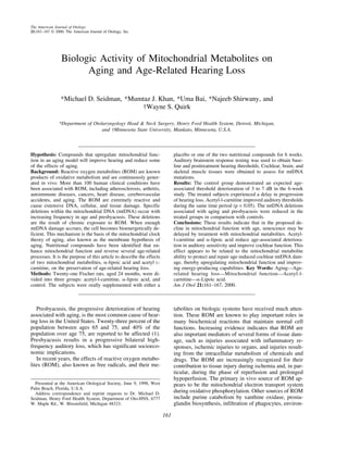
FIG. 2. Polymerase chain reaction. Gel A: Gel profile of ND1-16S rRNA amplified from tissue samples. Gel B: Gel profile of mtDNA4834
common aging deletion amplified from tissue samples. Lanes in both Gels A and B represent the following: 1. Control with no DNA. 2.
Brain. 3. Auditory nerve (control group). 4. Stria vascularis. 5. Brain. 6. Auditory nerve (group treated with -lipoic acid). 7. Stria vascularis.
8. Brain. 9. Auditory nerve (group treated with acetyl-l-carnitine). 10. Stria vascularis. 11. 100 bp ladder.
The process of aging is associated with many molecu- which are responsible for producing mitochondrial DNA
lar, biochemical, and physiologic changes, including in- damage, including mitochondrial DNA deletions. Spe-
creases in DNA damage, reduction in mitochondrial cific deletions are known to be directly proportional to
function, decreases in cellular water concentrations, aging, such as the common aging deletion in humans,
ionic changes, and decreased elasticity of cellular mem- which is 4977 bp in length (4834 bp in rats). When
branes. One contributing factor to this process is altered mtDNA del reach a certain level, the mitochondria be-
vascular characteristics, such as reduced flow and vas- come bioenergetically inefficient. Recent studies in our
cular plasticity as well as increased vascular permeability laboratory have shown that aged animals have reductions
(7). These age-related changes may result in reductions in auditory sensitivity with reductions in mitochondrial-
in oxygen and nutrient delivery and also waste elimina- associated function and increases in mtDNA deletions.
tion (2–5). These physiologic inefficiencies favor the These findings have been demonstrated with rat, mouse,
production of ROM. Additionally, there is support in the and human lymphocytes and human archival temporal
literature for age-associated reduction in endogenous en- bones (10,23).
zymes that protect from ROM damage, including super- An increasing body of evidence supports the role of
oxide dismutase, catalase, and glutathione (21,22). Col- supplementation with compounds that can upregulate
lectively, these changes enhance the generation of ROM, mitochondrial function. Specifically, we have called
these compounds mitochondrial metabolites. Mitochon-
drial metabolites have many diverse functions, as out-
lined earlier in this article. The primary mechanisms in-
volved in protection from aging appear to be multifac-
torial and would include their antioxidant properties,
enhanced ATP production, increased efficiency of CNS
receptors, and cell-membrane stability.
The apparent age-related deficits in mitochondrial
function could be slowed or reversed by ALCAR, a nor-
mal component of the inner mitochondrial membrane
that serves as a precursor of acetyl-CoA as well as the
neurotransmitter acetylcholine. ALCAR has been shown
FIG. 3. MtDNA deletion quantification: Ratio of deleted mtDNA to reverse the age-related decrease in the levels of the
to the total mtDNA represented after quantification with enzyme-
linked immunoassay (ELISA). Comparison of ratios in brain, au-
mitochondrial membrane phospholipid cardiolipin and
ditory nerve, and stria vascularis made between the control group the activity of the phosphate carrier in rat heart mito-
and the treated groups. chondria (24). Furthermore, the age-associated decrease
The American Journal of Otology, Vol. 21, No. 2, 2000
6. 166 M. D. SEIDMAN ET AL.
in mtDNA transcription is reversed rapidly by ALCAR. ergetic deficiency by supplementing with mitochondrial
Aged rat brain and heart are reported to possess a metabolites.
reduced steady-state level of mitochondrial transcripts
because of reduced RNA synthesis. Pretreatment of REFERENCES
senescent rats with ALCAR restores the levels of mito-
chondrial transcripts to adult levels in a time- and dose- 1. Gates GA, Caspery DM, Clark W, et al. Presbyacusis. Otolaryngol
dependent function (20). The effects of ALCAR on mi- Head Neck Surg 1986;100:266–71.
2. Kimura RS, Schuknecht HF. The ultra structure of the human stria
tochondrial function in the aging brain are supported by vascularis. Acta Otolaryngologica 1970;69:415–27.
its ability to create a shift in ATP production from gly- 3. Harkins SW. Effects of age and interstimulus interval on the brain
colytic pathways to the mitochondria (25). It is plausible stem auditory evoked potential. Int J Neurosci 1981;15:107–18.
that ALCAR can increase the metabolic efficiency of 4. Rosenhall U, Pederson K, Dotevall M. Effects of presbycusis and
compromised subpopulations of mitochondria and cause other types of hearing loss on auditory brain stem responses. Scand
Audiol 1986;15:179–85.
a redistribution of the metabolic workload, resulting in 5. Hoeffding V, Feldman ML. Changes with age in the morphology
increased cellular efficiency, and possibly decreases the of the cochlear nerve in rats: light microscopy. J Comp Neurol
rate at which mitochondria derived oxidants are pro- 1988;276:537–46.
duced. 6. Axelsson A. The cochlear blood vessels in guinea pigs of different
ages. Acta Otolaryngol (Stockh) 1971;72:172–81.
-Lipoic acid as an oral supplement is used for health 7. Seidman MD, Khan MJ, Dolan D, Quirk WS. Age-related differ-
benefits and has also been used as a therapeutic agent in ences in cochlear microcirculation and auditory brain stem re-
a variety of hepatic and neurologic disorders as well as in sponse. Arch Otolaryngol Head Neck Surg 1996;122:1221–6.
mushroom poisoning. Consideration has also been given 8. Hattori K, Tanaka M, Sugiyama S, et al. Age-dependent increase
in deleted mitochondrial DNA in the human heart: possible con-
to the use of -lipoic acid in the treatment of AIDS, tributory factor to presbycardia. Am Heart J 1991;121:1735–42.
atherosclerosis, and diabetes mellitus (17), in which de- 9. Wallace DC. Mitochondrial genetics: a paradigm for aging and
creased levels of -lipoic acid have been found. Inter- degenerative diseases? Science 1992;256:628–32.
estingly, a specific 10.4-kb mitochondrial DNA deletion 10. Seidman MD, Bai U, Khan MJ, et al. Association of mitochondrial
DNA deletions and cochlear pathology: a molecular biologic tool.
has been found in patients with diabetes mellitus and Laryngoscope 1996;106:777–83.
sensorineural hearing loss. Thus, it may also be supposed 11. Shigenaga MK, Hagen TM, Ames BN. Oxidative damage and
that patients with these disorders might benefit from a mitochondrial decay in aging. Proc Natl Acad Sci USA 1994;91:
diet supplemented with -lipoic acid. Dietary supple- 10771–8.
12. Sapolsky RM, Krey LC, McEwen BS. Prolonged glucocorticoid
mentation of -lipoic acid successfully prevents myocar- exposure reduces hippocampal neuron number: implications for
dial damage induced by ischemia-reperfusion injury aging. J Neurosci 1985;5:1222–7.
(26). Presently, its primary therapeutic use is for the 13. Imperato A, Ramacci TM, Angelucci L. Acetyl L-carnitine en-
treatment of diabetic polyneuropathy (17). hances acetylcholine release in the striatum and hippocampus of
Deafness has also been shown to have an association awake freely moving rats. Neurosci Lett 1989:107:251–5.
14. Ghirardi O, Milano S, Ramacci MT, Angelucci L. Effects of acetyl
with mtDNA del. It has been suggested that mitochon- L-carnitine chronic treatment on discrimination models in aged
drial diseases should be considered in cases of progres- rats. Physiol Behav 1988;44:769–73.
sive sensorineural hearing loss, especially with the co- 15. Caprioli A, Ghirardi O, Ramacci MT, Angelucci L. Age-dependent
existence of multisystem involvement (27,28). Other deficits in radial maze performance in the rat: effect of chronic
treatment with acetyl L-carnitine. Prog Neuropsychopharmacol
studies have identified mutations in the tRNA-Leu gene Biol Psychiatry 1990;14:359–69.
in a large pedigree with maternally inherited diabetes 16. Bast A, Haenen GRMM. Interlay between lipoic acid and gluta-
mellitus type II and deafness (29). Several human studies thione in the protection against microsomal lipid peroxidation. Bio-
have demonstrated an association of mitochondrial DNA chem Biophysiol Acta 1988;963:558–61.
17. Suzuki YJ, Aggarwal B, Packer L. Alpha-lipoic acid is a potent
mutations and presbyacusis, including a study showing inhibitor of NF-Kb activation in human T cells. Biochem Bio-
that older patients with presbyacusis had a higher fre- physiol Res Commun 1992;189:1709–15.
quency of the common aging deletion (4977 bp) than did 18. Kagan VE, Shvedova A, Serbinova E, et al. Dihydrolipoic acid: a
patients of similar age without presbyacusis (Veda N, et universal antioxidant both in the membrane and in the aqueous
phase. Biochem Pharmacol 1992;44:1637–49.
al. Unpublished data). More recently, it has been dem- 19. Devasagayam TP, Subramanian M, Pradhan DS, Sies H. Chem
onstrated by use of human archival temporal bones that Biol Int 1993;86:79–92.
14 of 17 aged patients with presbyacusis had the 4977-bp 20. Gadaleta MN, Petruzalla V, Daddabbo L, et al. Mitochondrial
deletion, compared with 8 of 17 control patients with DNA transcription and translation in aged rat: effect of acetyl-L-
normal hearing. carnitine. Ann NY Acad Sci 1994;717:150–60.
21. Semsei I, Szeszek F, Nagy I. In-vivo studies of the age dependent
In conclusion, it is becoming increasingly clear that decrease of the rates of total and mRNA synthesis in the brain
ROM production increases with aging. Concomitantly, cortex of rats. Arch Gerontol Geriatr 1982;1:29–42.
there is a significant reduction in the antioxidant protec- 22. Richardson A, Butler JA, Rutherford MS, et al. Effects of age and
tive enzymes. The combined effect leads to an excess of dietary restrictions on the expression of alpha-2 -microglobulin.
oxidative damage, which causes mitochondrial mutations J Biol Chem 1987;262:605–13.
23. Bai U, Seidman MD, Hinojosa R, Quirk WS. Mitochondrial DNA
with reductions in the capacity for OXPHOS, hence re- deletions associated with aging and possibly presbyacusis: a hu-
duced energy production. The current experiments pro- man archival temporal bone study. Am J Otol 1997;18:1–5.
vide a rationale to allow for improvement in this bioen- 24. Paradies G, Ruggiero FM, Petrosillo G, Gadaleta MN, Quaglieri-
The American Journal of Otology, Vol. 21, No. 2, 2000
7. MITOCHONDRIAL METABOLITES AND AGING 167
ello E. Carnitine-acylcarnitine translocase activity in cardiac mi- 27. Miyabayashi S, Hanamizu H, Endo H, et al. A new type of mito-
tochondria from aged rats: the effect of acetyl-L-carnitine. Mech chondrial DNA deletion in patients with encephalomyopathy. J
Aging Dev 1995;84:103–12. Inher Metab Dis 1991;14:805–12.
25. Aureli T, Miccheli A, Ricciolini R, et al. Aging brain: effect of 28. Ballinger SW, Shoffner JM, Hedaya EV, et al. Maternally trans-
acetyl L-carnitine treatment on rat brain energy and phospholipid mitted diabetes and deafness associated with a 10.4 kb mitochon-
metabolism. A study by 31P and 1H NMR spectroscopy. Brain Res drial DNA deletion. Natl Genet 1992;1:3–7.
1990;526:108–12. 29. Maassen JA, Van Den Ouweland JM, ’t Hart LM, Lemkes HH.
26. Sebinova E, Khwaja S, Reznick AZ, Packer L. Thioctic acid pro- Maternally inherited diabetes and deafness: a diabetic subtype as-
tects against ischemia-reperfusion injury in the isolated perfused sociated with a mutation in mitochondrial DNA. Hormone Met Res
Langendorff heart. Free Rad Res Commun 1994;17:49–58. 1997;29:50–55.
The American Journal of Otology, Vol. 21, No. 2, 2000