1. What is rheumatoid arthritis (RA)?
Rheumatoid arthritis (RA) is an autoimmune disease that causes chronic inflammation of the joints.
Autoimmune diseases are illnesses that occur when the body's tissues are mistakenly attacked by
their own immune system. The immune system contains a complex organization of cells and
antibodies designed normally to "seek and destroy" invaders of the body, particularly infections.
Patients with autoimmune diseases have antibodies in their blood that target their own body tissues,
where they can be associated with inflammation. While inflammation of the tissue around the joints
and inflammatory arthritis are characteristic features of rheumatoid arthritis, the disease can also
cause inflammation and injury in other organs in the body. Because it can affect multiple other
organs of the body, rheumatoid arthritis is referred to as a systemic illness and is sometimes called
rheumatoid disease. Rheumatoid arthritis that begins in people under 16 years of age is referred to
as juvenile rheumatoid arthritis.
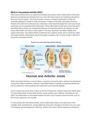
Picture of a joint with rheumatoid arthritis
While rheumatoid arthritis is a chronic illness, meaning it can last for years, patients may experience
long periods without symptoms. However, rheumatoid arthritis is typically a progressive illness that
has the potential to cause significant joint destruction and functional disability.
A joint is where two bones meet to allow movement of body parts. Arthritis means joint inflammation.
The joint inflammation of rheumatoid arthritis causes swelling, pain, stiffness, and redness in the
joints. The inflammation of rheumatoid disease can also occur in tissues around the joints, such as
the tendons, ligaments, and muscles.
In some people with rheumatoid arthritis, chronic inflammation leads to the destruction of the
cartilage, bone, and ligaments, causing deformity of the joints. Damage to the joints can occur early
in the disease and be progressive. Moreover, studies have shown that the progressive damage to
2. the joints does not necessarily correlate with the degree of pain, stiffness, or swelling present in the
joints.
Rheumatoid arthritis is a common rheumatic disease, affecting approximately 1.3 million people in
the United States, according to current census data. The disease is three times more common in
women as in men. It afflicts people of all races equally. The disease can begin at any age and even
affects children (juvenile rheumatoid arthritis), but it most often starts after 40 years of age and
before 60 years of age. Though uncommon, in some families, multiple members can be affected,
suggesting a genetic basis for the disorder.
What are causes and risk factors of rheumatoid arthritis?
The cause of rheumatoid arthritis is unknown. Even though infectious agents such as viruses,
bacteria, and fungi have long been suspected, none has been proven as the cause. The cause of
rheumatoid arthritis is a very active area of worldwide research. It is believed that the tendency to
develop rheumatoid arthritis may be genetically inherited (hereditary). Certain genes have been
identified that increase the risk for rheumatoid arthritis. It is also suspected that certain infections or
factors in the environment might trigger the activation of the immune system in susceptible
individuals. This misdirected immune system then attacks the body's own tissues. This leads to
inflammation in the joints and sometimes in various organs of the body, such as the lungs or eyes.
It is not known what triggers the onset of rheumatoid arthritis. Regardless of the exact trigger, the
result is an immune system that is geared up to promote inflammation in the joints and occasionally
other tissues of the body. Immune cells, called lymphocytes, are activated and chemical messengers
(cytokines, such as tumor necrosis factor/TNF, interleukin-1/IL-1, and interleukin-6/IL-6) are
expressed in the inflamed areas.
Environmental factors also seem to play some role in causing rheumatoid arthritis. For example,
scientists have reported that smoking tobacco, exposure to silica mineral, and chronic periodontal
disease all increase the risk of developing rheumatoid arthritis.
What are rheumatoid arthritis symptoms and signs?
RA symptoms come and go, depending on the degree of tissue inflammation. When body tissues
are inflamed, the disease is active. When tissue inflammation subsides, the disease is inactive (in
remission). Remissions can occur spontaneously or with treatment and can last weeks, months, or
years. During remissions, symptoms of the disease disappear, and people generally feel well. When
the disease becomes active again (relapse), symptoms return. The return of disease activity and
symptoms is called a flare. The course of rheumatoid arthritis varies among affected individuals, and
periods of flares and remissions are typical.
When the disease is active, RA symptoms can include fatigue, loss of energy, lack of appetite, low-
grade fever, muscle and joint aches, and stiffness. Muscle and joint stiffness are usually most
3. notable in the morning and after periods of inactivity. This is referred to as morning stiffness. Arthritis
is common during disease flares. Also during flares, joints frequently become warm, red, swollen,
painful, and tender. This occurs because the lining tissue of the joint (synovium) becomes inflamed,
resulting in the production of excessive joint fluid (synovial fluid). The synovium also thickens with
inflammation (synovitis).
Rheumatoid arthritis usually inflames multiple joints and affects both sides of the body. In its most
common form, therefore, it is referred to as a symmetric polyarthritis. Early symptoms may be subtle.
The small joints of both the hands and wrists are often involved. Early symptoms of rheumatoid
arthritis can be pain and prolonged stiffness of joints, particularly in the morning. Symptoms in the
hands with rheumatoid arthritis include difficulty with simple tasks of daily living, such as turning door
knobs and opening jars. The small joints of the feet are also commonly involved, which can lead to
painful walking, especially in the morning after arising from bed. Occasionally, only one joint is
inflamed. When only one joint is involved, the arthritis can mimic the joint inflammation caused by
other forms of arthritis, such as gout or joint infection. Chronic inflammation can cause damage to
body tissues, including cartilage and bone. This leads to a loss of cartilage and erosion
and weakness of the bones as well as the muscles, resulting in joint deformity, destruction, and loss
of function. Rarely, rheumatoid arthritis can even affect the joint that is responsible for the tightening
of our vocal cords to change the tone of our voice, the cricoarytenoid joint. When this joint is
inflamed, it can cause hoarseness of the voice. Symptoms in children with rheumatoid arthritis
include limping, irritability, crying, and poor appetite.
What are complications of rheumatoid disease?
Since rheumatoid arthritis is a systemic disease, its inflammation can affect organs and areas of the
body other than the joints. Inflammation of the glands of the eyes and mouth can cause dryness of
these areas and is referred to as Sjögren's syndrome. Dryness of the eyes can lead to corneal
abrasion. Inflammation of the white parts of the eyes (the sclerae) is referred to asscleritis and can
be very dangerous to the eye. Rheumatoid inflammation of the lung lining (pleuritis) causes chest
pain with deep breathing, shortness of breath, or coughing. The lung tissue itself can also become
inflamed and scarred, and sometimes nodules of inflammation (rheumatoid nodules) develop within
the lungs. Inflammation of the tissue (pericardium) surrounding the heart, called pericarditis, can
cause a chest pain that typically changes in intensity when lying down or leaning forward.
Rheumatoid arthritis is associated with an increased risk for heart attack. Rheumatoid disease can
reduce the number of red blood cells (anemia) and white blood cells. Decreased white cells can be
associated with an enlarged spleen(referred to as Felty's syndrome) and can increase the risk of
infections. The risk of lymph gland cancer (lymphoma) is higher in patients with rheumatoid arthritis,
especially in those with sustained active joint inflammation. Firm lumps or firm bumps under the skin
(subcutaneous nodules called rheumatoid nodules) can occur around the elbows and fingers where
there is frequent pressure. Even though these nodules usually do not cause symptoms, occasionally
they can become infected. Nerves can become pinched in the wrists to cause carpal tunnel
syndrome. A rare, serious complication, usually with longstanding rheumatoid disease, is blood
vessel inflammation (vasculitis). Vasculitis can impair blood supply to tissues and lead to tissue
4. death (necrosis). This is most often initially visible as tiny black areas around the nail beds or as leg
ulcers.
How do physicians diagnose rheumatoid arthritis?
There is no singular test for diagnosing rheumatoid arthritis. The diagnosis is based on the clinical
presentation. Ultimately, rheumatoid arthritis is diagnosed based on a combination of the
presentation of the joints involved, characteristic joint swelling and stiffness in the morning, the
presence of blood rheumatoid factor andcitrulline antibody, as well as findings of rheumatoid nodules
and radiographic changes (X-ray testing).
The first step in the diagnosis of rheumatoid arthritis is a meeting between the doctor and the
patient. The doctor reviews the history of symptoms, examines the joints for inflammation,
tenderness, swelling, and deformity, the skin for rheumatoid nodules (firm bumps under the skin,
most commonly over the elbows or fingers), and other parts of the body for inflammation. Certain
blood and X-ray tests are often obtained. The diagnosis will be based on the pattern of symptoms,
the distribution of the inflamed joints, and the blood and X-ray findings. Several visits may be
necessary before the doctor can be certain of the diagnosis. A doctor with special training in arthritis
and related diseases is called a rheumatologist.
It is the inflammation in the joint that helps to distinguish rheumatoid arthritis from common types of
arthritis that are not inflammatory, such as osteoarthritis or degenerative arthritis. The distribution of
joint inflammation is also important to the doctor in making a diagnosis. In rheumatoid arthritis, the
small joints of the hands and fingers, wrists, feet, and knees are typically inflamed in a symmetrical
distribution (affecting both sides of the body). When only one or two joints are inflamed, the
diagnosis of rheumatoid arthritis becomes more difficult. The doctor may then perform other tests to
exclude arthritis due to infection or gout. The detection of rheumatoid nodules (described above),
most often around the elbows and fingers, can suggest the diagnosis.
Abnormal antibodies can be found in the blood of people with rheumatoid arthritis with simple blood
testing. An antibody called "rheumatoid factor" (RF) can be found in 80% of patients with rheumatoid
arthritis. Patients who are felt to have rheumatoid arthritis and do not have positive rheumatoid factor
testing are referred to as having "seronegative rheumatoid arthritis." Citrulline antibody (also referred
to as anticitrulline antibody, anticyclic citrullinated peptide antibody, and anti-CCP antibody) is
present in 50%-75% people with rheumatoid arthritis. It is useful in the diagnosis of rheumatoid
arthritis when evaluating cases of unexplained joint inflammation. A test for citrulline antibodies is
especially helpful in looking for the cause of previously undiagnosed inflammatory arthritis when the
traditional blood test for rheumatoid arthritis, rheumatoid factor, is not present. Citrulline antibodies
have been felt to represent the earlier stages of rheumatoid arthritis in this setting. Citrulline
antibodies also have been associated with more aggressive forms of rheumatoid arthritis. Another
antibody called the "antinuclear antibody" (ANA) is also frequently found in people with rheumatoid
arthritis.
5. A blood test called the sedimentation rate (sed rate) is a crude measure of the inflammation of the
joints. The sed rate actually measures how fast red blood cells fall to the bottom of a test tube. The
sed rate is usually faster (high) during disease flares and slower (low) during remissions. Another
blood test that is used to measure the degree of inflammation present in the body is the C-reactive
protein. Blood testing may also reveal anemia, since anemia is common in rheumatoid arthritis,
particularly because of the chronic inflammation.
The rheumatoid factor, ANA, sed rate, and C-reactive protein tests can also be abnormal in other
systemic autoimmune and inflammatory conditions. Therefore, abnormalities in these blood tests
alone are not sufficient for a firm diagnosis of rheumatoid arthritis.
Joint X-rays may be normal or only demonstrate swelling of soft tissues early in the disease. As the
disease progresses, X-rays can reveal bony erosions typical of rheumatoid arthritis in the joints.
Joint X-rays can also be helpful in monitoring the progression of disease and joint damage over time.
Bone scanning, a procedure using a small amount of a radioactive substance, can also be used to
demonstrate the inflamed joints. MRI scanning can also be used to demonstrate joint damage.
The American College of Rheumatology has developed a system for classifying rheumatoid arthritis
that is primarily based upon the X-ray appearance of the joints. This system helps medical
professionals classify the severity of your rheumatoid arthritis with respect to cartilage, ligaments,
and bone.
Stage I
no damage seen on X-rays, although there may be signs of bone thinning
Stage II
on X-ray, evidence of bone thinning around a joint with or without slight bone damage
slight cartilage damage possible
joint mobility may be limited; no joint deformities observed
atrophy of adjacent muscle
abnormalities of soft tissue around joint possible
Stage III
on X-ray, evidence of cartilage and bone damage and bone thinning around the joint
joint deformity without permanent stiffening or fixation of the joint
6. extensive muscle atrophy
abnormalities of soft tissue around joint possible
Stage IV
on X-ray, evidence of cartilage and bone damage and osteoporosis around joint
joint deformity with permanent fixation of the joint (referred to as ankylosis)
extensive muscle atrophy
abnormalities of soft tissue around joint possible
Rheumatologists also classify the functional status of people with rheumatoid arthritis as follows:
Class I: completely able to perform usual activities of daily living
Class II: able to perform usual self-care and work activities but limited in activities outside of
work (such as playing sports, household chores)
Class III: able to perform usual self-care activities but limited in work and other activities
Class IV: limited in ability to perform usual self-care, work, and other activities
The doctor may elect to perform an office procedure called arthrocentesis. In this procedure, a sterile
needle and syringe are used to drain joint fluid out of the joint for study in the laboratory. Analysis of
the joint fluid in the laboratory can help to exclude other causes of arthritis, such as infection and
gout. Arthrocentesis can also be helpful in relieving joint swelling and pain. Occasionally, cortisone
medications are injected into the joint during the arthrocentesis in order to rapidly relieve joint
inflammation and further reduce symptoms.
What is the treatment for rheumatoid arthritis?
There is no known cure for rheumatoid arthritis. To date, the goal of treatment in rheumatoid arthritis
is to reduce joint inflammation and pain, maximize joint function, and prevent joint destruction and
deformity. Early medical intervention has been shown to be important in improving outcomes.
Aggressive management can improve function, stop damage to joints as monitored on X-rays, and
prevent work disability. Optimal RA treatment involves a combination of medications, rest, joint-
strengthening exercises, joint protection, and patient (and family) education. Treatment is
customized according to many factors such as disease activity, types of joints involved, general
health, age, and patient occupation. RA treatment is most successful when there is close
cooperation between the doctor, patient, and family members.
7. Two classes of medications are used in treating rheumatoid arthritis: fast-acting "first-line drugs" and
slow-acting "second-line drugs" (also referred to as disease-modifying antirheumatic drugs or
DMARDs). The first-line drugs, such as aspirin and cortisone (corticosteroids), are used to reduce
pain and inflammation. The slow-acting second-line drugs, such asmethotrexate (Rheumatrex,
Trexall, Otrexup), and hydroxychloroquine(Plaquenil), promote disease remission and prevent
progressive joint destruction.
The degree of destructiveness of rheumatoid arthritis varies among affected individuals. Those with
uncommon, less destructive forms of the disease or disease that has quieted after years of activity
("burned out" rheumatoid arthritis) can be managed with rest plus pain control and anti-inflammatory
medications alone. In general, however, function is improved and disability and joint destruction are
minimized when the condition is treated earlier with second-line drugs (disease-modifying
antirheumatic drugs), even within months of the diagnosis. Most people require more aggressive
second-line drugs, such as methotrexate, in addition to anti-inflammatory agents. Sometimes these
second-line drugs are used in combination. In some cases with severe joint deformity, surgery may
be necessary.
First-line" rheumatoid arthritis medications
Acetylsalicylate (aspirin), naproxen(Naprosyn), ibuprofen (Advil, Medipren, Motrin),
and etodolac (Lodine) are examples of nonsteroidal anti-inflammatory drugs (NSAIDs). NSAIDs are
medications that can reduce tissue inflammation, pain, and swelling. NSAIDs are not cortisone.
Aspirin, in doses higher than those used in treating headaches and fever, is an effective anti-
inflammatory medication for rheumatoid arthritis. Aspirin has been used for joint problems since the
ancient Egyptian era. The newer NSAIDs are just as effective as aspirin in reducing inflammation
and pain and require fewer dosages per day. Patients' responses to different NSAID medications
vary. Therefore, it is not unusual for a doctor to try several NSAID drugs in order to identify the most
effective agent with the fewest side effects. The most common side effects of aspirin and other
NSAIDs include stomach upset, abdominal pain, ulcers, and even gastrointestinal bleeding. In order
to reduce gastrointestinal side effects, NSAIDs are usually taken with food. Additional medications
are frequently recommended to protect the stomach from the ulcer effects of NSAIDs. These
medications include antacids, sucralfate(Carafate), proton-pump inhibitors (Prevacid and others),
and misoprostol (Cytotec). Newer NSAIDs include selective Cox-2 inhibitors, such
as celecoxib (Celebrex), which offer anti-inflammatory effects with less risk of stomach irritation and
bleeding risk.
Corticosteroid medications can be given orally or injected directly into tissues and joints. They are
more potent than NSAIDs in reducing inflammation and in restoring joint mobility and function.
Corticosteroids are useful for short periods during severe flares of disease activity or when the
disease is not responding to NSAIDs. However, corticosteroids can have serious side effects,
especially when given in high doses for long periods of time. These side effects include weight gain,
facial puffiness, thinning of the skin and bone, easy bruising,cataracts, risk of infection, muscle
wasting, and destruction of large joints, such as the hips. Corticosteroids also carry some increased
8. risk of contracting infections. These side effects can be partially avoided by gradually tapering the
doses of corticosteroids as the individual achieves improvement in symptoms. Abruptly discontinuing
corticosteroids can lead to flares of the disease or other symptoms of corticosteroid withdrawal and
is discouraged. Thinning of the bones due to osteoporosis may be prevented by calcium and vitamin
D supplements.
"Second-line" or "slow-acting" rheumatoid arthritis drugs (disease-
modifying anti-rheumatic drugs or DMARDs)
While "first-line" medications (NSAIDs and corticosteroids) can relieve joint inflammation and pain,
they do not necessarily prevent joint destruction or deformity. Rheumatoid arthritis requires
medications other than NSAIDs and corticosteroids to stop progressive damage to cartilage, bone,
and adjacent soft tissues. The RA medications needed for ideal management of the disease are also
referred to as disease-modifying antirheumatic drugs or DMARDs. They come in a variety of forms
and are listed below. These "second-line" or "slow-acting" medicines may take weeks to months to
become effective. They are used for long periods of time, even years, at varying doses. If maximally
effective, DMARDs can promote remission, thereby retarding the progression of joint destruction and
deformity. Sometimes a number of DMARD second-line medications are used together as
combination therapy. As with the first-line medications, the doctor may need to try different second-
line medications before treatment is optimal.
Research suggests that patients who respond to a DMARD with control of the rheumatoid disease
may actually decrease the known risk (small but real) of lymphoma (cancer of lymph nodes) that
exists from simply having rheumatoid arthritis. The various available DMARDs are reviewed next.
Hydroxychloroquine (Plaquenil) is related toquinine and has also been used in the treatment
of malaria. It is used over long periods for the treatment of rheumatoid arthritis. Possible side effects
include upset stomach, skin rashes, muscle weakness, and vision changes. Even though vision
changes are rare, people taking Plaquenil should be monitored by an eye doctor (ophthalmologist).
Sulfasalazine (Azulfidine) is an oral medication traditionally used in the treatment of mild to
moderately severe inflammatory bowel diseases, such asulcerative colitis and Crohn's colitis.
Azulfidine is used to treat rheumatoid arthritis in combination with anti-inflammatory medications.
Azulfidine is generally well tolerated. Common side effects include rash and upset stomach.
Because Azulfidine is made up of sulfa and salicylate compounds, it should be avoided by people
with known sulfa allergies.
Methotrexate has gained popularity among doctors as an initial second-line drug because of both its
effectiveness and relatively infrequent side effects. It also has an advantage in dose flexibility
(dosages can be adjusted according to needs). Methotrexate is an immunosuppressive drug. It can
affect the bone marrow and the liver, even rarely causing cirrhosis. All people taking methotrexate
9. require regular blood tests to monitor blood counts and liver function. Taking folic acid as a
supplement can reduce the risk of methotrexate side effects.
Gold salts have been used to treat rheumatoid arthritis throughout most of the past century. Gold
thioglucose (Solganal) and gold thiomalate(Myochrysine) are given by injection, initially on a weekly
basis, for months to years. Oral gold, auranofin (Ridaura), was introduced in the 1980s. Side effects
of gold (oral and injectable) include skin rash, mouth sores, kidney damage with leakage of protein in
the urine, and bone marrow damage with anemia and low white cell count. Those receiving gold
treatment are regularly monitored with blood and urine tests. Oral gold can causediarrhea. These
gold drugs have lost favor because of the availability of more effective treatments, particularly
methotrexate.
D-penicillamine (Depen, Cuprimine) can be helpful in selected cases of progressive forms of
rheumatoid arthritis. Side effects are similar to those of gold. They include fever, chills, mouth sores,
a metallic taste in the mouth, skin rash, kidney and bone marrow damage, stomach upset, and easy
bruising. People taking this medication require routine blood and urine tests. D-penicillamine can
rarely cause symptoms of other autoimmune diseases and is no longer commonly used for the
treatment of rheumatoid arthritis.
Immunosuppressive medicines are powerful medications that suppress the body's immune system.
A number of immunosuppressive drugs are used to treat rheumatoid arthritis. They include
methotrexate as described
above,azathioprine (Imuran), cyclophosphamide (Cytoxan), chlorambucil(Leukeran), and
cyclosporine (Sandimmune). Because of potentially serious side effects, immunosuppressive
medicines (other than methotrexate) are generally reserved for those who have very aggressive
disease or those with serious complications of rheumatoid inflammation, such as blood vessel
inflammation (vasculitis). The exception is methotrexate, which is not frequently associated with
serious side effects and can be carefully monitored with blood testing. Methotrexate has become a
preferred second-line medication as a result.
Immunosuppressive medications can depress bone-marrow function and cause anemia, a low white
cell count, and low platelet counts. A low white count can increase the risk of infections, while a low
platelet count can increase the risk of bleeding. Methotrexate rarely can lead to liver cirrhosis, as
described above, and allergic reactions in the lung. Cyclosporine can cause kidney damage
and high blood pressure. Because of potentially serious side effects, immunosuppressive
medications are used in low doses, usually in combination with anti-inflammatory agents.
Combinations of traditional DMARDs, including sulfasalazine, methotrexate, and
hydroxychloroquine, together have been shown by researchers to be another potent method of
stopping disease progression of rheumatoid arthritis.