This document summarizes a study that evaluated 25-hydroxy vitamin D (25(OH)D) as a potential novel biomarker for the diagnosis of hepatocellular carcinoma (HCC). The study measured serum 25(OH)D and alpha-fetoprotein (AFP) levels in 40 HCC patients and 40 healthy controls. Results found that 25(OH)D levels were significantly lower in HCC patients compared to controls. A cutoff level of <6.5 ng/ml for 25(OH)D diagnosed HCC with 88.5% sensitivity and 100% specificity. AFP had lower sensitivity of 55% at its standard cutoff of <37 ng/ml, though 100% specificity. The study concluded that
![Research In Cancer and Tumor 2014, 3(1): 19-25
DOI: 10.5923/j.rct.20140301.03
25-Hydroxy Vitamin D: Novel Biomarker for the
Diagnosis of Hepatocellular Carcinoma
Eman M. I. Youssef1,*
, Haneya A. A. Ali2
, Gehan H. Ewieda1
,
Wafaa Mohi El-deen Abd El-fatah1
, Nashwa El-Khouly3,4
1
Department of Medical Biochemistry, Faculty of Medicine (for girls), Al-Azhar University, Egypt
2
Department of Microbiology and Immunology, Faculty of Medicine (for girls), Al-Azhar University, Egypt
3
Department of Internal Medicine, Faculty of Medicine (for girls), Al-Azhar University, Cairo, Egypt
4
Department of Internal Medicine, Faculty of Medicine, Taibah University, KSA
Abstract Background: Serum concentration of 25-hydroxy vitamin D (25(OH) D) is the major circulating form of
vitamin D and the best indicator of assessment of vitamin D status. It reflects vitamin D obtained from food and supplements
and has a fairly long circulating half-life of 15 days. 25(OH)D levels do not indicate the amount of vitamin D stored in body
tissues. The non-classical actions of vitamin D which are anti-proliferation, pro-differentiation, pro-apoptosis,
anti-inflammation, and immune regulation, have received great attention during the past decade. Vitamin D insufficiency has
been associated with the occurrence of various types of cancer, the association between vitamin D status and hepatocellular
carcinoma (HCC) has not been well investigated. Objective: This study was designed to evaluate the role of serum vitamin D
concentration in the diagnosis of HCC and to consider its potential role as a novel marker for HCC compared with AFP.
Subjects and methods: This study was conducted on 80 Egyptian subjects and classified into two groups: HCC group (n =
40), in addition to forty age and sex matched healthy individuals as a control group. HCC was diagnosed histologically or by
imaging. Serum 25(OH)D and plasma AFP levels were quantitatively determined using ELISA technique for all participants
together with full clinical assessment, liver biochemical profile, viral markers, conventional ultrasound (US) and abdominal
triphasic CT scan Results: Serum 25(OH)D concentration was significantly lower in HCC patients (3ng/ml) than controls
(27.25 ng/ml) (P =0.001). Furthermore, the cutoff level of 25(OH) D for diagnosis of HCC in this study was <6.5 ng/ml, with
a sensitivity and specificity of 88.5% and 100% respectively. The diagnostic sensitivity of AFP at a cutoff of <37ng/ml was
55% and the specificity was 100%. Conclusions: Serum 25(OH)D could be a potential diagnostic marker for HCC and is
relatively comparative to AFP.
Keywords Hepatocellular carcinoma (HCC), 25-hydroxy vitamin D(25(OH) D), Alpha-fetoprotein (AFP)
1. Introduction
HCC is a highly aggressive carcinoma of the liver, the
fifth most common cancer worldwide, the fourth leading
cause of cancer related death [1, 2] and affects more than
half a million people annually [3]. In Egypt, chronic
hepatitis C virus (HCV) is the main cause of liver cirrhosis
and cancer [4]. Risk factors for HCC include; infection with
hepatitis B virus (HBV) [5] or HCV, alcoholic and
nonalcoholic cirrhosis and exposure to environmental toxins
such as aflatoxin [6].
HCC is usually diagnosed at late stages, and often has
very poor prognosis with limited treatment options [7, 8].
Early diagnosis of HCC is of great importance in order
to offer the possibility of curative treatment [9]. AFP and
* Corresponding author:
emyoussef4@gmail.com (Eman M. I. Youssef)
Published online at http://journal.sapub.org/rct
Copyright © 2014 Scientific & Academic Publishing. All Rights Reserved
ultrasonography are usually used as diagnostic tools. Serum
AFP is the only marker that has been widely used for
screening and diagnosis of HCC [10]. However,
development of false-negative rates with AFP was as high as
40% for patients with early hepatocellular carcinoma [11].
Thus the identification of novel biochemical markers for
HCC remains an important goal around the world [12].
Vitamin D is a prohormone with several active
metabolites and acts as a hormone. Vitamin D is a
fat-soluble vitamin whose principal biological action is to
regulate calcium, bone homeostasis [13] and cancer
prevention during the last decade due to presence of many
physiological processes, including cell growth and
differentiation, detoxification of xenobiotic and modulation
of adaptive and innate immunity [14, 15] and antineoplastic
effects throughanti-proliferative action and programmed cell
death [16]. Also, vitamin D can inhibit cancer cell invasion
by interfering with specific steps such as angiogenesis and
metastasis through decreasing the activity of certain
proteases which degrade extracellular matrix and basement](https://image.slidesharecdn.com/vitd-220327130033/85/vit-d-pdf-1-320.jpg)
![Research In Cancer and Tumor 2014, 3(1): 19-25
DOI: 10.5923/j.rct.20140301.03
25-Hydroxy Vitamin D: Novel Biomarker for the
Diagnosis of Hepatocellular Carcinoma
Eman M. I. Youssef1,*
, Haneya A. A. Ali2
, Gehan H. Ewieda1
,
Wafaa Mohi El-deen Abd El-fatah1
, Nashwa El-Khouly3,4
1
Department of Medical Biochemistry, Faculty of Medicine (for girls), Al-Azhar University, Egypt
2
Department of Microbiology and Immunology, Faculty of Medicine (for girls), Al-Azhar University, Egypt
3
Department of Internal Medicine, Faculty of Medicine (for girls), Al-Azhar University, Cairo, Egypt
4
Department of Internal Medicine, Faculty of Medicine, Taibah University, KSA
Abstract Background: Serum concentration of 25-hydroxy vitamin D (25(OH) D) is the major circulating form of
vitamin D and the best indicator of assessment of vitamin D status. It reflects vitamin D obtained from food and supplements
and has a fairly long circulating half-life of 15 days. 25(OH)D levels do not indicate the amount of vitamin D stored in body
tissues. The non-classical actions of vitamin D which are anti-proliferation, pro-differentiation, pro-apoptosis,
anti-inflammation, and immune regulation, have received great attention during the past decade. Vitamin D insufficiency has
been associated with the occurrence of various types of cancer, the association between vitamin D status and hepatocellular
carcinoma (HCC) has not been well investigated. Objective: This study was designed to evaluate the role of serum vitamin D
concentration in the diagnosis of HCC and to consider its potential role as a novel marker for HCC compared with AFP.
Subjects and methods: This study was conducted on 80 Egyptian subjects and classified into two groups: HCC group (n =
40), in addition to forty age and sex matched healthy individuals as a control group. HCC was diagnosed histologically or by
imaging. Serum 25(OH)D and plasma AFP levels were quantitatively determined using ELISA technique for all participants
together with full clinical assessment, liver biochemical profile, viral markers, conventional ultrasound (US) and abdominal
triphasic CT scan Results: Serum 25(OH)D concentration was significantly lower in HCC patients (3ng/ml) than controls
(27.25 ng/ml) (P =0.001). Furthermore, the cutoff level of 25(OH) D for diagnosis of HCC in this study was <6.5 ng/ml, with
a sensitivity and specificity of 88.5% and 100% respectively. The diagnostic sensitivity of AFP at a cutoff of <37ng/ml was
55% and the specificity was 100%. Conclusions: Serum 25(OH)D could be a potential diagnostic marker for HCC and is
relatively comparative to AFP.
Keywords Hepatocellular carcinoma (HCC), 25-hydroxy vitamin D(25(OH) D), Alpha-fetoprotein (AFP)
1. Introduction
HCC is a highly aggressive carcinoma of the liver, the
fifth most common cancer worldwide, the fourth leading
cause of cancer related death [1, 2] and affects more than
half a million people annually [3]. In Egypt, chronic
hepatitis C virus (HCV) is the main cause of liver cirrhosis
and cancer [4]. Risk factors for HCC include; infection with
hepatitis B virus (HBV) [5] or HCV, alcoholic and
nonalcoholic cirrhosis and exposure to environmental toxins
such as aflatoxin [6].
HCC is usually diagnosed at late stages, and often has
very poor prognosis with limited treatment options [7, 8].
Early diagnosis of HCC is of great importance in order
to offer the possibility of curative treatment [9]. AFP and
* Corresponding author:
emyoussef4@gmail.com (Eman M. I. Youssef)
Published online at http://journal.sapub.org/rct
Copyright © 2014 Scientific & Academic Publishing. All Rights Reserved
ultrasonography are usually used as diagnostic tools. Serum
AFP is the only marker that has been widely used for
screening and diagnosis of HCC [10]. However,
development of false-negative rates with AFP was as high as
40% for patients with early hepatocellular carcinoma [11].
Thus the identification of novel biochemical markers for
HCC remains an important goal around the world [12].
Vitamin D is a prohormone with several active
metabolites and acts as a hormone. Vitamin D is a
fat-soluble vitamin whose principal biological action is to
regulate calcium, bone homeostasis [13] and cancer
prevention during the last decade due to presence of many
physiological processes, including cell growth and
differentiation, detoxification of xenobiotic and modulation
of adaptive and innate immunity [14, 15] and antineoplastic
effects throughanti-proliferative action and programmed cell
death [16]. Also, vitamin D can inhibit cancer cell invasion
by interfering with specific steps such as angiogenesis and
metastasis through decreasing the activity of certain
proteases which degrade extracellular matrix and basement](https://image.slidesharecdn.com/vitd-220327130033/75/vit-d-pdf-1-2048.jpg)
![20 Eman M. I. Youssef et al.: 25-Hydroxy Vitamin D: Novel Biomarker
for the Diagnosis of Hepatocellular Carcinoma
membrane [17].
HCC is usually asymptomatic in the early stages and tends
to be invasive. Therefore, most patients are presented with an
incurable disease at the time of detection which makes its
early diagnosis critical for a good prognosis [18]. Hence, the
aim of this work was to focus on the potential role of serum
25(OH) D as a diagnostic biomarker for HCC and evaluation
of its diagnostic utilities in comparison to AFP.
2. Subjects and Methods
2.1. Subjects
This study was conducted on 40 HCC patientsrecruited
from the outpatient clinic of Internal Medicine Department
of Al-Zaharaa University Hospital, Cairo, Egypt, from
December 2012 till September 2013; in addition to 40
individualswith matched age and sex to the patients were
included into the study who served as a control group. All
studied individuals included in this study were evaluated by
history taking, thorough clinical examination, and laboratory
tests including liver functions (alanine aminotransferase
(ALT), aspartate aminotransferase (AST), totaland
directbilirubin, serum albumin (ALB), international
normalized ratio (INR), complete blood picture and viral
markers [hepatitis B surface antigen (HBs Ag) and hepatitis
C virus antibody (HCV Ab)]. In addition toradiological
investigations for diagnosis of HCC was based on one of the
following: Ultrasound and computed tomography (CT)] with
elevated AFP of more than 400 ng/ml, abdominal triphasic
computerized tomography (CT) scan and his to-pathological
assessment if needed. Other malignancies or autoimmune
diseases were excluded from the study. A verbal informed
consent was obtained from all subjects enrolled in the study.
Specimens Collection: Seven milliliters of venous blood
were obtained from each patient and control subjects with a
sterile syringe was drawn without anticoagulant, allowed to
stand for 2 hours at room temperature then centrifuged at
3000 rpm for 10 minute. Serum were collected and divided
into two aliquots, one for routine laboratory investigations
and the other aliquots were stored at -20 C till the time of use,
avoid repeated freezing/ thawing.
2.2. Methods
Measurement of serum AFP by ELISA Method
The Immunospec AFP is a quantitative solid phase
enzyme-linkedimmunosorbent assay (ELISA). AFP was
determined using ELISA kit (Catalog No.E1-205) supplied
from Immunospec Corporation, 7018 Owens mouth Ave.
Suite 103Canoga Park, CA, 91303, according to the
manufacturer’s instructions.
Detection of Human 25-OH-Vitamin-D by ELISA
Method
Enzyme immuno-assay for the quantitative determination
of serum 25-OH-vitamin-D concentrations in serum by
human enzyme-linked immunoassay 25-OH-Vitamin-D
(ELISA) kits (Catalog number: EA300/96, LOTVID1411)
[Digital-Life-Design (DLD)], Adlerhorst 15, D-22459
Hamburg, Germany, according to the manufacturer’s
instructions, this assay employs the calibrators and patient
samples are diluted with biotin-labeled 25-OH Vitamin D
and added to microplate wells coated with monoclonal
anti-25-OH Vitamin D antibodies. The standard curve from
which the 25-OH Vitamin D concentrations in the serum
samples can be taken is obtained by point-to-point plotting
of the extinction values measured for the 6 calibration sera
against the corresponding units (linear/log). Use “4-PL”
plotting for calculation of the standard curve by computer.
2.3. Statistical Analysis
Statistical presentation and analysis of the present study
was conducted using Chi-squared test, and that of 2
independent parametric data through Mann Whitney U test
by SPSS V 20. The following tests were applied: the X2
test
to compare qualitative variables between two independent
groups. ROCs were used to evaluate the diagnostic value of
25-OH Vitamin D and AFP and to identify the optimal
cut-off values. P-value was considered insignificant at the
level of > 0.05, significant at the level of ≤ 0.05 and highly
significant at the level of ≤ 0.01.
3. Results
The results obtained are presented in tables (1-4) and
figures (1, 2). HCC group, forty patients (33 males and 7
females) ranged from 28 to 70 years old with a mean age of
55.83 ± 8.20 years and forty healthy control subjects (36
males and 4 females) ranged from 47 to 58 years old with a
mean age of 52.70 ± 5.90. Characteristics of the patients with
HCC and healthy controls are presented in table (1). Detailed
laboratory data of all studied groups are shown in table (2).
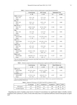
Table 1. Comparison of the demographic data between the studied groups
Control Group HCC Group Chi-square test
No. % No. % X2
/t p-value
Sex
Female 4 10.0% 7 17.5%
0.949 0.330
Male 36 90.0% 33 82.5%
Age (years)
Mean ± SD 52.70 ± 5.90 55.83 ± 8.20
1.429 0.160
Range 47 – 58 28 – 70](https://image.slidesharecdn.com/vitd-220327130033/85/vit-d-pdf-2-320.jpg)
![22 Eman M. I. Youssef et al.: 25-Hydroxy Vitamin D: Novel Biomarker
for the Diagnosis of Hepatocellular Carcinoma
Figure 1. Median of AFP and 25 (OH) D in two studied groups
Figure 2. ROC curve for AFP versus 25 (OH) D in diagnosis of HCC
Correlation of 25(OH) D concentration was made with
different laboratory parameters, it was found a significant
negative correlation between it and ALT, AST (-0.680,
P≤0.001) and (r = - 0.605, P ≤ 0.001) respectively) Also, a
negative correlation between it and AFP was found but not
significant (r = - 0.053, P = 0.843).
In receiver operator characteristic (ROC) curve analyses
to study the HCC diagnostic ability of both markers, 25 (OH)
D (ng/ml) at the best cut-off value (< 6.5ng/ml) had
sensitivity of 88.5%, specificity of 100%, positive predictive
value (PPV) of 100 %, and negative predictive value (NPV)
of 88.9% for detection of HCC patients (are aunder the curve
(AUC) = 0.93). The data of ROC analysis for AFB at a
cut-off value of <37 ng/ml had sensitivity of 55% and
specificity of100% (Table 4 and Figure 2).
4. Discussion
A number of studies investigated the relations between
serum vitamin D levels and various cancers such as cancer of
the colorectal [19], esophagus [20], lung and ovaries [21],
prostate [22] and pancreas [23], little epidemiologic data for](https://image.slidesharecdn.com/vitd-220327130033/85/vit-d-pdf-4-320.jpg)
![Research In Cancer and Tumor 2014, 3(1): 19-25 23
vitamin D and liver cancer are available, despite the
important role of the liver in metabolizing the circulating
form of vitamin D [24]. HCC is now a rather common
malignancy in Egypt which usually develops on top of liver
cirrhosis secondary to viral infection [25]. It is three times
more common in men than women, which could be
explained by differences in exposure to risk factors [26].The
results of the present study were higher than in a study done
in Egypt by Hammad et al. [27], who studied series of 1328
with HCC cases and they reported that HCC is significantly
higher in men than women (77.7 and 22.3%, respectively).
The present study demonstrated that 25 (OH) D levels
significantly differed between two groups, where median
serum 25 (OH) D levels was significantly lower in HCC
(3ng/ml) than healthy controls (27.25 ng/ml) (P ≤0.001). On
the contrary, there is no significant difference between both
groups as regard AFP. These results were similar to a recent
prospective cohort study done by Finkelmeier et al. [28].
25-hydroxyvitamin D3 (25(OH)D3) levels were
subsequently measured in two-hundred patients with HCC.
25(OH)D3 levels were compared to stages of cirrhosis and
HCC. The mean serum 25(OH)D3 concentration was 17 ± 13
ng/mL with a range of 1-72 ng/mL. 25(OH)D3 serum levels
negatively correlated with the stage of cirrhosis as well as
with stages of HCC. Patients with severe 25(OH)D3
deficiency had the highest mortality risk and high AFP levels
(>400 ng/mL). They concluded that 25(OH)D3 deficiency
was associated with advanced stages of HCC and it is a
prognostic indicator for a poor outcome. Also, our results
were in agreement with a study done by Hammad et al. [27],
who found a significant progressive decline in the active
vitamin D status was noted in all three clinical states
(patients with peri-hepatic fibrosis, hepatic cirrhosis, or
HCC) and these too were associated with progressive liver
disease.
Similarly, Lange et al. [26], who measured the largest
study of non-cirrhotic patients with chronic HCV infection.
Vitamin D status was assessed in a cohort of 468 patients.
The average 25(OH)D level was 17 ng/ml and 25% of the
patients had levels below 10 ng/ml. They concluded that the
prevalence of vitamin D deficiency was greater in patients
with more advanced fibrosis. Also they provided evidence
for a functionally relevant contribution of reduced 25(OH)D
serum levels to the risk of developing hepatitis C virus
(HCV)-related hepatocellular carcinoma (HCC).
Additionally, Mansoor et al. [29], they made a study of
prevalence and significance of vitamin D deficiency and
insufficiency among apparently healthy adults found high
prevalence of 25(OH) vitamin D deficiency 90% had low
serum 25(OH)D levels (69.9% were deficient and 21.1% had
insufficient levels of 25(OH)D among apparently healthy
adults, hospital staff and health care professionals. And also,
Fisher et al. [30] evaluated vitamin D levels in 100 patients
with liver disease, 51 with cirrhosis and 49 without cirrhosis,
including 38 patients with chronic hepatitis C. They
concluded that the prevalence of vitamin D deficiency was
significantly higher in cirrhotic than non-cirrhotic subjects
(86.3% versus 49.0%, p = 0.0001). Vitamin D insufficiency
has been previously linked to the development of HCC [31].
However, causal relationships remained mostly unclear
because the most studies were small or concentrated on the
assessment of 25(OH)D3 serum levels at the date of HCC,
which may result in false-positive associations due the
influence of impaired liver function on circulating
25(OH)D3 [32]. Another study done by Caputo et al. [33],
they found that the inhibitory effect of
1,25-dihydroxyvitamin D3 on growth of the human liver
cancer cell line which express functional receptors able to
specifically bind 1,25-(OH)(2)D3. The highest level of
functional receptor was found in the human liver cancer cell
line resulted from arrest in the G0/G1 phase of the cell cycle.
For the diagnosis of HCC in the current study, ROC curve
was made to detect a cutoff value for diagnosis of vitamin D
deficiency that was found to be equal or less than 6.5ng/ml.
Serum levels of 25-hydroxyvitamin D showed 88.5%
sensitivity, 100% specificity, 100% PPV and 88.9% NPV
and AUC 93%.In addition to, sensitivity and specificity of
AFP were 55% and 100%, respectively, at a cutoff value ≤
37 ng/ml. At this level, the AUC, PPV and NPV were59%,
100% and 69% respectively, which suggests that
determining 25 (OH) D levels might be superior to AFP
levels in diagnosis of HCC. These findings from our study in
accordance with previous study made by Falleti et al. [34],
found vitamin D deficiency in large proportion (55.8%) in
their patients with HCC where vitamin D deficiency was
diagnosed according to a cutoff value < 15ng/ml. They
concluded that the vitamin D receptor (VDR) genetic
polymorphisms are significantly associated with the
occurrence of HCC inpatients with liver cirrhosis. This
relationship is more specific for patients with an alcoholic
etiology. The most common used laboratory marker for
diagnosis is alpha-fetoprotein. However, it has a high rate of
false-negative and false positive results, as revealed by
Samir et al. [35], who stated that sensitivity and specificity
depend on the cut-off value chosen. In HCC-cirrhotic
patients, using a cut-off level of 20ng/mL, sensitivity is only
around 60% and specificity ranges from 80% to 94%. This
prompted the need of other reliable markers for this disease.
Also, Giannini et al. [11], who concluded that serum AFP
levels have no prognostic meaning in well-compensated
cirrhosis patients with single, small HCC.
5. Conclusions
In this study, it was concluded that the serum level of
25(OH) D is significantly lower in HCC patients in
comparison to controls. A receiver operating characteristics
(ROC) analysis suggests that serum 25 (OH) D could be a
biomarker for HCC.It is recommended to carry out further
studies with a larger sample size are mandatory to underline
the accuracy of our findings before their application at the
population level. Additional studies are necessary to gain
greater insight into the impact of 25(OH) D in the
pathogenesis of HCC and its potential usefulness in HCC](https://image.slidesharecdn.com/vitd-220327130033/85/vit-d-pdf-5-320.jpg)
![24 Eman M. I. Youssef et al.: 25-Hydroxy Vitamin D: Novel Biomarker
for the Diagnosis of Hepatocellular Carcinoma
patients. Further combining 25(OH) D with gene expression
or with other diagnostic markers might increase the
accuracy of HCC diagnosis. Studying of 25(OH) D in
different grades and various stages of HCC with follow up
of patients over several years will reveal the role of 25(OH)
D in predicting prognosis.Further clinical investigations on
the effect of vitamin D supplementation in treating CHC are
needed. Supplementation with daily vitamin D is
recommended to avoid hazards of vitamin D deficiency.
REFERENCES
[1] Ferlay J, Shin HR, Bray F, Forman D, Mathers C and Parkin
DM (2010): Estimates of worldwide burden of cancer in 2008:
GLOBOCAN 2008. Int J Cancer; 127: 2893–2917.
[2] Poustchi H., Sepanlou S.G and Esmaili S (2010):
Hepatocellular Carcinoma in the World and the Middle East.
Middle East Journal of Digestive Diseases; 2: 31-41.
[3] White DL and El-Serag HB (2011): Epidemiology of
Hepatocellular Carcinoma. In: Wang XW et al., eds.
Molecular Genetics of Liver Neoplasia, Cancer Genetics.
Springer Science and Business Media; LLC: 51-73.
[4] Lehan EM and Wilson ML (2009): Epidemiology of hepatitis
viruses among hepatocellular carcinoma cases and healthy
people in Egypt: A systematic review and meta-analysis. Int. J.
Cancer; 124: 690-697.
[5] Lin CL and Kao JH (2013): Risk stratification for hepatitis B
virus related hepatocellular carcinoma. J Gastroenterol
Hepatol; 28: 10-17.
[6] Bouchard MJ and Navas-Martin S (2011): Hepatitis B and C
virus hepatocarcinogenesis: Lessons learned and future
challenges. Cancer Letters; 305: 123–143.
[7] Frau M, Biasi F, Feo F and Pascale R (2010): Prognostic
markers and putative therapeutic targets for hepatocellular
carcinoma. Molecular Aspects of Medicine; 31: 179–193.
[8] Singhal A, Jayaraman M, Dhanasekaran DN and Kohli V
(2012): Molecular and serum markers in hepatocellular
carcinoma: predictive tools for prognosis and recurrence. Crit
Rev Oncol Hematol; 82 (2): 116-140.
[9] Wang HY and Ding J (2011): Molecular Signaling in
Hepatocellular Carcinoma. In: Wang XW et al., eds.
Molecular Genetics of Liver Neoplasia, Cancer Genetics.
Springer Science and Business Media; LLC: 373-396.
[10] Di Carlo I, Mannino M, Toro A, Ardiri A and Galia A (2012):
Persistent increase in alpha-fetoprotein level in a patient
without underlying liver disease who underwent curative
resection of hepatocellular carcinoma. A case report and
review of the literature. World J Surg Oncol., 6: 10:79.
[11] Giannini EG1, Marenco S, Borgonovo G, Savarino V,
Farinati F, Del Poggio P, Rapaccini GL, Anna Di Nolfo M,
Benvegnù L, Zoli M, Borzio F, Caturelli E,Chiaramonte M
and Trevisani F (2012): Alpha-fetoprotein has no prognostic
role in small hepatocellular carcinoma identified during
surveillance in compensated cirrhosis.Hepatology; 56(4):
1371-9.
[12] Bertino G, Ardiri A, Malaguarnera M, Malaguarnera G and
Bertino N (2012): Hepatocellualar carcinoma serum markers.
Semin Oncol., 39: 410-433.
[13] Stokes CS, Volmer DA, Grunhage Fand Lammert F (2013):
Vitamin D in chronic liver disease. Liver Int., 33:338-352.
[14] Kitson MT and Roberts SK (2012): D-livering the message:
the importance of vitamin D status in chronic liver disease. J
Hepatol., 57: 897-909.
[15] Chiang KC, Yeh CN, Chen MFand Chen TC (2011):
Hepatocellular carcinoma and vitamin D: a review. J
Gastroenterol Hepatol., 26: 1597–1603.
[16] Vanoirbeek E, Krishnan A, Eelen G, Verlinden L, Bouillon R,
Feldman D and Verstuyf A (2011) The anti-cancer and
anti-inflammatory actions of 1,25(OH)2D3. Best Pract Res
Clin Endocrinol Metab., 25: 593–604.
[17] Yuan-Ping Han, Ming Kong, Sujun Zheng, Yan Ren,
Longdon Zhu, Hongbo Shi and Zhongping Duan
(2013):Vitamin D in liver diseases: From mechanisms to
clinical trials .Journal of Gastroenterology and Hepatology
2013; 28 (Suppl. 1): 49–55.
[18] Forner A, Llovet JMand Bruix J (2012): Hepatocellular
carcinoma. Lancet., 379: 1245-1255.
[19] Jenab M, Bueno-de-Mesquita HB, Ferrari P, van Duijnhoven
FJ, Norat T, Pischon T, Jansen EH, Slimani N, Byrnes G,
Rinaldi S, Olsen A, Overvad K, Boutron-Ruault MC,
Clavel-Chapelon F, Morois S, Kaaks R, Linseisen J, Boeing
H, Bergmann MM, Trichopoulou A, Misirli G, Trichopoulos
D, Berrino F, Vineis P, Panico S, Palli D, Tumino R, Ros MM,
van Gils CH, Peeters PH, Brustad M, Lund E, Tormo MJ,
Ardanaz E, Rodrı´guez L, Sa´nchez MJ, Dorronsoro M,
Gonzalez CA, Hallmans G, Palmqvist R, Roddam A, Key TJ,
Khaw KT, Autier P, Hainaut Pand Riboli E (2010):
Association between pre-diagnostic circulating vitamin D
concentration and risk of colorectal cancer in European
populations: a nested case-control study. BMJ 340: b5500.
[20] Chen W, Dawsey SM, Qiao YL, Mark SD, Dong ZW, Taylor
PR, Zhao Pand Abnet CC (2007): Prospective study of serum
25(OH)-vitamin D concentration and risk of oesophageal and
gastric cancers. Br J Cancer; 97: 123–128.
[21] Abnet CC, Chen Y, Chow WH, Gao YT, Helzlsouer KJ, Le
Marchand L,McCullough ML, Shikany JM, Virtamo J,
Weinstein SJ, Xiang YB, Yu K, Zheng W, Albanes D, Arslan
AA, Campbell DS, Campbell PT, Hayes RB, Horst RL,
Kolonel LN, Nomura AM, Purdue MP, Snyder K and Shu XO
(2010): Circulating 25-hydroxyvitamin D and risk of
esophageal and gastric cancer: cohort consortium vitamin D
pooling project of rarer cancers. Am J Epidemiol., 172:
94–106.
[22] Ahn J, Peters U, Albanes D, Purdue MP, Abnet CC,
Chatterjee N, Horst RL, Hollis BW, Huang WY, Shikany JM
and Hayes RB (2008): Prostate, Lung, Colorectal, and
Ovarian Cancer Screening Trial Project Team Serum vitamin
D concentration and prostate cancer risk: a nested
case-control study. J Natl Cancer Inst., 100: 796–804.
[23] Stolzenberg-Solomon RZ, Jacobs EJ, Arslan AA, Qi D, Patel
AV, Helzlsouer KJ, Weinstein SJ, McCullough ML, Purdue
MP, Shu XO, Snyder K, Virtamo J, Wilkins LR, Yu K,
Zeleniuch-Jacquotte A, Zheng W, Albanes D, Cai Q, Harvey
C, Hayes R, Clipp S, Horst RL, Irish L, Koenig K, Le](https://image.slidesharecdn.com/vitd-220327130033/85/vit-d-pdf-6-320.jpg)
![Research In Cancer and Tumor 2014, 3(1): 19-25 25
Marchand L and Kolonel LN (2010): Circulating
25-hydroxyvitamin D and risk of pancreatic cancer: cohort
consortium vitamin D pooling project of rarer cancers. Am J
Epidemiol., 172: 81–93.
[24] Wang JB, Abnet CC, Chen W, Dawsey SM, Fan JH and Yin
LY (2013): Association between serum 25(OH) vitamin D,
incident liver cancer and chronic liver disease mortality in the
Linxian Nutrition Intervention Trials:a nested case-control
study. Br J Cancer; 109:1997-2004.
[25] Gomaa AI, Khan SA, Toledano MB, Waked I and
Taylor-Robinson SD (2009): Hepatocellular carcinoma:
Epidemiology, risk factors and pathogenesis. World J
Gastroenterol., 14(27): 4300-4308.
[26] Lange CM, Miki D, Ochi H, Nischalke H-D and Bojunga J
(2013): Genetic Analyses Reveal a Role for Vitamin D
Insufficiency in HCV-Associated Hepatocellular Carcinoma
Development. PLoS ONE; 8(5): e64053.
[27] Hammad LN, Abdelraouf SM, Hassanein FS, Mohamed WA
and Schaalan MF (2013): Circulating IL-6, IL-17 and vitamin
D in hepatocellular carcinoma: potential biomarkers for a
more favorable prognosis? J Immunotoxicol., 10: 380- 386.
[28] Finkelmeier F1, Kronenberger B, Köberle V, Bojunga J,
Zeuzem S, Trojan J, Piiper Aand Waidmann O (2014): Severe
25-hydroxyvitamin D deficiency identifies a poor prognosis
in patients with hepatocellular carcinoma - a prospective
cohort study. Aliment Pharmacol Ther.,9(10):1204-12.
[29] Mansoor S, Habib A , Ghani F , Fatmi Z, Badruddin S ,
Mansoor S, Siddiqui I and Jabbar A (2010): Prevalence and
significance of vitamin D deficiency and insufficiency among
apparently healthy adults. Clinical Biochemistry Journal;
CLB07508:5; 4c.
[30] Fisher L, Fisher A. Vitamin D and parathyroid hormone in
outpatients with noncholestatic chronic liver disease. Clin
Gastroenterol Hepatol. 2007; 5:513–520.
[31] Chiang KC, Yeh CN, Chen MF, Chen TC (2011)
Hepatocellular carcinoma and vitamin D: a review. J
Gastroenterol Hepatol 26: 1597–1603.
[32] Campbell FC, Xu H, El-Tanani M, Crowe P, Bingham V
(2010) The yin and yang of vitamin D receptor (VDR)
signaling in neoplastic progression: operational networks and
tissue-specific growth control. Biochem Pharmacol 79: 1–9
[33] Caputo A, Pourgholami MH, Akhter J and Morris DL (2003):
1, 25- Dihydroxyvitamin D(3) induced cell cycle arrest in the
human primary liver cancer cell line HepG2. Hepatol Res., 26:
34–39.
[34] Falleti E, Bitetto D, Fabris C, Cussigh A, Fontanini E,
Fornasiere E, Fumolo E, Bignulin S, Cmet S, Minisini R,
Pirisi M and Toniutto P (2010): Vitamin D receptor gene
polymorphisms and hepatocellular carcinoma in alcoholic
cirrhosis. World J Gastroenterol., 16(24): 3016–3024.
[35] Samir G, Stephen Band Jeffrey K (2003): Test characteristics
of alphafetoprotein for detecting hepatocellular carcinoma in
patients with hepatitis C: a systematic review and critical
analysis. Annals of Internal Medicine; 139(1): 46-50.](https://image.slidesharecdn.com/vitd-220327130033/85/vit-d-pdf-7-320.jpg)


![CTEV [ clubfoot] DR ARUN LAL ,DR MOHAMED ASHRAF travancore medical college k...](https://cdn.slidesharecdn.com/ss_thumbnails/ctevclubfootdrarunlaldrmohamedashraftravancoremedicalcollegekollamkeralaindia-260208063247-18fc466c-thumbnail.jpg?width=640&height=640&fit=bounds)


![PERI-PROSTHETIC FRACTURE NAIL-PLATE CONSTRUCT [NPC].pptx](https://cdn.slidesharecdn.com/ss_thumbnails/drarunkumardrmohamedashrafperiprostheticfrasturenail-plateconstructnpc-260209164459-7e9d15a1-thumbnail.jpg?width=640&height=640&fit=bounds)


