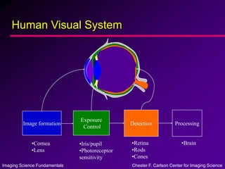
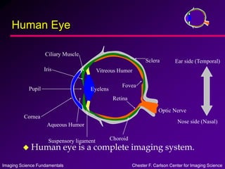
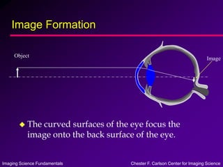
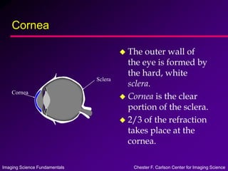
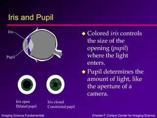
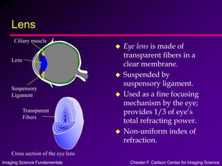
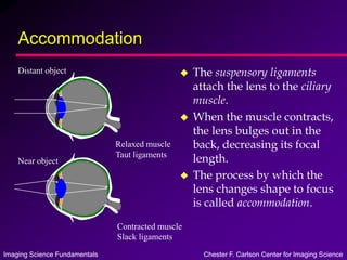
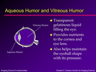
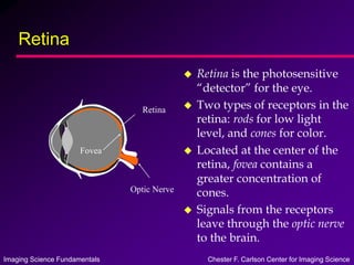
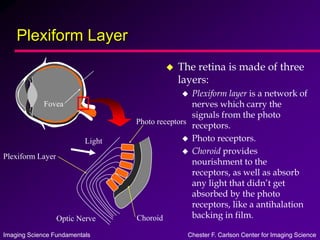
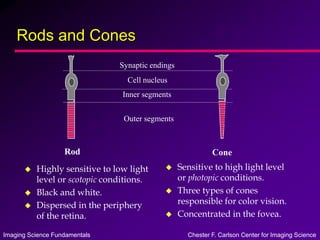
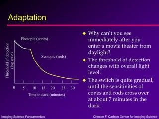
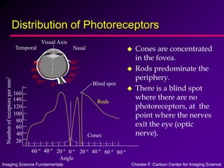
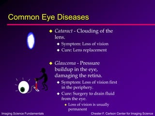
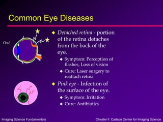
The human eye is a complex imaging system that forms images on the retina. Light enters through the cornea and lens, which focus the image onto the light-sensitive retina in the back of the eye. The retina contains two types of photoreceptor cells, rods and cones, which detect light and enable both low-light and color vision. Signals from the retina are transmitted to the brain via the optic nerve for image processing and interpretation. Several optical defects and diseases can impair vision, including myopia, hyperopia, astigmatism, cataracts, glaucoma, and retinal detachment.
Ophthalmoscopy
A clinical examination of the posterior segment by the means of an ophthalmoscope.
It is primarily done to assess the state of fundus and detect the opacities of ocular media.
Ophthalmoscope
An instrument that allows the ophthalmologist to look inside a person’s eye and see the details of the living retina
These lectures has prepared for postgraduate student (Ophthalmology) according to the curriculum of Bangladesh College of Physician and Surgeons (BCPS) and Bangabondhu Sheikh Mujib Medical University (BSMMU) Bangladesh
The human eye is one of the most valuable and sensitive sense organs in the human body. It enables us to see the wonderful world and colours around us.
Structure of eye:
The human eye has the following main parts:
Cornea: It is the transparent spherical membrane covering the front of the eye. Light enters the eye through this membrane.
Crystalline lens: The eyes lens is a convex lens made of a transparent, soft and flexible material like a jelly made of proteins.
Iris: It is a dark muscular diaphragm between the cornea and the lens. It controls the size of the pupil. It is the colour of the iris that we call as the colour of the eye.
Pupil: It is a small hole between the iris through which light enters the eye. In dim light, it opens up completely due to expansion of iris muscles, but in bright light it becomes very small due to contraction of iris muscles.
On this image, please label all marked structures Definedescribe t.pdflibowskymcinnisell69
On this image, please label all marked structures: Define/describe the following vocabulary
words pertaining to your Senses optic disc rightarrow Accommodation rightarrow Olfactory
Glomeruli rightarrow Photoreceptor Rods rightarrow Bone Deafness rightarrow Visual Acuity
rightarrow
Solution
1. Marked structures should be labeled as follows;
Left side marks (from top to bottom):
(a) Optic nerve
(b) Retina
(c) Choroid
(d) Sclera
Right side marks (from top to bottom)
(a) Lens
(b) Cornea
(c) Iris
(d) Ciliary body
2. Definitions of terminologies given;
(a) Optic Disc: Optic disc is the point of exit of ganglion cell axons from the eye. It is a blind
spot in the eye, as there are no rods and cone cells present on it. These ganglion cell axonsafter
leaving the eye form the optic nerve. Therefore, optic disc is also known as optic nerve head.
(b) Accomodation: Accomodation is considered as the reflex action of eye, i.e, the way one can
focus on nearby or distant object, thereby changing optical power frequently to maintain the
clear image of the object. In this manner, the shape of lens and and pupil size changes frequently.
(c) Olfactory glomeruli: Olfactory glomeruli is the point at which the information from the axons
of olfactory receptor neurons is received; the synapses are formed between the terminal end of
olfactory nerve with dendrites of mitral, tufted cells and periglomerular. It is a structure located
in the olfactory bulb in the mammalian central nervous system. Olfactory glomeruli are sperical
in shape.
(d) Photoreceptor rods: Phtotoreceptor rods are the cells in the retina of the eye that help in clear
sight in dim light. They are located towards the outer edges of the retina. They are present in
millions, and are more sensitive than cone cells.
(e) Bone deafness: Sond waves are conducted from outer ear, tympanic membrane to inner ear
through middle ear ossicles. In case of problems that lead blocking of conduction of sound waves
from outer to inner ear, leads to conductive hearing loss, also known as bone deafness. In such
cases, passage of sound across the ear drum and the inner ear is blocked.
(f) Visual Acuity: It refers to the \'clarity of vision\', which depends on both optical and neural
factors, such as focal capacity of retina, clarity of lens, interpretative functioning of the brain..
The Indian economy is classified into different sectors to simplify the analysis and understanding of economic activities. For Class 10, it's essential to grasp the sectors of the Indian economy, understand their characteristics, and recognize their importance. This guide will provide detailed notes on the Sectors of the Indian Economy Class 10, using specific long-tail keywords to enhance comprehension.
For more information, visit-www.vavaclasses.com
The Roman Empire A Historical Colossus.pdfkaushalkr1407
The Roman Empire, a vast and enduring power, stands as one of history's most remarkable civilizations, leaving an indelible imprint on the world. It emerged from the Roman Republic, transitioning into an imperial powerhouse under the leadership of Augustus Caesar in 27 BCE. This transformation marked the beginning of an era defined by unprecedented territorial expansion, architectural marvels, and profound cultural influence.
The empire's roots lie in the city of Rome, founded, according to legend, by Romulus in 753 BCE. Over centuries, Rome evolved from a small settlement to a formidable republic, characterized by a complex political system with elected officials and checks on power. However, internal strife, class conflicts, and military ambitions paved the way for the end of the Republic. Julius Caesar’s dictatorship and subsequent assassination in 44 BCE created a power vacuum, leading to a civil war. Octavian, later Augustus, emerged victorious, heralding the Roman Empire’s birth.
Under Augustus, the empire experienced the Pax Romana, a 200-year period of relative peace and stability. Augustus reformed the military, established efficient administrative systems, and initiated grand construction projects. The empire's borders expanded, encompassing territories from Britain to Egypt and from Spain to the Euphrates. Roman legions, renowned for their discipline and engineering prowess, secured and maintained these vast territories, building roads, fortifications, and cities that facilitated control and integration.
The Roman Empire’s society was hierarchical, with a rigid class system. At the top were the patricians, wealthy elites who held significant political power. Below them were the plebeians, free citizens with limited political influence, and the vast numbers of slaves who formed the backbone of the economy. The family unit was central, governed by the paterfamilias, the male head who held absolute authority.
Culturally, the Romans were eclectic, absorbing and adapting elements from the civilizations they encountered, particularly the Greeks. Roman art, literature, and philosophy reflected this synthesis, creating a rich cultural tapestry. Latin, the Roman language, became the lingua franca of the Western world, influencing numerous modern languages.
Roman architecture and engineering achievements were monumental. They perfected the arch, vault, and dome, constructing enduring structures like the Colosseum, Pantheon, and aqueducts. These engineering marvels not only showcased Roman ingenuity but also served practical purposes, from public entertainment to water supply.
Welcome to TechSoup New Member Orientation and Q&A (May 2024).pdfTechSoup
In this webinar you will learn how your organization can access TechSoup's wide variety of product discount and donation programs. From hardware to software, we'll give you a tour of the tools available to help your nonprofit with productivity, collaboration, financial management, donor tracking, security, and more.
Instructions for Submissions thorugh G- Classroom.pptxJheel Barad
This presentation provides a briefing on how to upload submissions and documents in Google Classroom. It was prepared as part of an orientation for new Sainik School in-service teacher trainees. As a training officer, my goal is to ensure that you are comfortable and proficient with this essential tool for managing assignments and fostering student engagement.
How to Make a Field invisible in Odoo 17Celine George
It is possible to hide or invisible some fields in odoo. Commonly using “invisible” attribute in the field definition to invisible the fields. This slide will show how to make a field invisible in odoo 17.
How to Split Bills in the Odoo 17 POS ModuleCeline George
Bills have a main role in point of sale procedure. It will help to track sales, handling payments and giving receipts to customers. Bill splitting also has an important role in POS. For example, If some friends come together for dinner and if they want to divide the bill then it is possible by POS bill splitting. This slide will show how to split bills in odoo 17 POS.
Ethnobotany and Ethnopharmacology:
Ethnobotany in herbal drug evaluation,
Impact of Ethnobotany in traditional medicine,
New development in herbals,
Bio-prospecting tools for drug discovery,
Role of Ethnopharmacology in drug evaluation,
Reverse Pharmacology.
Synthetic Fiber Construction in lab .pptxPavel ( NSTU)
Synthetic fiber production is a fascinating and complex field that blends chemistry, engineering, and environmental science. By understanding these aspects, students can gain a comprehensive view of synthetic fiber production, its impact on society and the environment, and the potential for future innovations. Synthetic fibers play a crucial role in modern society, impacting various aspects of daily life, industry, and the environment. ynthetic fibers are integral to modern life, offering a range of benefits from cost-effectiveness and versatility to innovative applications and performance characteristics. While they pose environmental challenges, ongoing research and development aim to create more sustainable and eco-friendly alternatives. Understanding the importance of synthetic fibers helps in appreciating their role in the economy, industry, and daily life, while also emphasizing the need for sustainable practices and innovation.
2. Imaging Science Fundamentals Chester F. Carlson Center for Imaging Science
In this section . . .
Anatomy of human eye
Image formation by human eye
Method of light detection
Retinal processing
Eye optical defects and diseases
3. Imaging Science Fundamentals Chester F. Carlson Center for Imaging Science
Human Visual System
Image formation
Exposure
Control
Detection Processing
•Cornea
•Lens
•Iris/pupil
•Photoreceptor
sensitivity
•Retina
•Rods
•Cones
•Brain
4. Imaging Science Fundamentals Chester F. Carlson Center for Imaging Science
Human Eye
Human eye is a complete imaging system.
Ear side (Temporal)
Nose side (Nasal)
Cornea
Aqueous Humor
Pupil
Iris
Ciliary Muscle
Sclera
Fovea
Retina
Optic Nerve
Vitreous Humor
Eyelens
Choroid
Suspensory ligament
5. Imaging Science Fundamentals Chester F. Carlson Center for Imaging Science
Image Formation
The curved surfaces of the eye focus the
image onto the back surface of the eye.
Object
Image
6. Imaging Science Fundamentals Chester F. Carlson Center for Imaging Science
Cornea
The outer wall of
the eye is formed by
the hard, white
sclera.
Cornea is the clear
portion of the sclera.
2/3 of the refraction
takes place at the
cornea.
Sclera
Cornea
7. Imaging Science Fundamentals Chester F. Carlson Center for Imaging Science
Iris and Pupil
Colored iris controls
the size of the
opening (pupil)
where the light
enters.
Pupil determines the
amount of light, like
the aperture of a
camera.
Iris
Iris open
Dilated pupil
Iris closed
Constricted pupil
Pupil
8. Imaging Science Fundamentals Chester F. Carlson Center for Imaging Science
Lens
Eye lens is made of
transparent fibers in a
clear membrane.
Suspended by
suspensory ligament.
Used as a fine focusing
mechanism by the eye;
provides 1/3 of eye’s
total refracting power.
Non-uniform index of
refraction.
Lens
Suspensory
Ligament
Ciliary muscle
Transparent
Fibers
Cross section of the eye lens
9. Imaging Science Fundamentals Chester F. Carlson Center for Imaging Science
Accommodation
The suspensory ligaments
attach the lens to the ciliary
muscle.
When the muscle contracts,
the lens bulges out in the
back, decreasing its focal
length.
The process by which the
lens changes shape to focus
is called accommodation.
Relaxed muscle
Taut ligaments
Distant object
Near object
Contracted muscle
Slack ligaments
10. Imaging Science Fundamentals Chester F. Carlson Center for Imaging Science
Aqueous Humor and Vitreous Humor
Transparent
gelatinous liquid
filling the eye.
Provides nutrients
to the cornea and
eye lens.
Also helps maintain
the eyeball shape
with its pressure.
Vitreous Humor
Aqueous Humor
11. Imaging Science Fundamentals Chester F. Carlson Center for Imaging Science
Retina
Retina is the photosensitive
“detector” for the eye.
Two types of receptors in the
retina: rods for low light
level, and cones for color.
Located at the center of the
retina, fovea contains a
greater concentration of
cones.
Signals from the receptors
leave through the optic nerve
to the brain.
Retina
Optic Nerve
Fovea
12. Imaging Science Fundamentals Chester F. Carlson Center for Imaging Science
Plexiform Layer
The retina is made of three
layers:
Plexiform layer is a network of
nerves which carry the
signals from the photo
receptors.
Photo receptors.
Choroid provides
nourishment to the
receptors, as well as absorb
any light that didn’t get
absorbed by the photo
receptors, like a antihalation
backing in film.
Fovea
Plexiform Layer
Photo receptors
Choroid
Optic Nerve
Light
13. Imaging Science Fundamentals Chester F. Carlson Center for Imaging Science
Rods and Cones
Highly sensitive to low light
level or scotopic conditions.
Black and white.
Dispersed in the periphery
of the retina.
Synaptic endings
Cell nucleus
Inner segments
Outer segments
Rod Cone
Sensitive to high light level
or photopic conditions.
Three types of cones
responsible for color vision.
Concentrated in the fovea.
14. Imaging Science Fundamentals Chester F. Carlson Center for Imaging Science
Adaptation
Why can’t you see
immediately after you
enter a movie theater from
daylight?
The threshold of detection
changes with overall light
level.
The switch is quite gradual,
until the sensitivities of
cones and rods cross over
at about 7 minutes in the
dark.
Photopic (cones)
Scotopic (rods)
0 5 10 15 20 25 30
Time in dark (minutes)
Threshold
of
detection
(log
scale)
15. Imaging Science Fundamentals Chester F. Carlson Center for Imaging Science
Distribution of Photoreceptors
Cones are concentrated
in the fovea.
Rods predominate the
periphery.
There is a blind spot
where there are no
photoreceptors, at the
point where the nerves
exit the eye (optic
nerve).
20 º
40 º
60 º
80 º
20 º
40 º
60 º
80º
0 º
Angle
0 º
20 º
40 º
60 º 80 º
60 º
40 º
20 º
20
40
60
80
100
120
140
160
Number
of
receptors
per
mm
2
Rods
Cones
Blind spot
Visual Axis
Nasal
Temporal
16. Imaging Science Fundamentals Chester F. Carlson Center for Imaging Science
Human Vision
Human Cone Response to Color
three cone types (S,I,L) correspond to B,G,R
400 460 530 650
600 700
500
Wavelength (nm)
Relative
response
Blue Cyan Green Red
490
I L
S
17. Imaging Science Fundamentals Chester F. Carlson Center for Imaging Science
Retina
The retina is made of network of nerve cells.
The network works together to reduce the amount of
information in a process called lateral inhibition.
To optic nerve
Light Cones
Rods
Bipolar
cells
Amicrine
cells
Ganglion
cells
Horizontal
cells
18. Imaging Science Fundamentals Chester F. Carlson Center for Imaging Science
Hermann Grid
Illustrates lateral inhibition.
19. Imaging Science Fundamentals Chester F. Carlson Center for Imaging Science
Hermann Grid
Point A looks darker because there are 4 inhibitory
inputs
Point B looks lighter because there are only 2 inhibitory
A B
20. Imaging Science Fundamentals Chester F. Carlson Center for Imaging Science
Mach Bands
Actual
brightness
Perceived
by you
21. Imaging Science Fundamentals Chester F. Carlson Center for Imaging Science
Eye Defects
Image focuses on the
retina for a normal eye.
Distant objects look
blurry for a myopic (near
sighted) eye.
Near objects look blurry
for a hyperopic (far
sighted) eye.
Normal
Myopic
Hyperopic
Object at infinity
Eyes at relax state.
22. Imaging Science Fundamentals Chester F. Carlson Center for Imaging Science
Myopia - Near sightedness
Distant objects look
blurry because the eye
cannot relax any farther
so that the image is
focused before the
retina.
Near object in focus
without accommodation.
Corrected with a
negative lens.
Myopic eye relaxed
Blurry
Myopia corrected
with a negative lens
Far object
The virtual image from the diverging lens appears to be closer.
Near object
Myopic eye relaxed
In focus
Far object
23. Imaging Science Fundamentals Chester F. Carlson Center for Imaging Science
Hyperopia - Far sightedness
Near objects look
blurry because the eye
cannot accommodate
enough for near
objects.
Far object in focus.
Corrected with a
positive lens.
Hyperopic eye
Partially accommodated
In focus
Hyperopia
corrected with a
positive lens
Far object
Light from the converging lens looks as though it is coming from the distance.
Hyperopic eye
Fully accommodated
Blurry
Near object
Near object
24. Imaging Science Fundamentals Chester F. Carlson Center for Imaging Science
Contact Lens
Contact lens is an
alternative to
corrective lenses.
Changes the
curvature of the
cornea by adhering
to the surface with
some fluid.
Cornea
Contact lens
Fluid
25. Imaging Science Fundamentals Chester F. Carlson Center for Imaging Science
Presbyopia - “Old eye”
Lens hardens with age.
Eye cannot adequately
accommodate near
objects.
Bifocals (lens with two
focal lengths) contains
a concave lens for
distance (if needed)
and a convex lens for
near objects.
Near objects magnified
Far objects
Concave for distance
correction (if needed)
Convex for near object
correction
26. Imaging Science Fundamentals Chester F. Carlson Center for Imaging Science
Astigmatism
The cornea is not
spherical; Focal length
different from one
plane to a
perpendicular plane.
F’ horizontal
F’ Vertical
Object
Image at F’ Vertical
Image at F’ Horizontal
Cornea
Direction of blur
27. Imaging Science Fundamentals Chester F. Carlson Center for Imaging Science
Astigmatism
Correction of astigmatism
is done through the use
of a cylindrical lens.
Cylindrical lens converge
rays in one plane but not
the perpendicular plane.
Cylindrical lens
Rays in the vertical
plane are undeviated
Rays in the
horizontal plane are
focused
28. Imaging Science Fundamentals Chester F. Carlson Center for Imaging Science
Common Eye Diseases
Cataract - Clouding of the
lens.
Symptom: Loss of vision
Cure: Lens replacement
Glaucoma - Pressure
buildup in the eye,
damaging the retina.
Symptom: Loss of vision first
in the periphery.
Cure: Surgery to drain fluid
from the eye.
Loss of vision is usually
permanent
29. Imaging Science Fundamentals Chester F. Carlson Center for Imaging Science
Common Eye Diseases
Detached retina - portion
of the retina detaches
from the back of the
eye.
Symptom: Perception of
flashes, Loss of vision
Cure: Laser surgery to
reattach retina
Pink eye - Infection of
the surface of the eye.
Symptom: Irritation
Cure: Antibiotics
Ow!
30. Imaging Science Fundamentals Chester F. Carlson Center for Imaging Science
Your eye care
Go see a doctor if you think
there is something wrong with
your eye-
Early detection is essential to
keeping damage low and
preventing permanent loss
of your vision.