1. Optimization of Adeno-Associated Viral Delivery into a
Machado-Joseph Disease/SCA3 Mouse Model
Tabtila Chowdhury & Hayley McLoughlin
Department of Neurology
ABSTRACT
Machado-Joseph Disease, also known as Spinocerebellar ataxia type 3 (SCA3), is a
dominantly inherited neurodegenerative disease that leads to loss of muscle control
and coordination in both the upper and lower body, dystonia, fasciculations of the
face or tongue, neuropathy, problems regarding the autonomic nervous system and
urination. SCA3 is caused by an expansion of the polyglutamine (CAG) region of the
human ataxin-3 gene, leading to mutant protein misfolding and cellular toxicity.
Previous research has described the use of viral delivery of gene knockdown. The
goal of our research is to identify the optimal viral serotype delivery routes for use in
SCA3 disease therapy. Specifically, we assessed two viral serotypes known for their
broad spread in the brain, adeno-associated viral serotypes 2/9 and 2/5 (AAV2/9 and
AAV2/5). Viruses expressing enhanced green fluorescent protein (eGFP) for
visualization of viral spread, were directly injected into mice brains and analyzed to
assess which viral serotype exhibited the greatest viral delivery to affected brain
regions in SCA3. Each viral serotype was delivered in equal amounts into the deep
cerebellar nuclei (DCN) in two ways: one injection into the central DCN or two
injections into the lateral/medial DCN region per hemisphere. Through our research
we wish to discover which delivery route and viral serotype shows most optimal
targeting of SCA3 affected regions, these regions are specifically the cerebellum,
brainstem, and the thalamus. Our study found that the AAV2/9 viral serotype with a
single central DCN injection per hemisphere provided us with the optimal viral
spread.
HYPOTHESIS
We hypothesize that injection of AAV2/9 into the lateral/medial deep cerebellar nuclei
region will produce a greater viral spread across the SCA3 affected brain region.
METHODS
• Each viral serotype, AAV2/9 and AAV2/5, was delivered in equal amounts into the
deep cerebellar nuclei (DCN) of wildtype mice brains in two ways: 1 injections into
the central DCN per hemisphere or 2 injections into the lateral/medial DCN region per
hemisphere.
• There was 4 replicates of each condition.
• The mice brains were perfused while in the body and then extracted and embedded in
sucrose solution to allow the brain to keep its composure.
• Brains were sagittally sectioned into 40 µm slices on a microtome.
• Sections placed on slides for imaging of viral spread under a fluorescent microscope.
CONCLUSIONS
We conclude that AAV2/9 injected into the central DCN has the greatest viral
spread across the SCA3 affected brain regions.
• AAV2/9 showed a more robust transduction to glial and neuronal cell types
in regions of SCA3 clinical importance. Relative to AAV2/5.
• A single injection into the central DCN may have had better spread than
two injections into the lateral/medial DCN region per hemisphere due to a
single central DCN injection saturating more DCN cells over the
lateral/medial injection.
FUTURE DIRECTIONS
• Our findings confirmed correct viral serotype and injection route for future
RNAi therapeutic studies.
• We will also explore additional injection routes for AAV2/9 including:
• Facial Vein
• Jugular Vein
• Tail Vein Delivery
BACKGROUND
• Adeno-associated viruses (AAVs) can
infect both dividing and dormant cells
without integrating into the species’
genome.
• AAV2/5 and AAV2/9 serotypes have
broad tropism in the brain, thus
allowing for greatest viral expression
in SCA3 affected regions.
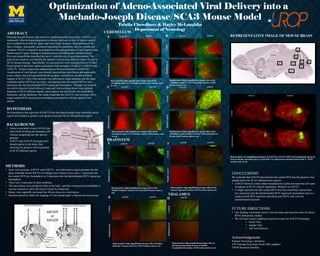
REPRESENTATIVE IMAGE OF MOUSE BRAIN
BRAINSTEM
THALAMUS
CEREBELLUM
Acknowledgments
Paulson Neurology Laboratory
T32 Training Neurology Grant (McLoughlin)
UROP Research Scholars
AAV2/5 AAV2/9
CentralMedial/Lateral
Representative (4x magnification) images of AAV2/5 or AAV2/9 eGFP viral transduction (green) in
8-week old mice microinjected at a central DCN or split between medial and lateral DCN. DAPI
nuclear stain in red.
A AAV2/5 AAV2/9
CentralMedial/Lateral
Representative high magnification images (20x) of the
cerebellum region. 8th Cerebellar Lobule. DAPI nuclear stain
in red.
B AAV2/5 AAV2/9
CentralMedial/Lateral
Representative high magnification images (20x) of the
cerebellum region. 4th & 5th Cerebellar Lobules. DAPI
nuclear stain in red.
C AAV2/5 AAV2/9
CentralMedial/Lateral
Representative high magnification images (20x) of the
cerebellum region. 3rd Cerebellar Lobule. DAPI nuclear stain
in red.
D AAV2/5 AAV2/9
CentralMedial/Lateral
Representative high magnification images (20x) of the
cerebellum region. Medial Cerebral Nuclei, Dorsolateral
Protub. DAPI nuclear stain in red.
E AAV2/5 AAV2/9
CentralMedial/Lateral
Representative high magnification images (20x) of the
Medial Vestibular Nucleus, Parvicell. DAPI nuclear stain in
red.
F AAV2/5 AAV2/9
CentralMedial/Lateral
Representative high magnification images (20x) of the
Lateral Tegmental Nucleus. DAPI nuclear stain in red.
G AAV2/5 AAV2/9
CentralMedial/Lateral
Representative high magnification images (20x) of Pontine
Reticular Nucleus, Oral Part. DAPI nuclear stain in red.
H AAV2/9
CentralMedial/Lateral
Representative high magnification images (20x) of
the Rostral Interstitial Nucleus of Medial
Longitudinal Fasciculus. DAPI nuclear stain in red.