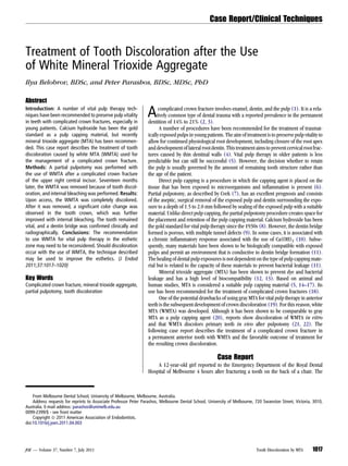
This case report describes the treatment of tooth discoloration caused by the use of white mineral trioxide aggregate (WMTA) during a partial pulpotomy procedure to treat a complicated crown fracture. Seventeen months after the initial procedure, the tooth exhibited discoloration from the WMTA. The WMTA was removed and internal bleaching was performed, which improved the color of the tooth. The removal of the discolored WMTA alone provided significant improvement in tooth color. This case highlights the potential for WMTA to cause tooth discoloration and suggests its use in the esthetic zone may need reconsideration.
Clinical Replacement Therapy and the Immediate Post-extraction Dental ImplantAbu-Hussein Muhamad
Immediate dental implants have greatly reduced the treatment time and the number of surgical intervene tions. Recently it has been noted that this treatment modality can be used in aesthetically demanding cases especially the anterior maxilla. The aim of this article is to describe a clinical case in which a fractured maxillary canine was replaced by an osseointegrated implant using a simplified technique in a patient who was a smoker and presented poor oral hygiene. The technique adopted permits a reduction of the number of implant components and consequently a lower cost of treatment, while at the same time maintaining acceptable aesthetic and functional outcomes.
The permanent teeth with open apex and large periapical lesion are diffcult to treat as a traditional root canal procedure, therefore calcium hydroxide place an important role in reducing the periapical infl ammation. Management of open apex can be done using mineral trioxide aggregate (MTA) which can be placed in apical 3-4 mm. The aim of this This case report describes the use of mineral trioxide aggregate (MTA) for management of a periapically compromised immature tooth.
DENTAL AVULSION- IMMEDIATE REPLANTATION: 8- YEAR FOLLOW UP CASEAbu-Hussein Muhamad
Avulsion of permanent front teeth is a rare accident , mostly affecting children between seven and nine year s of age.
Replanted and splinted, these teeth often develop inflammat ion, severe resorption or ankylosis affect ing alveolar bone
development and have to be extracted sooner or later . This repor t proposes a discussion on the var ious pecul iar ities of a
tooth avulsion case with immediate replantation, such as a long retent ion per iod, root canal fil ling with MTA, or thodontic
treatment.
Avulsion of permanent front teeth is a rare accident, mostly affecting children between seven and nine years of age. Replanted and splinted, these teeth often develop inflammation, severe resorption or ankylosis affecting alveolar bone development and have to be extracted sooner or later. This report proposes a discussion on the various peculiarities of a tooth avulsion case with immediate replantation, such as a long retention period, root canal filling with MTA, orthodontic treatment .
Surgery of Labially Impacted Canine & Orthodontic Management – A Case ReportAbu-Hussein Muhamad
Maxillary canines are one of the most common teeth that are impacted among patients seeking orthodontic treatment. Depending on the position of these impacted teeth, various surgical techniques have been employed for their exposure. His primary goal of surgical phase is to provide the means for correct position of orthodontic anchorage. Additionally, the technique used must ensure favorable tissue anatomy that will permit long-term maintenance of periodontal health. In the present case, a labially impacted maxillary left canine was surgically exposed using an apically positioned flap. Orthodontic extrusion was carried out further.
Single step apexification with mineral trioxide aggregateAbu-Hussein Muhamad
Abstract: The completion of root development and closure of the apex occurs up to 3 years after the eruption of the tooth. The treatment of pulpal injury during this period provides a significant challenge for the clinician. The most commonly advocated medicament is calcium hydroxide, although recently considerable interest has been expressed in the use of mineral trioxide aggregate (MTA). We report a case with MTA were used successfully for one step apexification in teeth with open apex. Key words: Immature teeth, one visit apexification, Mineral Trioxide Aggregate, monoblock, artificial barrier
Vital pulp therapy plays important role in preserving tooth and tooth vitality in both primary and permanent teeth.
Direct pulp capping, indirect pulp capping, pulpotomy has been covered in this presentation. All materials possibly useful in vital pulp therapy as well as recent advances have been included with all the evidences.
MANAGEMENT OF OPEN APEX IN PERMANENT TEETH WITH CALCIUM HYDROXIDE PASTEAbu-Hussein Muhamad
Calcium hydroxide is a multi purpose agent, and there have been an increasing number of indications for its use in endodontics. Some of its indications include inter-appointment intracanal medicaments, endodontic sealers, pulp capping agents, apexification, pulpotomy and weeping canals. The aim of this study was to report the 10 year follow-up data of an apexification treatment applied to a permanent incisor of a young patient treated with calcium hydroxide.
Management of Open Apex in Permanent Teeth with BiodentineAbu-Hussein Muhamad
Biodentine is new calcium silicate based cement that exhibits physical and chemical properties similar to those described for certain Portland cement derivatives. This article demonstrates the use of the newer material, Biodentine as an apical matrix barrier in root end apexification procedure. This case reports present apexification and successful healing with the use of Biodentine as an apical barrier matrix.
The Indian Dental Academy is the Leader in continuing dental education , training dentists in all aspects of dentistry and
offering a wide range of dental certified courses in different formats.
Osseo-integrated dental implants have been widely used for the rehabilitation of tooth loss. Although dental implants
are considered an available treatment in the paradigm shift from traditional dental therapies, such as fixed dental bridges and
removable dentures, the fundamental problems must be overcome prior to their clinical use in young patients who are still
undergoing jawbone growth. A bio-engineered functional bio-hybrid implant that is combined with adult-derived periodontal
tissue and attached with bone tissue can act as a substitute for cementum. This bio-hybrid implant was successfully engrafted
and it restored physiological function, including bone remodelling, regeneration and appropriate responsiveness to noxious
stimuli. Thus, this article reviews the functional bio-hybrid implant’s potential for clinical use as a next-generation dental
implant using adult-derived tissues.
Partial or complete edentulism has multiple implications in relation to function, esthetics and future rehabilitative treatment. This case report illustrates the management of a patient with extreme consequences of partial edentulism in the maxillary arch and total edentulism in the mandibular arch. The main clinical findings were unopposed remaining teeth, over eruption of the remaining teeth, loss of vertical dimension of occlusion, and significant disfigurement of the occlusal plane. Following the diagnostic procedure, a well-coordinated prosthodontic treatment involving liaison with other dental disciplines was indicated. The management involved an innovative combination of fixed and removable prostheses in conjunction with intentional root canal therapy of the remaining natural teeth. Series of provisional prostheses were applied to facilitate the transition to the final treatment.
Key-words: Edentulism, Vertical dimension, Provisional Restoration, Fixed and Removable prosthesis
Single-Step Apexification with Mineral Trioxide Aggregate (MTA) –Case ReportsAbu-Hussein Muhamad
The completion of root development and closure of the apex occurs up to 3 years after the eruption of
the tooth. The treatment of pulpal injury during this period provides a significant challenge for the clinician.
The most commonly advocated medicament is calcium hydroxide, although recently considerable interest has
been expressed in the use of mineral trioxide aggregate (MTA). We report a case with MTA were used
successfully for one step apexification in teeth with open apex
Autogenous tooth fragment reattachment ; a 12 years follow-upAbu-Hussein Muhamad
The fractures of the anterior teeth are a common form of dental trauma that mainly affects children and adolescents. One of the therapeutic options for managing coronal tooth fractures when the tooth fragment is available and there is no or minimal violation of the biological width is the Autogenous reattachment of the dental fragment.. Reattachment of fractured fragment can provide good and long lasting esthetics. This is a report of a 12 -year follow-up of a coronal fracture case successfully treated using tooth fragment reattachment.
Keywords: Composite resins, coronal fracture, fragment reattachment
Zirconium Dental Implants And Crown for Congenitally Missing Maxillary Latera...Abu-Hussein Muhamad
Zirconia implants were familiarized into dental implantology. Zirconia appears
to be an appropriate implant material due to its low plaque affinity, tooth like color, biocompatibility and mechanical properties. The following a case presentations will show how the acid-etched zirconia Implant can be used to functionally and aesthetically replace congenitally missing left lateral incisor tooth germ in the maxilla, and achieve optimal soft tissues and health.
Crestal bone loss around dental implants after implantation of Tricalcium pho...Dr. Anuj S Parihar
Background and Aims: Bone loss around dental implants is generally measured by monitoring changes in marginal bone level using radiographs. After the first year of implantation, an implant should have <0.2 mm annual loss of marginal bone level to satisfy the criteria of success. However, the success rate of dental implants depends on the amount of the crestal bone around the implants. The main aim of this study was to evaluate and compare the crestal bone loss around implants placed with particulate β‑Tricalcium Phosphate Bone Graft and platelet concentrates. Methods: 50 individuals received hundred dental implants. Each individual received one dental implant in the edentulous site filled with β‑Tricalcium Phosphate Bone Graft along (β‑TCP) with Platelet‑ Rich Plasma (PRP)
(Group A) and another in edentulous site filled only with β‑Tricalcium Phosphate Bone Graft (Group B) in the posterior edentulous region. All the 100 implants were prosthetically loaded after a healing period of three months. Crestal bone loss was measured on mesial, distal, buccal and lingual side of each implant using periapical radiographs 3 months, 6 months and 9 months after implant placement. Results: The average crestal bone loss 9 months after the implants placement in Group A and Group B was 2.75 mm and 2.23 mm respectively, the value being statistically significant (P < 0.05). In both Group A and Group B, the average crestal bone loss was maximum on the lingual side followed by buccal, distal and mesial sides. Conclusion: β‑TCP is a promising biomaterial for clinical
situations requiring bone augmentation. However, the addition of PRP results in decreased bone loss around the dental implants.
Modern Treatment for Congenitally Missing Teeth : A Multidisciplinary Appro...Abu-Hussein Muhamad
The maxillary lateral incisor is the second most common congenitally absent tooth. There are several treatment options for replacing the missing maxillary lateral incisor, including canine substitution, tooth-supported restoration, or single-tooth implant. Dental implants are an appropriate treatment option for replacing missing maxillary lateral incisor teeth in adolescents when their dental and skeletal development is complete. This case report presents the treatment of a patient with congenitally missing maxillary lateral incisors using dental implants. Finally, the importance of interdisciplinary team treatment planning is emphasized as a requirement for achieving optimal final esthetics
Prosthetic rehabilitation of a xerostomia patient with a mandibular split salivary reservoir denture
Xerostomia is defined as dryness of the mouth due to lack of normal secretions of saliva
Mucosal lubricants eg. wet mouth (ICPA Health Products Ltd),aqwet (Cipla Ltd)
Salivary stimulants eg. colgate dry mouth relief.
Healthy diet intake.
Salivary substitutes ( liquid or gel form ).
Chewing gums.
Proper water intake.
Salivary reservoirs
Similar to Treatment-of-tooth-discoloration-after-the-use-of-white-mineral-trioxide-aggregate (20)
1. Treatment of Tooth Discoloration after the Use
of White Mineral Trioxide Aggregate
Ilya Belobrov, BDSc, and Peter Parashos, BDSc, MDSc, PhD
Abstract
Introduction: A number of vital pulp therapy tech-
niques have been recommended to preserve pulp vitality
in teeth with complicated crown fractures, especially in
young patients. Calcium hydroxide has been the gold
standard as a pulp capping material, but recently
mineral trioxide aggregate (MTA) has been recommen-
ded. This case report describes the treatment of tooth
discoloration caused by white MTA (WMTA) used for
the management of a complicated crown fracture.
Methods: A partial pulpotomy was performed with
the use of WMTA after a complicated crown fracture
of the upper right central incisor. Seventeen months
later, the WMTA was removed because of tooth discol-
oration, and internal bleaching was performed. Results:
Upon access, the WMTA was completely discolored.
After it was removed, a significant color change was
observed in the tooth crown, which was further
improved with internal bleaching. The tooth remained
vital, and a dentin bridge was confirmed clinically and
radiographically. Conclusions: The recommendation
to use WMTA for vital pulp therapy in the esthetic
zone may need to be reconsidered. Should discoloration
occur with the use of WMTA, the technique described
may be used to improve the esthetics. (J Endod
2011;37:1017–1020)
Key Words
Complicated crown fracture, mineral trioxide aggregate,
partial pulpotomy, tooth discoloration
Acomplicated crown fracture involves enamel, dentin, and the pulp (1). It is a rela-
tively common type of dental trauma with a reported prevalence in the permanent
dentition of 14% to 21% (2, 3).
A number of procedures have been recommended for the treatment of traumat-
ically exposed pulps in young patients. The aim of treatment is to preserve pulp vitality to
allow for continued physiological root development, including closure of the root apex
anddevelopment oflateralrootdentin. Thistreatment aimsto prevent cervicalrootfrac-
tures caused by thin dentinal walls (4). Vital pulp therapy in older patients is less
predictable but can still be successful (5). However, the decision whether to retain
the pulp is usually governed by the amount of remaining tooth structure rather than
the age of the patient.
Direct pulp capping is a procedure in which the capping agent is placed on the
tissue that has been exposed to microorganisms and inflammation is present (6).
Partial pulpotomy, as described by Cvek (7), has an excellent prognosis and consists
of the aseptic, surgical removal of the exposed pulp and dentin surrounding the expo-
sure to a depth of 1.5 to 2.0 mm followed by sealing of the exposed pulp with a suitable
material. Unlike direct pulp capping, the partial pulpotomy procedure creates space for
the placement and retention of the pulp capping material. Calcium hydroxide has been
the gold standard for vital pulp therapy since the 1930s (8). However, the dentin bridge
formed is porous, with multiple tunnel defects (9). In some cases, it is associated with
a chronic inflammatory response associated with the use of Ca(OH)2 (10). Subse-
quently, many materials have been shown to be biologically compatible with exposed
pulps and permit an environment that is conductive to dentin bridge formation (11).
The healing of dental pulp exposures is not dependent on the type of pulp capping mate-
rial but is related to the capacity of these materials to prevent bacterial leakage (11).
Mineral trioxide aggregate (MTA) has been shown to prevent dye and bacterial
leakage and has a high level of biocompatibility (12, 13). Based on animal and
human studies, MTA is considered a suitable pulp capping material (5, 14–17). Its
use has been recommended for the treatment of complicated crown fractures (18).
One of the potential drawbacks of using gray MTA for vital pulp therapy in anterior
teeth is the subsequent development of crown discoloration (19). For this reason, white
MTA (WMTA) was developed. Although it has been shown to be comparable to gray
MTA as a pulp capping agent (20), reports show discoloration of WMTA in vitro
and that WMTA discolors primary teeth in vivo after pulpotomy (21, 22). The
following case report describes the treatment of a complicated crown fracture in
a permanent anterior tooth with WMTA and the favorable outcome of treatment for
the resulting crown discoloration.
Case Report
A 12-year-old girl reported to the Emergency Department of the Royal Dental
Hospital of Melbourne 4 hours after fracturing a tooth on the back of a chair. The
From Melbourne Dental School, University of Melbourne, Melbourne, Australia.
Address requests for reprints to Associate Professor Peter Parashos, Melbourne Dental School, University of Melbourne, 720 Swanston Street, Victoria, 3010,
Australia. E-mail address: parashos@unimelb.edu.au
0099-2399/$ - see front matter
Copyright ª 2011 American Association of Endodontists.
doi:10.1016/j.joen.2011.04.003
Case Report/Clinical Techniques
JOE — Volume 37, Number 7, July 2011 Tooth Discoloration by MTA 1017
2. intraoral examination revealed a complicated crown fracture of the
maxillary right central incisor (tooth #8). The fractured coronal frag-
ment had been placed into a glass of milk. All teeth in the anterior
segment of maxillary and mandibular arches responded to CO2 pulp
testing at the time of presentation. Radiographic examination
(Fig. 1A) confirmed the clinical findings that tooth #8 had substantial
coronal tooth structure remaining for restoration by reattachment of
the coronal fragment or by a direct bonded resin composite restoration.
After the administration of local anesthesia, a partial pulpotomy
was performed under rubber dam on tooth #8 using a high-speed
size 2 round diamond bur with copious water coolant. The coronal
pulp stump was rinsed with 1% sodium hypochlorite for 2 minutes until
hemostasis was achieved. ProRootWMTA (DentsplyTulsaDental,Tulsa,
OK) was placed onto the pulp followed by Vitrebond (3M Dental Prod-
ucts Division, St Paul, MN), etching, Adper Single Bond Plus Adhesive
(3M Espe, St Paul, MN), and flowable composite (Revolution Formula
2; Kerr, Orange, CA) to bond the coronal tooth fragment. The enamel
surface at the fracture line was beveled, and a direct resin composite
restoration (Tetric N-Ceram; Ivoclar Vivadent, Schaan, Liechtenstein)
was placed to reinforce the two fragments (Fig. 1B). The patient was
subsequently reviewed in the Endodontics Unit of the Royal Dental
Hospital of Melbourne and did not report any immediate postoperative
discomfort.
The patient was reviewed at 1, 5, and 17 months. Throughout the
follow-up period, the maxillary anterior teeth remained asymptomatic
and responsive to CO2 pulp testing. At 1 month, a slight gray discolor-
ation couldbe seen justapical to the fracture line (Fig. 2A). At 5 months,
there was further evidence of crown discoloration (Fig. 2B).
At 17 months, the patient and her parents were concerned with the
crown discoloration (Fig. 2C). The tooth responded to CO2 pulp testing,
and a hard tissue bridge and continued apical root maturation were
Figure 1. (A) A periapical radiograph of tooth #8 upon presentation. (B) The
immediate postoperative radiograph showing the placement of WMTA and the
reattachment of the coronal fragment. (C) A radiograph at 17 months showing
the formation of hard-tissue bridge and root maturation. (D) A radiograph
post-bleaching and restoration of the access cavity.
Figure 2. (A) An intraoral photograph at the 1-month follow-up. Note the slight gray discoloration apical to the fracture line. (B) Further crown discoloration at
the 5-month recall. (C) Distinctly noticeable crown discoloration 17 months after partial pulpotomy with WMTA. (D) The removal of discolored WMTA.
Case Report/Clinical Techniques
1018 Belobrov and Parashos JOE — Volume 37, Number 7, July 2011
3. radiographically evident (Fig. 1C). After discussion with the parents and
the patient concerning internal bleaching, a local anesthetic was admin-
istered. Under a rubber dam, the pulp chamber was reaccessed. Using
the operating microscope, a high-speed size 2 round stainless steel bur
with water coolant was used tocompletelyremovethe discolored WMTA
(Fig. 2D), exposing the hard-tissue bridge (Fig. 3A). The removal of
WMTA was attempted using ultrasonics but was proved unsuccessful
because of cutting inefficiency; therefore, the use of a high-speed bur
with water cooling under a microscope was used, which was a quicker
and just as conservative an option.
Considerable improvement in the color of the tooth was seen
immediately after the removal of the discolored WMTA (Fig. 3B). At
the same appointment, a mixture of sodium perborate and saline was
placed in the access cavity to internally bleach the crown. No adverse
pulpal effects were anticipated because of the lack of porosity in the
dentine bridge formed after the use of MTA (10). Then, the access
opening was sealed with Cavit W (3M, Dental Products Division, St
Paul, MN).
The patient returned 1 week later asymptomatic, and the tooth was
still responding to CO2 pulp testing. Under rubber dam isolation, the
tooth was reaccessed, and the internal bleaching paste was removed
(Fig. 3C). The access cavity was subsequently restored with a polycar-
boxylate cement (Durelon, 3M ESPE) and resin composite restoration
(Tetric N-Ceram). The labial composite was also replaced with a trans-
lucent (4 Seasons medium enamel value, Ivoclar Vivadent) composite
resin restoration to match the patients improved esthetics (Figs. 1D and
3D). Because there was no noticeable improvement in the patient’s oral
hygiene, further oral hygiene instructions were also provided. The
patient was recalled 1 month later, and there was no change in the color
of the crown; the tooth continued to respond to CO2 pulp testing.
Discussion
Thiscaseconfirmedthatset WMTAwasresponsible forthe gradual
discoloration of tooth #8. The intraoral photograph in Figure 2D clearly
distinguishes between the discolored WMTA and the surrounding
dentin. The careful removal of WMTA under the operating microscope
ensured that no additional tooth structure was removed. Although
internal bleaching was used in this case, most of the discoloration
was within the WMTA (Figs. 2D and 3A) and not in the dentin. Consider-
able improvement in crown color was achieved after the removal of the
WMTA (Figs. 2C and 3B). Only a minor improvement was seen in the
color of the dentin internally (Fig. 3A and C) after internal bleaching.
Therefore, internal bleaching may not be required.
Although the biologic and the esthetic goals of treatment were
achieved in this case, it may be reasonable to reconsider using
WMTA for vital pulp therapy in the esthetic zone. Should discoloration
occur after the use of WMTA in the esthetic zone, careful removal of set
WMTA may be attempted after confirmation of dentin bridge formation.
Reattachment of the coronal fragment provides several advantages
including exact restoration of crown morphology, reduced chair time,
excellent esthetics, similar wear rate to opposing teeth, and a positive
emotional response from the patient (23). When simple reattachment
(ie, no preparation) is used, the use of resin composite in the adhesive
Figure 3. (A) All of the discolored WMTA was removed until a hard-tissue bridge was reached. (B) A noticeable improvement in the color of the crown could be
seen immediately after the removal of discolored WMTA. (C) The dentin color after 1 week of internal bleaching. (D) The excellent esthetic result after the removal
of discolored WMTA and internal bleaching.
Case Report/Clinical Techniques
JOE — Volume 37, Number 7, July 2011 Tooth Discoloration by MTA 1019
4. interface significantly increases the fracture strength compared with
bonding agents only (24). Beveling of the margins further increases
the fracture resistance and has been shown to have a better retention
and esthetic prognosis than direct composite resin restoration (25).
MTA rather than calcium hydroxide was used for pulp capping
after the partial pulpotomy. The main advantages of MTA are that it
provides a good protective barrier against bacterial penetration (26)
and is biocompatible (27). Furthermore, the presence of blood has
little impact on the degree of leakage (12). It has also been shown
that the bioactive property of MTA is superior (when compared with
Ca[OH]2 and other materials) in dentin bridge formation after pulp
capping and pulpotomies (10, 17). Hence, the use of MTA has been
recommended as the material of choice for vital pulp therapy (28, 29).
Because of the potential staining of tooth structure in an area in
which esthetics is important, a white powder form of MTA was manu-
factured (30). However, in vitro studies have reported a gray discolor-
ation of WMTA used in single-rooted human teeth and plastic blocks
after the setting reaction (21, 31). In both studies, the material was
affected in depth, which led the authors to speculate that a chemical
reaction was responsible for the discoloration.
Histologically, MTA forms a superior dentin bridge with less
underlying inflammation than Ca(OH)2 (10, 14). It also has greater
success for direct pulp capping carious exposures when there is
bacterial presence (32). However, no clinical study has compared
the use of MTA and Ca(OH)2 after traumatic exposure of the pulp.
Cvek (7) reported 96% success for partial pulpotomies using
Ca(OH)2. Therefore, the use of WMTA in the esthetic zone after trau-
matic exposure of the pulp, with its potential for discoloration of tooth
structure, must be questioned.
Acknowledgments
The authors deny any conflicts of interest related to this study.
References
1. Glendor U, Marcenes W, Jo A. Classification, epidemiology and Etiology. In:
Andreasen J, Andreasen F, Andersson L, eds. Textbook and Color Atlas of Traumatic
Injuries to the Teeth. 4th ed. Oxford: Blackwell Munksgaard; 2007:217–54.
2. Davis GT, Knott SC. Dental trauma in Australia. Aust Dent J 1984;29:217–21.
3. Onetto JE, Flores MT, Garbarino ML. Dental trauma in children and adolescents in
Valparaiso, Chile. Endod Dent Traumatol 1994;10:223–7.
4. Cvek M. Prognosis of luxated non-vital maxillary incisors treated with calcium
hydroxide and filled with gutta-percha. A retrospective clinical study. Endod Dent
Traumatol 1992;8:45–55.
5. Bogen G, Kim JS, Bakland LK. Direct pulp capping with mineral trioxide aggregate:
an observational study. J Am Dent Assoc 2008;139:305–15.
6. Cvek M, Cleaton-Jones PE, Austin JC, Andreasen JO. Pulp reactions to exposure after
experimental crown fractures or grinding in adult monkeys. J Endod 1982;8:391–7.
7. Cvek M. A clinical report on partial pulpotomy and capping with calcium hydroxide
in permanent incisors with complicated crown fracture. J Endod 1978;4:232–7.
8. Cox CF, Hafez AA. Biocomposition and reaction of pulp tissues to restorative treat-
ments. Dent Clin North Am 2001;45:31–48.
9. Cox CF, Subay RK, Suzuki S, Suzuki SH, Ostro E. Biocompatibility of various dental
materials: pulp healing with a surface seal. Int J Periodontics Restorative Dent 1996;
16:240–51.
10. Nair PN, Duncan HF, Pitt Ford TR, Luder HU. Histological, ultrastructural and quan-
titative investigations on the response of healthy human pulps to experimental
capping with mineral trioxide aggregate: a randomized controlled trial. Int Endod
J 2008;41:128–50.
11. Cox CF, Keall CL, Keall HJ, Ostro E, Bergenholtz G. Biocompatibility of surface-sealed
dental materials against exposed pulps. J Prosthet Dent 1987;57:1–8.
12. Torabinejad M, Higa RK, McKendry DJ, Pitt Ford TR. Dye leakage of four root end
filling materials: effects of blood contamination. J Endod 1994;20:159–63.
13. Torabinejad M, Parirokh M. Mineral trioxide aggregate: a comprehensive literature
review—part II: leakage and biocompatibility investigations. J Endod 2010;36:
190–202.
14. Aeinehchi M, Eslami B, Ghanbariha M, Saffar AS. Mineral trioxide aggregate (MTA)
and calcium hydroxide as pulp-capping agents in human teeth: a preliminary report.
Int Endod J 2003;36:225–31.
15. Ramachandran Nair PNR, Duncan HF, Pitt Ford TRP, Luder HU. Histological, ultra-
structural and quantitative investigations on the response of healthy human pulps to
experimental capping with mineral trioxide aggregate: a randomized controlled
trial. Int Endod J 2008;41:128–50.
16. Accorinte MLR, Loguercio AD, Reis A, et al. Evaluation of two mineral trioxide aggre-
gate compounds as pulp-capping agents in human teeth. Int Endod J 2009;42:
122–8.
17. Pitt Ford T, Torabinejad M, Abedi H, Bakland L, Kariyawasam S. Using mineral
trioxide aggregate as a pulp-capping material. J Am Dent Assoc 1996;127:1491.
18. Flores MT, Andersson L, Andreasen JO, et al. Guidelines for the management of trau-
matic dental injuries. I. Fractures and luxations of permanent teeth. Dent Traumatol
2007;23:66–71.
19. Karabucak B, Li D, Lim J, Iqbal M. Vital pulp therapy with mineral trioxide aggregate.
Dent Traumatol 2005;21:240–3.
20. Parirokh M, Asgary S, Eghbal MJ, et al. A comparative study of white and grey
mineral trioxide aggregate as pulp capping agents in dog’s teeth. Dent Traumatol
2005;21:150–4.
21. Boutsioukis C, Noula G, Lambrianidis T. Ex vivo study of the efficiency of two tech-
niques for the removal of mineral trioxide aggregate used as a root canal filling
material. J Endod 2008;34:1239–42.
22. Maroto M, Barberıa E, Planells P, Garcıa Godoy F. Dentin bridge formation after
mineral trioxide aggregate (MTA) pulpotomies in primary teeth. Am J Dent 2005;
18:151–4.
23. Reis A, Loguercio AD, Kraul A, Matson E. Reattachment of fractured teeth: a review of
literature regarding techniques and materials. Oper Dent 2004;29:226–33.
24. Pusman E, Cehreli ZC, Altay N, Unver B, Saracbasi O, Ozgun G. Fracture resistance of
tooth fragment reattachment: effects of different preparation techniques and adhe-
sive materials. Dent Traumatol 2010;26:9–15.
25. Andreasen FM, Noren JG, Andreasen JO, Engelhardtsen S, Lindh-Stromberg U. Long-
term survival of fragment bonding in the treatment of fractured crowns: a multicenter
clinical study. Quintessence Int 1995;26:669–81.
26. Torabinejad M, Rastegar AF, Kettering JD, Pitt Ford TR. Bacterial leakage of mineral
trioxide aggregate as a root-end filling material. J Endod 1995;21:109–12.
27. Torabinejad M, Hong CU, Pitt Ford TR, Kettering JD. Cytotoxicity of four root end
filling materials. J Endod 1995;21:489–92.
28. Parirokh M, Torabinejad M. Mineral trioxide aggregate: a comprehensive literature
review—part III: clinical applications, drawbacks, and mechanism of action.
J Endod 2010;36:400–13.
29. Witherspoon DE. Vital pulp therapy with new materials: new directions and treat-
ment perspectives—permanent teeth. J Endod 2008;34:S25–8.
30. Glickman GN, Koch KA. 21st-century endodontics. J Am Dent Assoc 2000;
131(suppl):39S–46S.
31. Watts JD, Holt DM, Beeson TJ, Kirkpatrick TC, Rutledge RE. Effects of pH and mixing
agents on the temporal setting of tooth-colored and gray mineral trioxide aggregate.
J Endod 2007;33:970–3.
32. Mente J, Geletneky B, Ohle M, et al. Mineral trioxide aggregate or calcium hydroxide
direct pulp capping: an analysis of the clinical treatment outcome. J Endod 2010;36:
806–13.
Case Report/Clinical Techniques
1020 Belobrov and Parashos JOE — Volume 37, Number 7, July 2011