Transgenic Mouse Methods And Protocols 2nd Edition Marten H Hofker Auth
1.
Transgenic Mouse MethodsAnd Protocols 2nd
Edition Marten H Hofker Auth download
https://ebookbell.com/product/transgenic-mouse-methods-and-
protocols-2nd-edition-marten-h-hofker-auth-2448464
Explore and download more ebooks at ebookbell.com
2.
Here are somerecommended products that we believe you will be
interested in. You can click the link to download.
Transgenic Mouse Methods And Protocols Marten H Hofker Auth
https://ebookbell.com/product/transgenic-mouse-methods-and-protocols-
marten-h-hofker-auth-4285098
Transgenic Mouse Methods And Protocols Melissa A Larson
https://ebookbell.com/product/transgenic-mouse-methods-and-protocols-
melissa-a-larson-11157262
Transgenic Mouse 1st Melissa A Larson
https://ebookbell.com/product/transgenic-mouse-1st-melissa-a-
larson-47712492
Transgenic Mouse 1st Edition Marten H Hofker Jan Deursen
https://ebookbell.com/product/transgenic-mouse-1st-edition-marten-h-
hofker-jan-deursen-1346230
3.
Whats Wrong WithMy Mouse Behavioral Phenotyping Of Transgenic And
Knockout Mice 2nd Edition Jacqueline N Crawley Phd
https://ebookbell.com/product/whats-wrong-with-my-mouse-behavioral-
phenotyping-of-transgenic-and-knockout-mice-2nd-edition-jacqueline-n-
crawley-phd-2178118
Transgenic Plants Recent Developments Recent Developments 1st Edition
Shen Yao Zhu Jiang Lo Hu
https://ebookbell.com/product/transgenic-plants-recent-developments-
recent-developments-1st-edition-shen-yao-zhu-jiang-lo-hu-51369244
Transgenic Plants And Crops 1st George C Khachatourians Alan Mchughen
https://ebookbell.com/product/transgenic-plants-and-crops-1st-george-
c-khachatourians-alan-mchughen-2024556
Transgenic Maize Methods And Protocols 1st Edition Dong Liu Auth
https://ebookbell.com/product/transgenic-maize-methods-and-
protocols-1st-edition-dong-liu-auth-2111604
Transgenic Fish In Developing Countries Vol 3 Methodologies For
Transgenic Fish First Ar Kapuscinski
https://ebookbell.com/product/transgenic-fish-in-developing-countries-
vol-3-methodologies-for-transgenic-fish-first-ar-kapuscinski-2143376
6.
Me t ho d s i n Mo l e c u l a r Bi o l o g y ™
Series Editor
John M. Walker
School of Life Sciences
University of Hertfordshire
Hatfield, Hertfordshire, AL10 9AB, UK
For other titles published in this series, go to
www.springer.com/series/7651
8.
Transgenic Mouse Methods
andProtocols
Second Edition
Edited by
Marten H. Hofker
UniversityMedicalCenterGroningen,UniversityofGroningen
DepartmentsofPathology&MedicalBiology
MedicalBiologySection,MolecularGenetics,Groningen,TheNetherlands
and
Jan M. van Deursen
DepartmentsofPediatrics,BiochemistryandMolecularBiology
MayoCollegeofMedicine,RochesterMN,USA
v
Preface
Modern biomedical researchis gradually tightening its grip on the genetic basis of com-
mon diseases by studying complete genomes, transcriptomes, and other complex biologi-
cal components. Pivotal to this progress in building complex networks is the detailed
knowledge of the individual components. Hence, functional studies of individual genes
will remain crucial. To obtain this functional information, genetically modified mice are
likely to stay at the center stage for the years to come. An important reason for this role is
that mice are genetically very similar to man. Moreover, gene function studies in mice are
in the context of a whole organism, and therefore provide information of gene–gene and
gene–environment interaction. This information offers excellent insight in the contribu-
tion of individual genes to the system. Moreover, virtually all human genes are conserved
in the mouse. The second edition of “Transgenic Mouse Methods and Protocols” covers
the production and analysis of transgenic and knockout mice. Much progress has been
made to facilitate the generation of genetically modified mice, and also to make the mouse
models more precise. The latter improvement involves a superior control over the timing,
level, and location of gene expression or gene disruption.
Many researchers played a crucial role in developing mouse technology to the excel-
lent state of art that has now been achieved. Landmarks include the generation of
(1) transgenic mice, (2) pluripotent embryonic stem (ES) cell cultures, (3) gene knockout
mice, (4) tissue-specific knockouts, and (5) systems for inducible gene expression mice.
Most of these landmarks have not been achieved in other mammalian systems with a com-
parable efficiency. In part, this is attributable to the availability of hundreds of different
inbred mouse strains, which allowed researchers to choose from a wide range of strains
while establishing these technologies. Transgenic Mouse Methods and Protocols have
essentially the same format as previous volumes of the series Methods in Molecular Biology.
Since mouse technology offers a wide range of possibilities, most chapters provide the
rationale for choosing the given protocol, which is then described in step-by-step detail.
The book can be roughly divided into three parts: a general introduction describing how
to deal with mice and how to generate transgenic mouse models; a part describing the
generation of conditional and induced knockout and transgenic mice, and a final section
offering alternative routes to study gene function in mice. We would like to thank the
authors for their excellent contributions and Ingrid van der Strate and Marijke Schreurs
for editorial assistance. We are very grateful to Be Wieringa, Anton Berns, and Robin
Lovell Badge for leading us into the world of gene-targeting and ES cell technology.
Marten H. Hofker
Jan M. van Deursen
ix
Contributors
Jérôme Artus •Developmental Biology Program, Sloan-Kettering Institute,
New York, NY, USA
Daren J. Baker • Department of Pediatrics and Adolescent Medicine,
Mayo Clinic College of Medicine, Rochester, MN, USA
Ruben van Boxtel • Hubrecht Institute and University Medical Center Utrecht,
Royal Netherlands Academy of Sciences, Utrecht, The Netherlands
Edwin Cuppen • Hubrecht Institute and University Medical Center Utrecht,
Royal Netherlands Academy of Sciences, Utrecht, The Netherlands
Jan M. van Deursen • Department of Pediatrics and Adolescent Medicine,
Mayo Clinic of Medicine, Rochester, MN, USA
Frits J. Fallaux • Utrecht Holdings, University of Utrecht, Utrecht,
The Netherlands
Roland H. Friedel • Mount Sinai School of Medicine, New YorkNY, USA
Yu Fu • Department of Reconstructive Sciences, School of Dental Medicine,
University of Connecticut Health Center, Farmington, CT, USA
George Gaitanaris • Omeros Corporation, Seattle,WA, USA
Marion J.J. Gijbels • Department of Molecular Genetics,
Cardiovascular Research Institute Maastricht, University of Maastricht,
Maastricht, The Netherlands
Philip L.S.M. Gordts • Laboratory for Experimental Mouse Genetics,
Center for Human Genetics, K.U. Leuven, Leuven, Belgium
Alexander Gragerov • Omeros Corporation, Seattle, WA, USA
Anna-Katerina Hadjantonakis • Developmental Biology Program,
Sloan-Kettering Institute, New York, NY, USA
Jurre Hageman • Laboratory of Pediatrics, Digestive and Metabolic Diseases,
Center for Liver, University Medical Center Groningen, University of Groningen,
Groningen, The Netherlands
Peter Heeringa • Department of Pathology & Medical Biology, Medical Biology
Section, University Medical Center Groningen, University of Groningen,
Groningen, The Netherlands
Marten H. Hofker • Department of Pathology & Medical Biology, Medical Biology
Section, Molecular Genetics, University Medical Center Groningen, University of
Gronignen, Groningen, The Netherlands
Casper C. Hoogenraad • Department of Neuroscience, Erasmus Medical Center,
Rotterdam, The Netherlands
Kyoji Horie • Omeros Corporation, Seattle,WA, USA
Ralf Kühn • Institute of Developmental Genetics, Helmholtz Center Munich –
German Research Center for Environmental Health, Neuherberg/Munich,
Germany
15.
x Contributors
Kyriakos E.Kypreos • Pharmacology Unit, Department of Medicine,
University of Patras Medical School, Rio, Greece
Ming Li • Department of Pediatrics and Adolescent Medicine, Mayo Clinic College of
Medicine, Rochester, MN, USA
Chunhong Liu • Genetics Program and Department of Cancer Genetics,
Roswell Park Cancer Institute, New York State Center of Excellence in
Bioinformatics and Life Sciences Buffalo, New York, NY, USA
Liviu A. Malureanu • Department of Pediatrics and Adolescent Medicine,
Mayo Clinic College of Medicine, Rochester, MN, USA
Peter Maye • Department of Reconstructive Sciences, School of Dental Medicine,
University of Connecticut Health Center, Farmington, CT, USA
Naomi Nakagata • Division of Reproductive Engineering, Center for Animal
Resources & Development (CARD), Kumamoto University, Kumamoto, Japan
Terunaga Nakagawa • Department of Chemistry and Biochemistry,
University of California, San Diego, CA, USA
Janine van Ree • Department of Pediatrics and Adolescent Medicine,
Mayo Clinic College of Medicine, Rochester, MN, USA
Sara Reekmans • Laboratory for Experimental Mouse Genetics, Center for Human
Genetics, K.U. Leuven, Leuven, Belgium
Anton J.M. Roebroek • Laboratory for Experimental Mouse Genetics,
Center for Human Genetics, K.U. Leuven, Leuven, Belgium
Thomas L. Saunders • University of Michigan, Transgenic Animal Model Core,
Ann Arbor, MI, USA
Bart van de Sluis • Department of Pathology & Medical Biology, Medical Biology
Section, Molecular Genetics, University Medical Center Groningen, University
of Gronignen, Groningen, The Netherlands
Paul F. Szurek • Genetics Program and Department of Cancer Genetics,
Roswell Park Cancer Institute, New York State Center of Excellence in
Bioinformatics and Life Sciences Buffalo, New York, NY, USA
Jan Willem Vonken • Department of Molecular Genetics, Maastricht University
Medical Center, Maastricht, The Netherlands
Benedikt Wefers • Institute of Developmental Genetics, Helmholtz Center
Munich – German Research Center for Environmental Health,
Neuherberg/Munich, Germany
Ko Willems van Dijk • Departments of Human and Clinical Genetics,
Leiden University Medical Center, Leiden, The Netherlands
Menno P.J. de Winther • Department of Molecular Genetics,
Cardiovascular Research Institute Maastricht, Maastricht University,
Maastricht, The Netherlands
Wolfgang Wurst • Institute of Developmental Genetics, Helmholtz Center
Munich – German Research Center for Environmental Health, Neuherberg/Munich,
Germany;
Max-Planck-Institute of Psychiatry, Molecular Neurogenetics, Munich, Germany
16.
Contributors xi
Y. EugeneYu • Genetics Program and Department of Cancer Genetics,
Roswell Park Cancer Institute, New York State Center of Excellence
in Bioinformatics and Life Sciences Buffalo, New York, NY, USA
Wei Zhou • Department of Pediatrics and Adolescent Medicine,
Mayo Clinic College of Medicine, Rochester, MN, USA
2 Hofker
gene modificationin the germ line. Some of these advances originate
from the fact that mice are easy to breed and have short genera-
tion times. Moreover, because of their small size, mice can be
housed in large numbers, which keeps the costs of experiments
within an affordable range.
In many cases, transgenic studies follow the “candidate gene
approach.” Such studies start upon obtaining evidence for a par-
ticular role of the gene in disease. This evidence may come from
human genetic studies. Transgenic mice are generated to confirm
the role of a disease gene and will help to unravel the underlying
molecular and biochemical mechanisms. In addition, the ensuing
disease model will help in designing novel therapeutic strategies.
It should be noted that for disease models, a thorough screen
through the existing resources is advised, before considering the
generation of a particular mouse model. The Jackson Laboratory
can provide a large number of different mouse models for diseases
(see http:/
/jaxmice.jax.org) that emerged from transgenic stud-
ies, or were obtained after spontaneous or induced random
mutation events. Additional resources have also proven to be impor
tant, including the Mutant Mouse Regional Resource Centers
(http:/
/www.mmrrc.org), and the European Mutant Mouse Archive
(http:/
/www.emmanet.org).
Transgenic studies can also initiate on the basis of the pre-
dicted gene function. Such predictions come from homology
between the mouse gene and genes in other organisms. One of
the most striking examples is in the field of developmental biol-
ogy regarding the analysis of the homeobox genes. Homeobox
genes were first discovered in D. melanogaster. Subsequently,
their evolutionarily conserved biological function was shown in
the mouse.
Following the elucidation of the complete genome sequences
of mice, humans, and many other organisms, it becomes increas-
ingly likely that transgenic studies will be initiated on the basis of
genomic studies. Via genomics, large numbers of genes or pro-
teins are studied in parallel. Interesting loci will emerge that
require functional analysis. In many cases, the gene function is
unknown. Often, however, even a homozygous null mutation in
a well-conserved gene will not show a phenotype. Aside from
functional redundancy, this may occur because the laboratory
mouse has not been under any selective pressure since the early
1900s. The impressive history and specific characteristics of the
laboratory mouse has been well documented (1). In the relatively
safe lab environment, and in the absence of natural stress condi-
tions, one might need to study different mouse phenotypes exten-
sively and may come up with only subtle effects. The best option
for discovering gene function in such cases is to increase the
stress on the system, for which there are numerous approaches.
Bycrossingthenovelmousemodelontoasensitizedstrain,increased
20.
3
Introduction: Strategies forDeveloping Genetically Modified Mice
genetic pressure can be achieved. An excellent example of this
strategy involves the role of putative “atherosclerosis” genes.
Novel mutant mouse strains can be crossed with a known model
for atherosclerosis. This breeding is crucial because common
inbred mouse strains are generally resistant to atherosclerosis.
Many genes with a role in atherosclerosis will not be recognized
because mice have a very healthy lipoprotein profile offering a
strong protection against the development of fatty streaks and
subsequent atherosclerotic plaques. Therefore, it is important to
make use of an athero-susceptible background. The apolipopro-
tein E-deficient mouse shows very high levels of atherogenic lipo-
proteins and has been used extensively as a susceptible background
to expose the role of other genes. Alternatively, changing the
environmental conditions may increase the susceptibility of the
mouse to atherosclerosis. High-fat diets containing cholesterol
are commonly used to induce atherosclerosis, thereby exposing
the role of genetic factors.
The differences between mouse and human are huge, but looking
through the eyes of a molecular geneticist some differences are
difficult to appreciate. For instance, virtually all genes are highly
conserved between mouse and human. Larger differences exist at
the DNA sequence level in noncoding regions. However, it is in
most cases possible to substitute entire mouse gene regions with
a full-length human gene and observe no differences in gene reg-
ulation and gene function. It can even be observed that a human-
specific tissue-specific expression is faithfully reproduced in the
mouse. Interestingly, mice and man show considerable metabolic
differences when examining lipoprotein metabolism. Due to these
differences, the mouse is virtually resistant to diet-induced ath-
erosclerosis. However, only very few mechanistic differences in
lipid metabolism between mouse and human have been discov-
ered. When these differences are being “repaired” or compen-
sated for, the lipoprotein metabolism changes into a “human-like”
metabolism.
From a medical perspective, disease processes in mice are
likely to be different from man. First, the mouse progresses rap-
idly through development and has a short life span. Therefore,
diseases that emerge during a specific developmental stage have
only a short window of opportunity. Also, with a lifespan of only
2 years, mice will age rapidly, and chronic human diseases only
have months to develop in a mouse. An example is the absence of
tumors in mice heterozygous for a mutation in the retinoblas-
toma gene. In the heterozygous state, this tumor suppressor gene
2. Mouse–Human
Differences
21.
4 Hofker
leads inalmost all cases to tumors in humans. However, in general,
the short time frame is not a major problem in mouse studies on
cancer, neurodegenerative, and cardiovascular disease. Second,
the physiological properties of a mouse are tuned to its small
size. With an average weight of only 40 g, it is clear that many
diseases will have a completely different course in a mouse, when
size and body mass are important. For example, a mutation in the
dystrophin gene, causing Duchenne’s muscular dystrophy in
humans, does not have a severe outcome in the mouse. Moreover,
a model for human atherosclerotic plaque rupture in the mouse
is difficult to obtain, which may be in part related to the fact that
the arteries differ in size by approximately 100-fold. The main
strength of the mouse models is that at the molecular and cellu-
lar levels, they correspond well to the human. This similarity pro-
vides a unique opportunity to study human diseases in a small
and quickly reproducing mammal. Therefore, one should accept
the fact that a mouse will never develop a disease exactly as it
occurs in a human.
Since the publication of one of the landmark papers on “conven-
tional transgenics” in 1982 by Palmiter and colleagues (2), the
technology for generating conventional transgenics using micro-
injection of fertilized oocytes has not altered much. Because many
constructs at that time were based on intronless cDNA clones and
not much insight was available regarding gene regulation, it has
been difficult to create good quality mouse models. It was diffi-
cult to express transgenes at the right moment, level, and loca-
tion. At present, however, knowledge of regulation of gene
expression has dramatically improved, leading to many innovative
methods that is introduced below.
From a geneticist’s viewpoint, conventional transgenic mice
represent gain-of-function models because one could only add a
gene and thereby gain a function. However, human genetic dis-
ease is often characterized by the loss of function or by homozy-
gous recessive gene mutations, which were both beyond the reach
of conventional transgenesis. More recently, transgenesis using
constructs based on RNA-interference (RNAi) has changed this
situation. This technology was invented in the late 1990s (3) and
allows reducing expression levels of a target gene. However, it is
difficult to completely silence genes. Hence, the RNAi technol-
ogy should be regarded as complementary to the knockout
approach described below.
The advancements in the late 1980s that allowed generating
the loss of function models were an enormous leap forward.
3. Transgenic
Technology: “Gain
of Function”
Versus “Loss
of Function”
22.
5
Introduction: Strategies forDeveloping Genetically Modified Mice
For this seminal work, the Nobel Price of Medicine was awarded
to Martin Evans, Mario Capecchi, and Oliver Smithies in 2007.
This technology was made possible due to progress in mouse
embryonic stemcell (ES) biology (4) and genetic engineering
using homologous recombination in mammalian cells (5). Then,
in 1988, an arbitrary gene was targeted in mouse ES cells (6)
demonstrating the wide applicability of the technology. This tech-
nology became known as gene targeting via homologous recombi-
nation in ES cells and was primarily used to generate knockout
mice. Importantly, the strategy for successful gene targeting did
not require an in vitro selection method to detect the loss of the
gene product of the targeted gene. In principle, every gene could
now be silenced using gene targeting (7). Hence, the mouse became
the species of choice for biomedical research. At present, it is
generally feasible to generate both loss-of-function and gain-of-
function models and also to restrict these changes to specific cell
types and developmental stages (discussed below).
Although the practices and principles of generating genetically
modified mice have been well established during the last two
decades, many innovations have been introduced. In this book,
we provide the background and the detailed protocols for mouse
technology, engineering constructs for generating transgenic
and knockout mice and providing alternative approaches for
establishing mouse models for the study of gene function. The
19 chapters are briefly introduced below.
Chapter 2 is devoted to general mouse technology, including
mouse husbandry and microinjections. The use of pronuclear
injection of DNA in fertilized oocytes as well as the injection of
embryonic stem cells in blastocysts is explained. Alternatively,
Chapter 3 demonstrates how embryonic stem cells can be used to
generate aggregation embryos. The advantage of this approach is
that chimeric mice, which can be heterogeneous with respect to
the cellular distribution of a specific mutation, can be efficiently
generated. This technology is also very useful to study mutations
that interfere with reproduction. In Chapter 4, protocols are pro-
vided on the preservation of mouse strains by cryopreservation of
sperm and subsequent in vitro fertilization. These reliable proto-
cols for cryopreservation are suitable for a wide application.
Cryopreservation is crucial for preventing overcrowding of the
mouse house. Further, the exchange of sperm may also turn out
to be more convenient than shipping live mice to other laboratories.
The important advantage would be that, except for (vertically
4. Contents of This
Book
4.1. General Mouse
Technology
23.
6 Hofker
transmitted) viruses,the receiving lab does not have to worry
about introducing unwanted contaminants, such as parasites and
bacteria. Chapter 5 provides the protocols for autopsy and histol-
ogy. Often, novel mouse models are being studied for only some
key organs. The more general approach for studying the entire
animal described here is justified, as it is crucial to identify pheno-
typic changes that may fall outside of the scope of the scientist.
Chapter 6 deals with the generation of conventional transgenic
mice by pronuclear injection of fertilized oocytes. A detailed
overview is given on construct design and the preparation of
DNA suitable for pronuclear microinjection.
Chapters 7 and 8 elaborate on several different strategies for
transgene design and generating transgenic mice. Chapter 7 pres-
ents the use of tetracycline-inducible transgenes, in particular in
combination with Cre recombinase. The tetracycline system is
currently the best system for inducible gene expression available.
Other systems exist as well. The applications of inducible promot-
ers (metallothionein, interferon) have been reported. However,
these promoters need endogenous ligands for activation, which
also leads to the simultaneous activation of other endogenous
genes. An exception is the use of an insect promoter activated by
ecdyson. However, the costs of ecdyson prohibit its routine labo-
ratory use in the mouse.
A recent strategy for the efficient generation of transgenic
mice is the use of lentiviral gene transduction (see Chapter 8).
This approach circumvents the need for laborious microinjections
into the pronucleus of fertilized oocytes. Lentivirus can be injected
in the zona pellucida, which is technically less demanding, and
therefore leading to a higher success rate.
Chapter 9 addresses the caveat of conventional transgenesis
that concerns the limited control over gene expression levels. This
problem forces the characterization of many founder lines.
Alternatively, transgenic mice can be generated upon the transfec-
tion of ES cells, which can be tested in vitro prior to generating
the transgenic mice. Two methods are being described. The first
allows for the induction of tissue-specific transgene expression
based on LoxP-Cre recombinase. The second approach makes
use of doxycycline-mediated induction of transgene expression.
Chapter 9 also summarizes the most essential protocols for culturing
ES cells. Since the technology has now been around for more
than a decade, most of the pitfalls have been eliminated, which
guarantees that mutated pluripotent ES Cell can be produced
with high efficiency.
Furthermore, Chapter 10 describes the procedures for gener-
ating ES-lines with transgenic constructs for the generation of
genetically modified mice. This approach addresses a major caveat
4.2. Conventional
Transgenesis
4.3.
Gene Targeting
24.
7
Introduction: Strategies forDeveloping Genetically Modified Mice
of conventional transgenics. The caveat is that initially a set of
founder mice are being generated, that will differ among each
other with respect to the expression level and pattern of the trans-
gene. Hence, it is necessary to carry out a large breeding program
for the selection of suitable transgenic lines. When generating a
“transgenic ES line” the ES cells can be tested prior to the gen-
eration of the mouse model and only a few promising cell-lines
can be used for embryo generation. This approach facilitates an
important application, which is the use of a vector allowing to
generate a “conditional transgenic mouse.” In combination with
mouse strains with tissue-specific expression of Cre recombinase,
this approach offers generating a series of different inducible
transgenic mouse models.
Chapter 10 and 11 explain the use of large bacterial artificial
chromosome (BAC) clones for the generation of gene-targeting
constructs and constructs for the generation of conventional
transgenic mice. For generating transgenic mice, large-insert
clones offer the best guarantee that most regulatory sequences
are included in the construct and native expression pattern can be
reproduced in the mouse. For the generation of gene-targeting
clones, the approaches provide an efficient cloning system cir-
cumventing laborious cloning procedures, which are usually
depended on the location of restriction enzyme cleavage sites.
The approaches make use of homologous recombination in
Escherichia coli. This strategy is required because long-insert
clones cannot be handled in more conventional cloning proce-
dures. Two methods are provided. Chapter 10 describes the use
of the RecA system in E. coli. A complementary approach based
on phage l is provided in Chapter 11.
Despite the enormous advances provided by the knockout
mice, there are also some caveats. One is the fact that the knock-
out mutations are transmitted via germ line cells. Hence, all cells
of the body inevitably carry the induced mutation. In some cases,
however, it is desirable to study cell type-specific knockout mice.
The Cre-LoxP system (see Chapter 12) has been developed,
allowing cell type-specific knockouts. In fact, LoxP-mediated
recombination upon the induction of Cre has opened up a vast
range of new possibilities, including the generation of larger chro-
mosome rearrangements and knockin mutations replacing the
mouse gene with a gene of choice. The use of tissue-specific
inducible Cre expression allows control of the gene mutation in
time and place. Hence, acquired somatic mutations can be repro-
duced at any stage of development.
Chapter 13 describes a method to generate hypomorphic
alleles. Such alleles are of high interest in case the complete knock-
out induces a lethal phenotype and in particular, when the knockout
mutation reduces cell viability. In that case, generating a hypomor-
phic allele allows one to disrupt the function of genes to a
25.
8 Hofker
lesser degreeand bypass the lethality caused by the knockout gene
mutations. This chapter complements the preceding chapters by
offering an approach to engineer a type of mutation that may be
very common in predisposition to common diseases in man.
Vector generation is a time consuming step toward developing
mouse models. Chapter 14 describes a resource named “MICER”
after the Mutagenic Insertion and Chromosome Engineering
Resource. MICER consists of an extremely large series targeting
vectors. These vectors are a feature of the ENSEMBL mouse
genome browser, which facilitates the selection of these clones.
The chapter provides the necessary background information and
methods that greatly facilitate the utilization of MICER. Chapters
15 and 16 describe a procedure to efficiently generate an allelic
series of targeted mutations. As most genes are currently studied
in knockout mice, it becomes more interesting to study some key
genes in greater detail. For instance, one would like to carry out
structure–function studies in vivo, by producing different muta-
tions in a single gene. This chapter elaborates on the generation
of knockin mice and focuses on recombinase-mediated cassette
exchange (RMCE), which is highly suitable to repeatedly target a
specific gene with high efficiency.
Apart from transgenic technology, there are several other
approaches toward studying gene function in the mouse that may
gain in importance because they are compatible with the desire to
combine functional research in the mouse with the demands of
genome-wide high-throughput research. There are many strate-
gies for generating large series of mutations, ranging from the
random insertion of DNA fragments to point mutations induced
by N-ethyl-N-nitrosourea (ENU).
Chapter 17 provides a method using randomly targeted ES
cells for the generation of mouse models. Key is to make use of a
vector that allows unbiased integration in the mouse genome to
obtain randomly mutated genes. Using pooling strategies and
sequencing, the appropriate clones can be identified and used for
the generation of mouse models for the gene of interest. Although
the initial steps require a relatively large investment, resources
have been established that ultimately lead to a wealth of different
targeted mutations that can be archived and stored in biobanks
until these clones are needed.
Chapter 18 describes the use of ENU-induced mutations.
The ENU method is unique in generating truly random muta-
tions. The only disadvantage of ENU is that the affected genes
more difficult to find because ENU induces point mutations. By
comparison, other mutations induced by transposons, (pro)viral
insertions, or other DNA fragments have larger changes that are
easier to identify. The protocol involves ENU-mutated male ani-
mals that are mated with untreated females. The subsequent F1
4.4. Other Genetic
Approaches
26.
9
Introduction: Strategies forDeveloping Genetically Modified Mice
population carries ENU-induced mutations. One can make use of
a phenotypic or a genotype driven screen. Due to the recent
advancements in genetic technologies, the identification of muta-
tions in a genotype driven screen has become more convenient
and is frequently used to identify a set of mutations within a spe-
cific gene.
Chapter 19 provides a protocol for bone marrow transplanta-
tion. At present, the use of bone marrow transplantation is not
only restricted to immunologists, who are able to substitute part
of the immune system of live mice. The replacement of the
hematopoietic system is of great interest, for instance, for research
into the role of the innate immune system in cardiovascular and
metabolic disease. The main draw to use this technology is the
fact that in order to generate genetic deficiencies specifically in
the hematopoietic system, the bone marrow transplantation is
convenient. Typically, bone marrow using cells from a donor with
a particular knockout mutation is transplanted to another mouse
strain. This experiment is relatively easy to organize. Alternative
protocols using conditional knockout mice are usually much more
time-consuming, especially if the background strain is another
type of knockout mice, the mouse breeding may take around
1 year to complete.
Chapter 20 provides the method of choice for generating
adenovirus vectors. The advantage of adenoviruses is that the
protocol is not complicated, and adenoviruses can easily be grown
to high titers. Moreover, adenoviruses can infect most nondivid-
ing cells and can be used to assess gene function readily in vivo.
Moreover, it should be noted that viral transduction can be used
for local delivery of Cre recombinase. Hence, there is no need to
set up transgenic mouse strains expressing Cre and to spend time
generating a breeding program to make the appropriate Cre-
LoxP combinations.
To conclude, I want to stress the importance of performing a
broad analysis of the newly generated mouse models. For exam-
ple, my own scientific background is in the field of atherosclero-
sis. At the time the first transgenic mouse was born in our
laboratory, we had a specific desire to understand the role of a
particular apolipoprotein mutant (APOE3Leiden) in lipoprotein
metabolism. We initially underestimated the usefulness of this
mouse model because our attention was exclusively focused on
the effects of APOE3Leiden on plasma lipid levels. It should be
clear, however, that transgenic mouse models force researchers to
stretch scientific horizons because one studies the impact of the
5. Closing
Remarks
27.
10 Hofker
mutant alleleon the entire animal, rather than on one particular
aspect of metabolism. Some 10 years later, the APOE3Leiden
mouse model developed for lipoprotein metabolism was central
to our studies on atherogenesis in the mouse, and the
APOE3Leiden model also plays a role in studying diabetes and
obesity. To appreciate the full potential of mouse models, it is
advised to embark on these studies with a multidisciplinary team.
It should be remembered that the actual generation of the model
takes much less effort than the subsequent research that may
originate from it.
References
1. Silver LM (1995). Mouse Genetics: Concepts
and Applications, Oxford University Press,
Oxford, UK.
2. Palmiter RD, Brinster RL, Hammer RE, et al.
(1982). Dramatic growth of mice that develop
from eggs microinjected with metallothionein-
growth hormone fusion genes. Nature 300,
611–615.
3. Fire A (1999). RNA-triggered gene silencing.
Trends Genet. 5, 358–363.
4. EvansMJ,KaufmanMH(1981).Establishment
in culture of pluripotential cells from mouse
embryos. Nature 292, 154–156.
5. Doetschman T, Gregg RG, Maeda N, Hooper
ML, Melton DW, Thompson S, Smithies O
(1987). Targeted correction of a mutant
HPRT gene in mouse embryonic stem-cells.
Nature 330, 576–578.
6. Mansour SL, Thomas KR, Capecchi MR
(1988). Disruption of the proto-oncogene
int-2 in mouse embryo-derived stem cells: a
general strategy for targeting mutations to
non-selectable genes. Nature 336, 348–352.
7. Somia N, Verma IM (2000). Gene therapy:
trials and tribulations. Nat. Rev. Genet. 1,
91–99.
12 Voncken
This topicand some of its exciting applications are described in
Chapters 8–10. In contrast to embryonic stem cell technology,
there is no need for homology between the injected DNA and the
host genome in this technique. Transgenic animals are generated
by (retro)viral transduction of early embryos, introduction of
transgenes in embryonic stem cells, or, more commonly, by
microinjection of DNA directly into one of the pronuclei of a
fertilized mouse egg (1–5). Typically, microinjected DNA will
integrate at one site within the genome, often as a concatamer
(a multi-copy insertion), arranged in a head-to-tail fashion.
Transgenesis may be used to study overexpression or ectopic
expression of a gene of interest. Alternatively, the effect of mutation
of a gene may be the subject of studies. In both instances, the basis
of study is analysis of the resulting altered phenotype. Depending
on the choice of regulatory sequences directing transgene expres-
sion (see Chapter 4), the expression of a transgene may follow the
expression pattern of its endogenous counterpart or be limited to
distinct cell types or particular developmental stages. Alternatively,
transgene expression may occur in cell types where the endogenous
gene is normally inactive (ectopic gene expression). Transgenes,
which hold mutant forms of genes, either spontaneously occurring
or genetically engineered, may exert dominant effects. Although
genetic manipulation is possible in tissue culture, the interaction of
transgenes with other genes, proteins, and other components of the
intact organism provides a much more complete and physiologically
relevant picture of the transgene’s function than could be achieved
in any other way. In addition to studies on gene function and pathol-
ogy, transgenesis, therefore, represents an important and biologi-
cally relevant tool to complement in vitro gene expression studies
aimed at, e.g., delineation of signal transduction pathways or iden-
tification of tissue-specific regulatory elements.
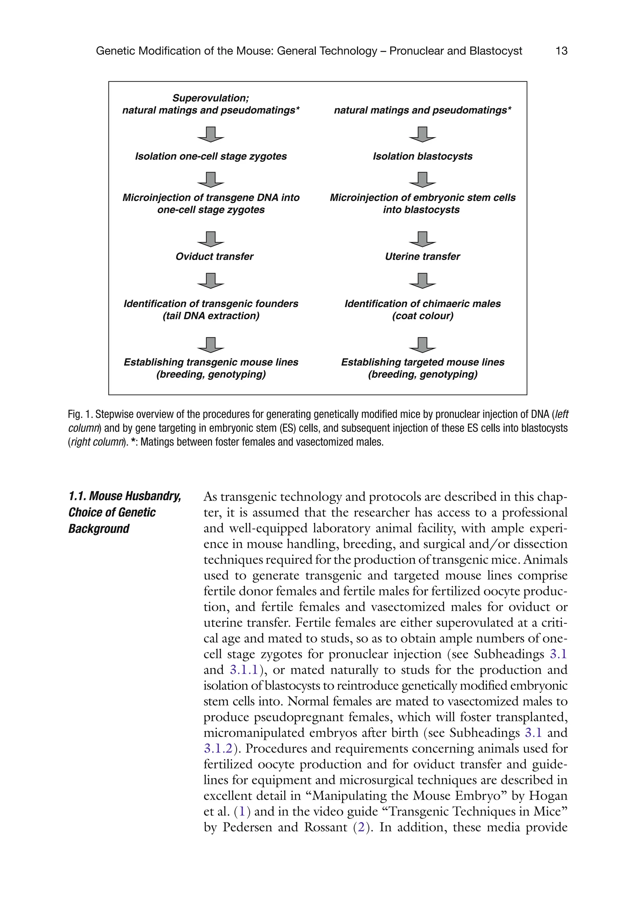
In Figure 1 below, an overview of the different experimental
aspects involved in making genetically modified animals via con-
ventional transgenic technology (i.e., pronuclear injection of one-
cell stage embryos) or gene targeting (i.e., embryonic stem cell
injection into blastocysts) is presented. In the following sections,
some basic guidelines for the production of transgenic mice will
be provided, including superovulation, microinjection of one-cell
stage zygotes, and identification of transgenic founder mice. Since
the animal and equipment use for embryonic stem cell injection
into blastocysts are in essence very similar to microinjection of
fertilized eggs, blastocyst injection, and uterine transfer are dis-
cussed in this chapter as well (see Figure 1 and Subheadings 3.2–
3.4). For basic molecular cloning techniques and strategies, and
molecular detection methods, such as polymerase chain reaction
(PCR)-based or Southern blot analysis, we recommend additional
readingin“MolecularCloning”bySambrooketal.(6).Transgenic
construct design is discussed in Chapter 4.
30.
13
Genetic Modification ofthe Mouse: General Technology – Pronuclear and Blastocyst
As transgenic technology and protocols are described in this chap-
ter, it is assumed that the researcher has access to a professional
and well-equipped laboratory animal facility, with ample experi-
ence in mouse handling, breeding, and surgical and/or dissection
techniques required for the production of transgenic mice. Animals
used to generate transgenic and targeted mouse lines comprise
fertile donor females and fertile males for fertilized oocyte produc-
tion, and fertile females and vasectomized males for oviduct or
uterine transfer. Fertile females are either superovulated at a criti-
cal age and mated to studs, so as to obtain ample numbers of one-
cell stage zygotes for pronuclear injection (see Subheadings 3.1
and 3.1.1), or mated naturally to studs for the production and
isolation of blastocysts to reintroduce genetically modified embryonic
stem cells into. Normal females are mated to vasectomized males to
produce pseudopregnant females, which will foster transplanted,
micromanipulated embryos after birth (see Subheadings 3.1 and
3.1.2). Procedures and requirements concerning animals used for
fertilized oocyte production and for oviduct transfer and guide-
lines for equipment and microsurgical techniques are described in
excellent detail in “Manipulating the Mouse Embryo” by Hogan
et al. (1) and in the video guide “Transgenic Techniques in Mice”
by Pedersen and Rossant (2). In addition, these media provide
1.1. Mouse Husbandry,
Choice of Genetic
Background
Superovulation;
natural matings and pseudomatings*
Isolation one-cell stage zygotes
Microinjection of transgene DNA into
one-cell stage zygotes
Oviduct transfer
Identification of transgenic founders
(tail DNA extraction)
Establishing transgenic mouse lines
(breeding, genotyping)
natural matings and pseudomatings*
Isolation blastocysts
Microinjection of embryonic stem cells
into blastocysts
Uterine transfer
Identification of chimaeric males
(coat colour)
Establishing targeted mouse lines
(breeding, genotyping)
Fig. 1. Stepwise overview of the procedures for generating genetically modified mice by pronuclear injection of DNA (left
column) and by gene targeting in embryonic stem (ES) cells, and subsequent injection of these ES cells into blastocysts
(right column). *: Matings between foster females and vasectomized males.
31.
14 Voncken
comprehensive informationon historical and genetic backgrounds
of in- and out-bred strains, on mouse embryology, and dissection
of specific developmental stages.
The need for genetic standardization of experimental animal
models in experimental and applied research has been historically
one of the reasons why inbred strains were established. A defined
genetic context is important, for instance, to establish the genetics
of cancer susceptibility, for studies on polygenic diseases, or for
immunological studies. In these instances, inbred mice are pre-
ferred to generate transgenic mouse models, because of their strain
homogeneity. A unique collection of inbred mouse strains is avail-
able worldwide. By definition, an inbred strain is derived by 20
generations of brother-to-sister matings and is essentially homozy-
gous at all genetic loci (1). The choice of genetic background is
determined by the aim of the experimental model. Sometimes a
reason for widespread application is simply a historical one (i.e.,
best studied strain in a given context), while in other instances,
there may be a clear advantage in using a particular strain because
of a certain predisposition, although the exact underlying genetic
cause (e.g., modifier loci, QTLs) is not always known. Although
there is considerable choice in inbred strains, the most widely
used strain is C57BL/6J, also known by the acronym B6. The
C57BL/6J strain, for instance, appears more sensitive to diet-
induced atherosclerosis, which makes this strain particularly valu-
able in cardiovascular research. The same inbred mouse strain is
used frequently in immunological and behavioral studies as well.
A common disadvantage of inbred strains, however, is their
reduced reproductive capacity and relative poor yield of one-cell
stage zygotes (fertilized eggs) upon superovulation compared to
that in F1 hybrids. Furthermore, “inbred” zygotes often have an
attenuated viability in vitro, microinjection, and transplantation.
An exception may be the recently introduced FVB/N inbred
strain, which does superovulate well yielding reasonable numbers
of fertilized eggs (7). In most other instances, however, F1 hybrids
are used to generate fertilized eggs for microinjection (sometimes
up to 30 or more). A relatively large fraction of F1 hybrid-derived
zygotes will develop to term. One of the most often used F1
hybrids is C57BL/6J×CBA (BCBA). Other F1-strain hybrids
applied are C57BL/6J×SJL, C3H/HeJ×C57BL/6J, C3H/
HeJ×DBA/2J, and C57BL/6J×DBA/2J (1).
1. Light-cycle controlled mouse room.
2. Female mice (4–6 weeks of age).
3. Fertile male mice (8–12 weeks up to 8 months of age).
2.
Materials
2.1. Superovulation:
Natural Matings and
Pseudomatings
32.
15
Genetic Modification ofthe Mouse: General Technology – Pronuclear and Blastocyst
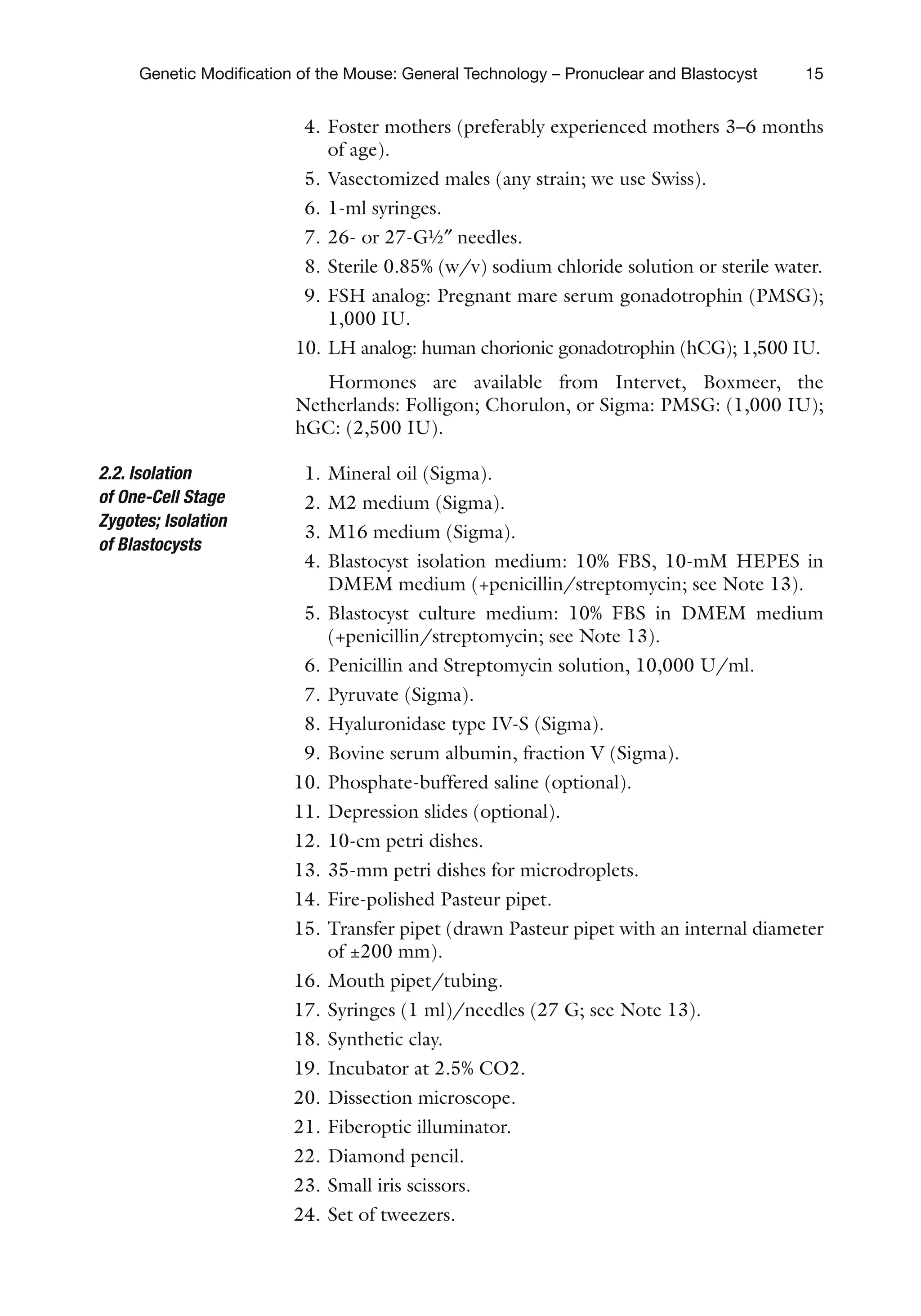
4. Foster mothers (preferably experienced mothers 3–6 months
of age).
5. Vasectomized males (any strain; we use Swiss).
6. 1-ml syringes.
7. 26- or 27-G½″ needles.
8. Sterile 0.85% (w/v) sodium chloride solution or sterile water.
9. FSH analog: Pregnant mare serum gonadotrophin (PMSG);
1,000 IU.
10. LH analog: human chorionic gonadotrophin (hCG); 1,500 IU.
Hormones are available from Intervet, Boxmeer, the
Netherlands: Folligon; Chorulon, or Sigma: PMSG: (1,000 IU);
hGC: (2,500 IU).
1. Mineral oil (Sigma).
2. M2 medium (Sigma).
3. M16 medium (Sigma).
4. Blastocyst isolation medium: 10% FBS, 10-mM HEPES in
DMEM medium (+penicillin/streptomycin; see Note 13).
5. Blastocyst culture medium: 10% FBS in DMEM medium
(+penicillin/streptomycin; see Note 13).
6. Penicillin and Streptomycin solution, 10,000 U/ml.
7. Pyruvate (Sigma).
8. Hyaluronidase type IV-S (Sigma).
9. Bovine serum albumin, fraction V (Sigma).
10. Phosphate-buffered saline (optional).
11. Depression slides (optional).
12. 10-cm petri dishes.
13. 35-mm petri dishes for microdroplets.
14. Fire-polished Pasteur pipet.
15. Transfer pipet (drawn Pasteur pipet with an internal diameter
of ±200 mm).
16. Mouth pipet/tubing.
17. Syringes (1 ml)/needles (27 G; see Note 13).
18. Synthetic clay.
19. Incubator at 2.5% CO2.
20. Dissection microscope.
21. Fiberoptic illuminator.
22. Diamond pencil.
23. Small iris scissors.
24. Set of tweezers.
2.2. Isolation
of One-Cell Stage
Zygotes; Isolation
of Blastocysts
33.
16 Voncken
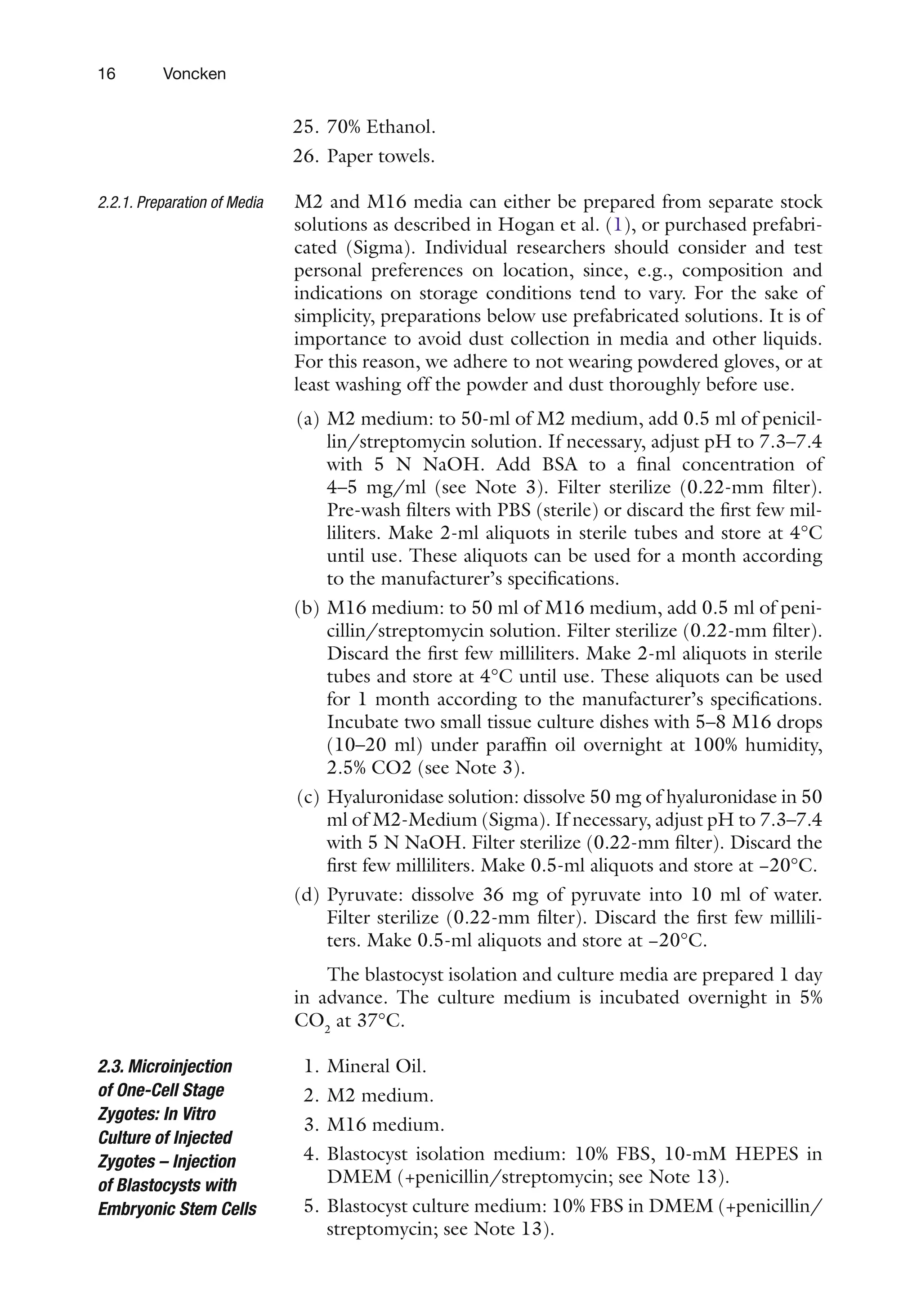
25. 70%Ethanol.
26. Paper towels.
M2 and M16 media can either be prepared from separate stock
solutions as described in Hogan et al. (1), or purchased prefabri-
cated (Sigma). Individual researchers should consider and test
personal preferences on location, since, e.g., composition and
indications on storage conditions tend to vary. For the sake of
simplicity, preparations below use prefabricated solutions. It is of
importance to avoid dust collection in media and other liquids.
For this reason, we adhere to not wearing powdered gloves, or at
least washing off the powder and dust thoroughly before use.
(a) M2 medium: to 50-ml of M2 medium, add 0.5 ml of penicil-
lin/streptomycin solution. If necessary, adjust pH to 7.3–7.4
with 5 N NaOH. Add BSA to a final concentration of
4–5 mg/ml (see Note 3). Filter sterilize (0.22-mm filter).
Pre-wash filters with PBS (sterile) or discard the first few mil-
liliters. Make 2-ml aliquots in sterile tubes and store at 4°C
until use. These aliquots can be used for a month according
to the manufacturer’s specifications.
(b) M16 medium: to 50 ml of M16 medium, add 0.5 ml of peni-
cillin/streptomycin solution. Filter sterilize (0.22-mm filter).
Discard the first few milliliters. Make 2-ml aliquots in sterile
tubes and store at 4°C until use. These aliquots can be used
for 1 month according to the manufacturer’s specifications.
Incubate two small tissue culture dishes with 5–8 M16 drops
(10–20 ml) under paraffin oil overnight at 100% humidity,
2.5% CO2 (see Note 3).
(c) Hyaluronidase solution: dissolve 50 mg of hyaluronidase in 50
ml of M2-Medium (Sigma). If necessary, adjust pH to 7.3–7.4
with 5 N NaOH. Filter sterilize (0.22-mm filter). Discard the
first few milliliters. Make 0.5-ml aliquots and store at −20°C.
(d) Pyruvate: dissolve 36 mg of pyruvate into 10 ml of water.
Filter sterilize (0.22-mm filter). Discard the first few millili-
ters. Make 0.5-ml aliquots and store at −20°C.
The blastocyst isolation and culture media are prepared 1 day
in advance. The culture medium is incubated overnight in 5%
CO2
at 37°C.
1. Mineral Oil.
2. M2 medium.
3. M16 medium.
4. Blastocyst isolation medium: 10% FBS, 10-mM HEPES in
DMEM (+penicillin/streptomycin; see Note 13).
5. Blastocyst culture medium: 10% FBS in DMEM (+penicillin/
streptomycin; see Note 13).
2.2.1. Preparation of Media
2.3. Microinjection
of One-Cell Stage
Zygotes: In Vitro
Culture of Injected
Zygotes – Injection
of Blastocysts with
Embryonic Stem Cells
34.
17
Genetic Modification ofthe Mouse: General Technology – Pronuclear and Blastocyst
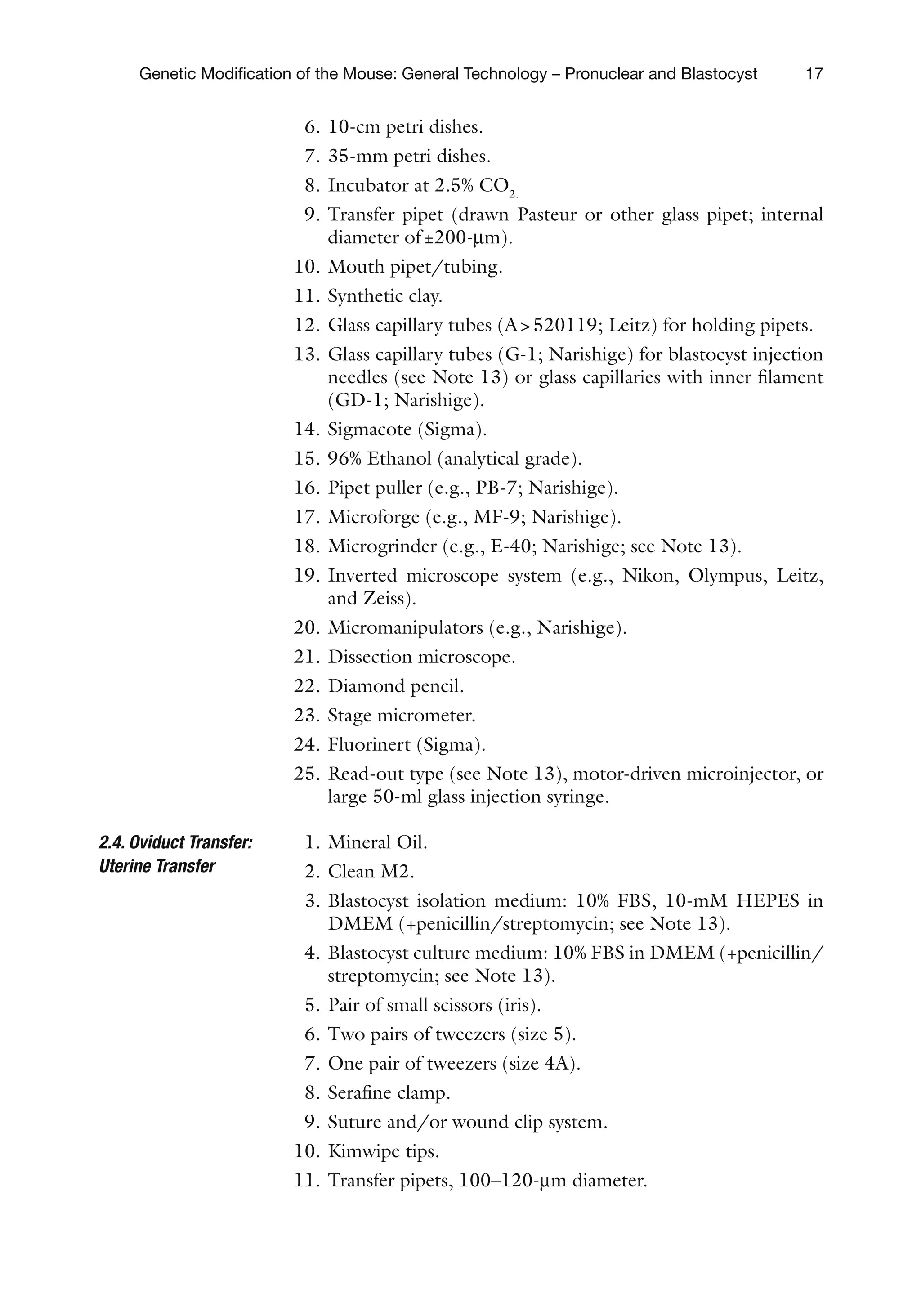
6. 10-cm petri dishes.
7. 35-mm petri dishes.
8. Incubator at 2.5% CO2.
9. Transfer pipet (drawn Pasteur or other glass pipet; internal
diameter of±200-mm).
10. Mouth pipet/tubing.
11. Synthetic clay.
12. Glass capillary tubes (A520119; Leitz) for holding pipets.
13. Glass capillary tubes (G-1; Narishige) for blastocyst injection
needles (see Note 13) or glass capillaries with inner filament
(GD-1; Narishige).
14. Sigmacote (Sigma).
15. 96% Ethanol (analytical grade).
16. Pipet puller (e.g., PB-7; Narishige).
17. Microforge (e.g., MF-9; Narishige).
18. Microgrinder (e.g., E-40; Narishige; see Note 13).
19. Inverted microscope system (e.g., Nikon, Olympus, Leitz,
and Zeiss).
20. Micromanipulators (e.g., Narishige).
21. Dissection microscope.
22. Diamond pencil.
23. Stage micrometer.
24. Fluorinert (Sigma).
25. Read-out type (see Note 13), motor-driven microinjector, or
large 50-ml glass injection syringe.
1. Mineral Oil.
2. Clean M2.
3. Blastocyst isolation medium: 10% FBS, 10-mM HEPES in
DMEM (+penicillin/streptomycin; see Note 13).
4. Blastocyst culture medium: 10% FBS in DMEM (+penicillin/
streptomycin; see Note 13).
5. Pair of small scissors (iris).
6. Two pairs of tweezers (size 5).
7. One pair of tweezers (size 4A).
8. Serafine clamp.
9. Suture and/or wound clip system.
10. Kimwipe tips.
11. Transfer pipets, 100–120-mm diameter.
2.4. Oviduct Transfer:
Uterine Transfer
35.
18 Voncken
12. Syntheticclay.
13. Inhalation anesthetic (see Note 5).
14. Small desiccator.
15. Injection sedative (see Note 5).
16. Syringes (1 ml)/needles (27 G; see Note 13)
17. 96% Ethanol.
18. Dissection microscope.
19. Fiberoptic illuminator.
20. Operation platform
1. Tail mix: 100-mM Tris–HCl, pH 8.5, 5-mM EDTA, 0.2%
SDS, and 200-mM NaCl.
2. Proteinase K: fresh stock solution: 20 mg/ml in TE buffer.
3. TE buffer: 10-mM Tris–HCl, pH 7.2–7.6, 1-mM EDTA.
4. Pasteur pipets, flame-polished.
5. Reaction tubes, 1.5 ml.
6. 55°C oven.
7. Rotator.
1. Tail mix: 50-mM Tris–HCl, pH 8.6, 100-mM EDTA, 1%
SDS, 100-mM NaCl.
2. Protease K: fresh stock solution: 10-mg/ml TE buffer
3. RNase: 10 mg/ml (heat inactivated; 10 min at 100°C).
4. Phenol–chloroform–isoamylalcohol (24:24:1; Phenol:
saturated with demiwater (autoclaved) adjusted to pH 7.0
with 1-M Tris–HCl, pH 8.0.
5. TE buffer:10-mM Tris–HCl, pH 7.2–7.6, 1-mM EDTA.
6. Pasteur pipets, flame-polished.
7. Reaction tubes, 1.5 ml.
8. 55°C oven.
9. Rotator.
10. Bench or wrist shaker.
11. 37°C waterbath.
Animals have access to water and standard chow ad libitum and
are housed under a 12-h day/night time regimen, most often
comprising a 6 a.m. to 6 p.m. light period (see Note 1). We have
2.5. Identification
of Transgenic
Founders: Tail DNA
Extraction
2.5.1. Rapid Procedure
Tail-Tip DNA Extraction
2.5.2. Standard Procedure
Tail-Tip DNA Extraction
3. Methods
3.1. Superovulation:
Natural Matings and
Pseudomatings
36.
19
Genetic Modification ofthe Mouse: General Technology – Pronuclear and Blastocyst
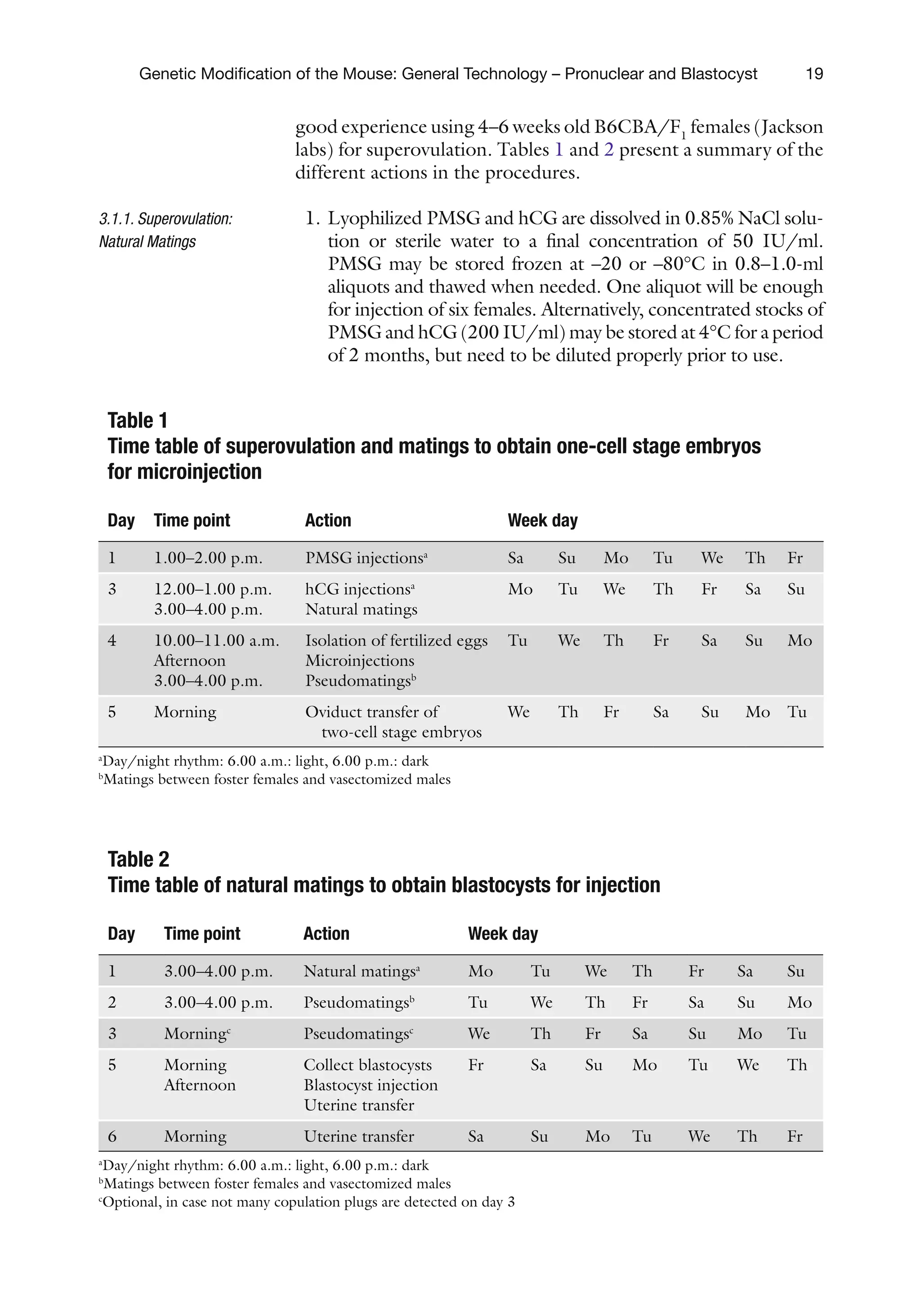
good experience using 4–6 weeks old B6CBA/F1
females (Jackson
labs) for superovulation. Tables 1 and 2 present a summary of the
different actions in the procedures.
1. Lyophilized PMSG and hCG are dissolved in 0.85% NaCl solu-
tion or sterile water to a final concentration of 50 IU/ml.
PMSG may be stored frozen at –20 or –80°C in 0.8–1.0-ml
aliquots and thawed when needed. One aliquot will be enough
for injection of six females. Alternatively, concentrated stocks of
PMSG and hCG (200 IU/ml) may be stored at 4°C for a period
of 2 months, but need to be diluted properly prior to use.
3.1.1. Superovulation:
Natural Matings
Table 1
Time table of superovulation and matings to obtain one-cell stage embryos
for microinjection
Day Time point Action Week day
1 1.00–2.00 p.m. PMSG injectionsa
Sa Su Mo Tu We Th Fr
3 12.00–1.00 p.m.
3.00–4.00 p.m.
hCG injectionsa
Natural matings
Mo Tu We Th Fr Sa Su
4 10.00–11.00 a.m.
Afternoon
3.00–4.00 p.m.
Isolation of fertilized eggs
Microinjections
Pseudomatingsb
Tu We Th Fr Sa Su Mo
5 Morning Oviduct transfer of
two-cell stage embryos
We Th Fr Sa Su Mo Tu
a
Day/night rhythm: 6.00 a.m.: light, 6.00 p.m.: dark
b
Matings between foster females and vasectomized males
Table 2
Time table of natural matings to obtain blastocysts for injection
Day Time point Action Week day
1 3.00–4.00 p.m. Natural matingsa
Mo Tu We Th Fr Sa Su
2 3.00–4.00 p.m. Pseudomatingsb
Tu We Th Fr Sa Su Mo
3 Morningc
Pseudomatingsc
We Th Fr Sa Su Mo Tu
5 Morning
Afternoon
Collect blastocysts
Blastocyst injection
Uterine transfer
Fr Sa Su Mo Tu We Th
6 Morning Uterine transfer Sa Su Mo Tu We Th Fr
a
Day/night rhythm: 6.00 a.m.: light, 6.00 p.m.: dark
b
Matings between foster females and vasectomized males
c
Optional, in case not many copulation plugs are detected on day 3
37.
20 Voncken
2. Thawa vial of PMSG and inject 4–6 weeks old females with
100-ml of PMSG (5 IU) between 1.00 and 2.00 p.m. on the
first day.
3. At day 3, 46–48 h after PMSG injection, thaw a vial of hCG
and inject the same females with 100 ml of hCG (5 IU;
12.00–1.00 p.m.).
4. The females are either transferred directly to fertile studs or in
the afternoon (see Note 2). Use one male per female. To
ensure maximum number of fertilized eggs, these male mice
are used only once a week. At 8 months of age, or when the
plugging ratio drops below 70%, the male mice are replaced.
5. Check for copulation plugs the next morning.
If natural matings are carried out for blastocyst production,
use females of 2–4 months of age. In terms of numbers of mice
used, the same guidelines apply as for “pseudomatings,” i.e., 2–3
females per male (see Subheading 3.1.2). Average yield per
“plugged” female (C57blk/6J) is 5–7 blastocysts. To produce
reasonable amounts of blastocysts for injection, superovulation is
sometimes used in different laboratories. The quality of blasto-
cysts obtained in this manner may, however, vary considerably.
Mating females with vasectomized (or genetically sterile) males
will generate pseudopregnant females required for re-implanta-
tion of microinjected zygotes or blastocysts (1). Basically, any
genetic background may be used to generate pseudopregnant
foster females, provided the females from this strain are known as
“good mothers.” Most F1 hybrids or outbred strains can be used;
for practical reasons we use females from the same background
(B6CBA/F1) as those used for superovulation. If possible, expe-
rienced mothers are preferred. The females should be at least 2
months old (i.e., 20 g body weight) but should not weigh over
30 g: the older and heavier the females, the more problems can be
expected in terms of fat accumulation, which can seriously ham-
per oviduct transfers. Since the females are mated in natural estrus,
obtaining enough pseudopregnant females for oviduct transfer
can be problematic. It is advisable to mate at least 5–6 females per
intended oviduct transfer. To increase the chances of mating a
female in estrus, several females (2–4) are placed in with one
vasectomized male, in contrast to 1-on-1 matings between super-
ovulated females and fertile studs. Experience in judging whether
females are in estrus can be helpful in obtaining sufficient plugged
pseudopregnant females. Females are placed in a cage with a resi-
dent male, not vice versa. Vasectomized males are housed sepa-
rately for at least 1–2 weeks (see Note 2). Oviduct transfers take
place on the day of copulation plug detection. The surplus of
plugged, pseudopregnant fosters can be re-used after 2 weeks.
The vasectomized male mice can be used twice a week for matings.
The foster females are checked regularly for signs of pregnancy.
3.1.2. Pseudopregnant
Females
38.
21
Genetic Modification ofthe Mouse: General Technology – Pronuclear and Blastocyst
Depending on the strain, the litter will be delivered around 19–21
days after the oviduct transfer.
When microinjections are planned, the order of procedures should
have some (chrono)logic to it. Although the planning of a micro-
injection session is highly personal, some suggestions are presented
below in Table 3. Day/night rhythm is as in Table 1(see Note 4).
1. On the day of isolation, add 20 ml of the pyruvate stock to
2 ml of M2 and M16 aliquots and mix by swirling gently.
2. Prepare the depression slides: one with 0.2 ml of M2, two
depression slides with 0.2 ml of hyaluronidase solution, and
three depression slides with 0.2 ml of M16 medium. Place
each depression slide in a 9-cm petri dish and incubate them
briefly at 37°C in the incubator. Alternatively, droplets of
media may be prepared in 35-mm dishes.
3. Collect females with a clear copulation plug. Kill the mice by
CO2
asphyxiation or cervical dislocation and transfer the ani-
mals to the location where zygotes are isolated.
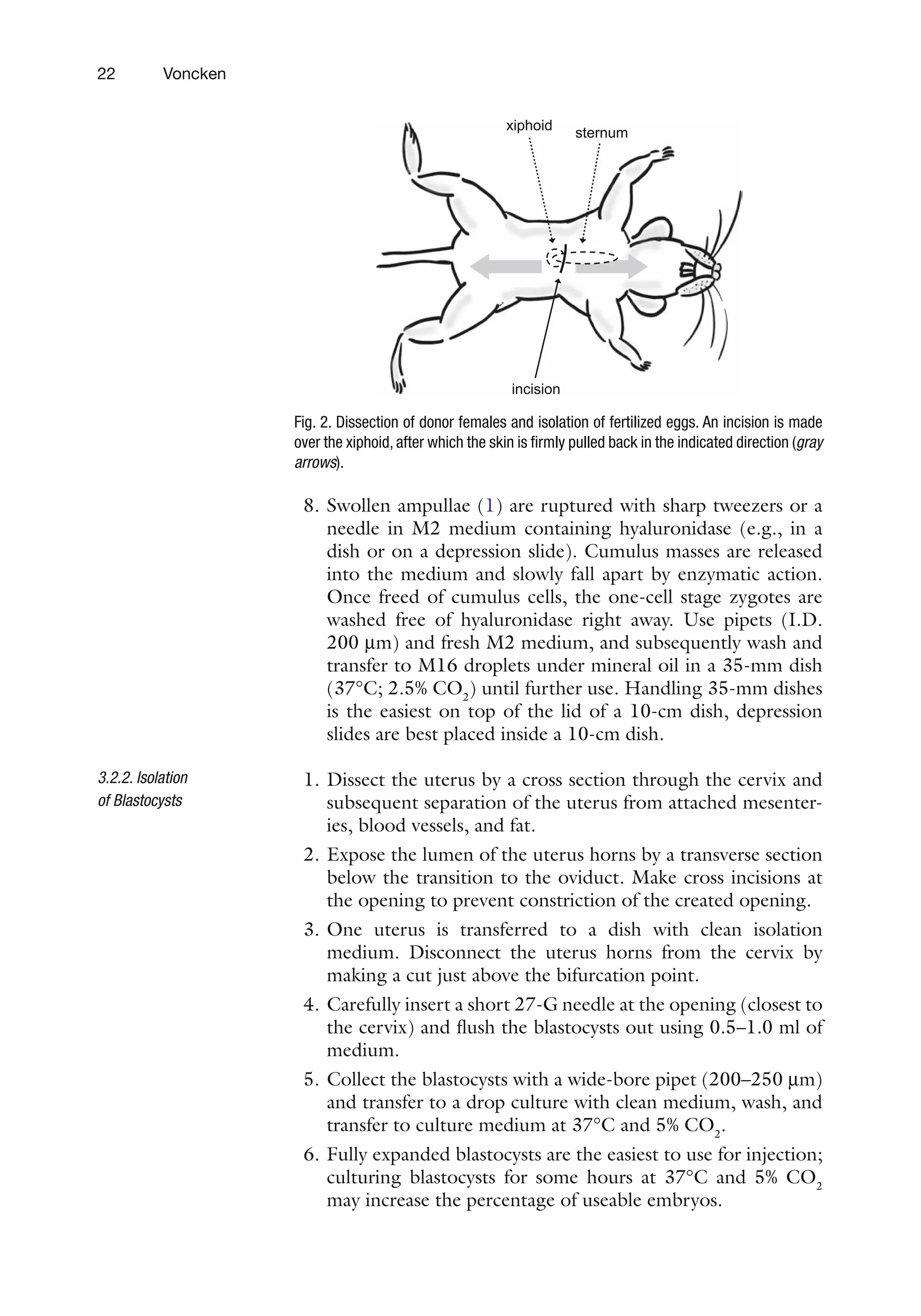
4. Place the females’ abdomen upward on a paper towel, wet the
abdomen with 70% ethanol, and make an incision over the
xiphoid (see Figure 2).
5. Grasp the skin with both hands and firmly pull back in oppo-
site directions (rostrally and caudally), essentially skinning the
mouse completely (see Figure 2).
6. Make incisions in body wall by lifting it with tweezers and
cutting it with scissors, and expose complete body cavity.
7. Move intestines aside gently and grab ovaries by fat pad,
remove ovaries plus oviducts by cutting through the transi-
tion to the uterus horn, and transfer to a dish with PBS to
wash off blood or debris.
3.2. Isolation
of One-Cell Stage
Zygotes: Isolation
of Blastocysts
3.2.1. Isolation of Fertilized
Oocytes
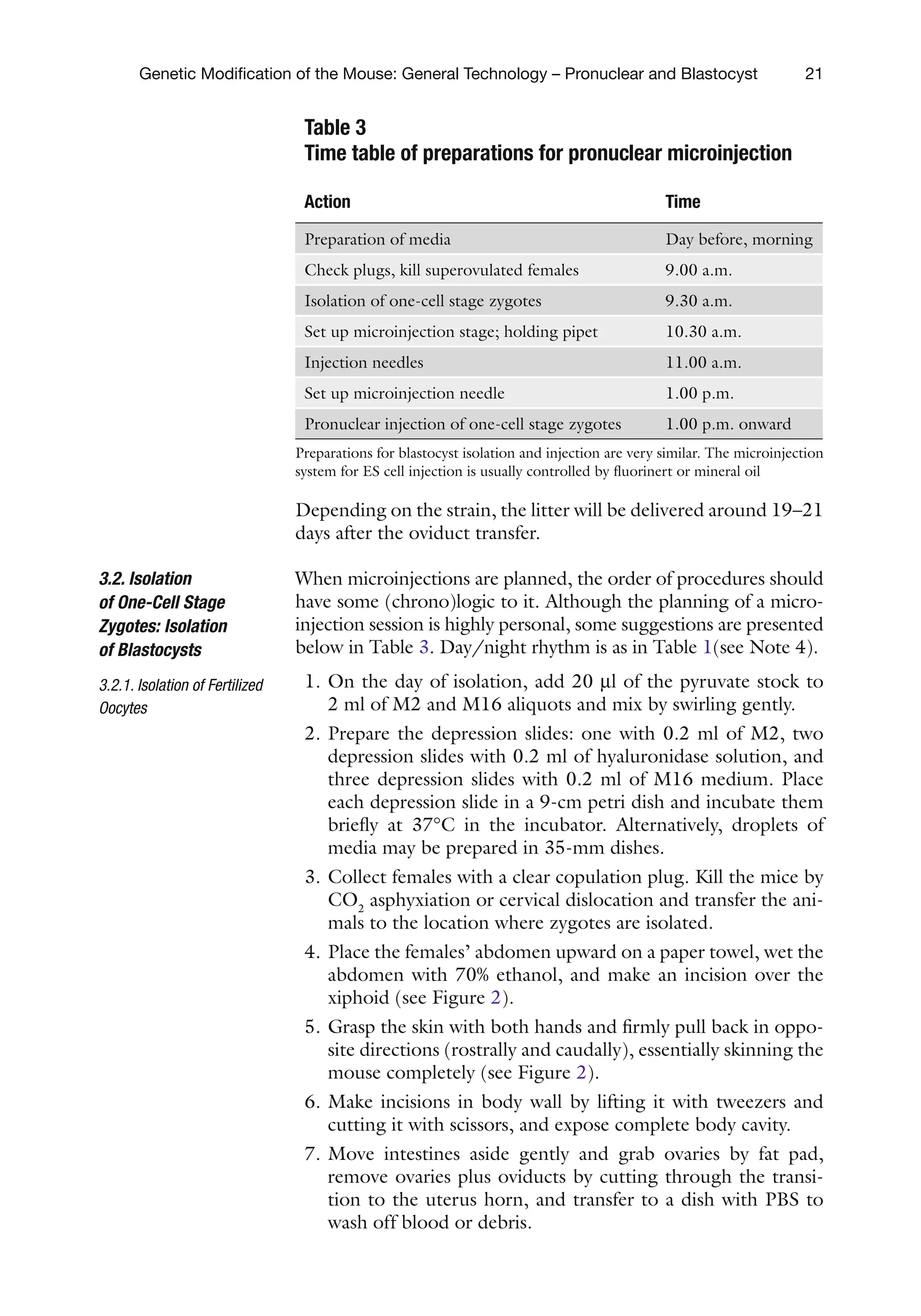
Table 3
Time table of preparations for pronuclear microinjection
Action Time
Preparation of media Day before, morning
Check plugs, kill superovulated females 9.00 a.m.
Isolation of one-cell stage zygotes 9.30 a.m.
Set up microinjection stage; holding pipet 10.30 a.m.
Injection needles 11.00 a.m.
Set up microinjection needle 1.00 p.m.
Pronuclear injection of one-cell stage zygotes 1.00 p.m. onward
Preparations for blastocyst isolation and injection are very similar. The microinjection
system for ES cell injection is usually controlled by fluorinert or mineral oil
39.
22 Voncken
8. Swollenampullae (1) are ruptured with sharp tweezers or a
needle in M2 medium containing hyaluronidase (e.g., in a
dish or on a depression slide). Cumulus masses are released
into the medium and slowly fall apart by enzymatic action.
Once freed of cumulus cells, the one-cell stage zygotes are
washed free of hyaluronidase right away. Use pipets (I.D.
200 mm) and fresh M2 medium, and subsequently wash and
transfer to M16 droplets under mineral oil in a 35-mm dish
(37°C; 2.5% CO2
) until further use. Handling 35-mm dishes
is the easiest on top of the lid of a 10-cm dish, depression
slides are best placed inside a 10-cm dish.
1. Dissect the uterus by a cross section through the cervix and
subsequent separation of the uterus from attached mesenter-
ies, blood vessels, and fat.
2. Expose the lumen of the uterus horns by a transverse section
below the transition to the oviduct. Make cross incisions at
the opening to prevent constriction of the created opening.
3. One uterus is transferred to a dish with clean isolation
medium. Disconnect the uterus horns from the cervix by
making a cut just above the bifurcation point.
4. Carefully insert a short 27-G needle at the opening (closest to
the cervix) and flush the blastocysts out using 0.5–1.0 ml of
medium.
5. Collect the blastocysts with a wide-bore pipet (200–250 mm)
and transfer to a drop culture with clean medium, wash, and
transfer to culture medium at 37°C and 5% CO2
.
6. Fully expanded blastocysts are the easiest to use for injection;
culturing blastocysts for some hours at 37°C and 5% CO2
may increase the percentage of useable embryos.
3.2.2. Isolation
of Blastocysts
sternum
xiphoid
incision
Fig. 2. Dissection of donor females and isolation of fertilized eggs. An incision is made
over the xiphoid, after which the skin is firmly pulled back in the indicated direction (gray
arrows).
40.
23
Genetic Modification ofthe Mouse: General Technology – Pronuclear and Blastocyst
The availability of an operational microinjection set-up is considered
a prerequisite to apply transgenic technology successfully. If no
microinjection unit is available, several types of microscopes,
micromanipulators, injectors, and peripheral equipment, to make
injection needles and holding pipets for either pronuclear or blas-
tocyst injection (see Chapter 8), such as needle pullers, micro-
forges, and grinders, are commercially available (Leitz, Narishige,
Nikon, Olympus, Sutter, and Zeiss). We refer to Hogan et al. (1)
foradetaileddescriptionofamicroinjectionset-up.Microinjection
of one-cell stage zygotes and subsequent transplantation are
essentially carried out as described in ref. (1). It is highly recom-
mended to consult the video guide “transgenic techniques in mice”
by Pedersen and Rossant (2) for a visual reference to the proto-
cols and procedures outlined in this chapter. In practice, one will
see that slight deviations from an existing protocol are possible
and sometimes necessary in order to make things work for the
individual user. Therefore, the protocol below only presents some
of the most essential steps in the microinjection procedure.
Use dust-free gloves when preparing holding pipets and injec-
tion needles. Holding pipets are heat-polished until an opening
of about 10–20 mm remains. Holding pipets and depression slides
can be siliconized and rinsed extensively with clean, dust-free 96%
ethanol; siliconized holding pipets can be re-used. Also, injection
needles can be treated similarly: dip the needle tip into silicon
solution and rinse it with alcohol before the needle is opened.
Several types of injection chambers may be used for microin-
jection. These chambers may consist of a slide and a Perspex ring
(outer diameter slightly smaller than the width of the slide;
1.5 mm in height), which is fixed in position on a siliconized
microscopic slide with 2% agarose. Alternatively, a depression
slide can be used as an injection chamber. The injection chamber
contains a droplet of 10 ml of M2 under light mineral oil.
Depending on the manner in which DNA is loaded into the injec-
tion system, a 3–5-ml DNA droplet may be positioned next to the
M2 droplet (also under oil), or a needle with an inner filament is
back-filled. The chambers are prepared just before use and kept in
a 90-mm petri dish in the CO2
incubator until use. Several injec-
tion chambers may be set up in parallel in this fashion and be
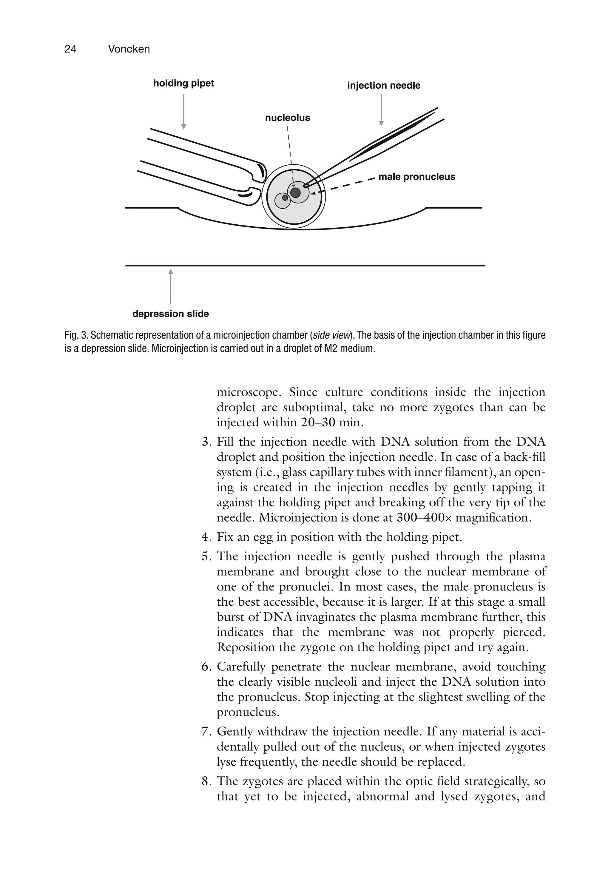
used alternately. Figure 3 depicts an example of a microinjection
set-up.
1. Place the microinjection chamber on the microscope. Position
the holding pipet. Positioning both the holding pipet and the
needle is usually carried out at a magnification of 100×.
2. Just before injection, a number of one-cell stage zygotes (20–40)
are transferred into the M2 droplet of the injection chamber.
Transferring one-cell stage zygotes between injection cham-
bers and culture droplets is best done under a dissection
3.3. Microinjection
of One-Cell Stage
Zygotes: In Vitro
Culture of Injected
Zygotes – Injection
of Blastocysts with
Embryonic Stem Cells
3.3.1. Preparations
3.3.2. Pronuclear Injection
41.
24 Voncken
microscope. Sinceculture conditions inside the injection
droplet are suboptimal, take no more zygotes than can be
injected within 20–30 min.
3. Fill the injection needle with DNA solution from the DNA
droplet and position the injection needle. In case of a back-fill
system (i.e., glass capillary tubes with inner filament), an open-
ing is created in the injection needles by gently tapping it
against the holding pipet and breaking off the very tip of the
needle. Microinjection is done at 300–400× magnification.
4. Fix an egg in position with the holding pipet.
5. The injection needle is gently pushed through the plasma
membrane and brought close to the nuclear membrane of
one of the pronuclei. In most cases, the male pronucleus is
the best accessible, because it is larger. If at this stage a small
burst of DNA invaginates the plasma membrane further, this
indicates that the membrane was not properly pierced.
Reposition the zygote on the holding pipet and try again.
6. Carefully penetrate the nuclear membrane, avoid touching
the clearly visible nucleoli and inject the DNA solution into
the pronucleus. Stop injecting at the slightest swelling of the
pronucleus.
7. Gently withdraw the injection needle. If any material is acci-
dentally pulled out of the nucleus, or when injected zygotes
lyse frequently, the needle should be replaced.
8. The zygotes are placed within the optic field strategically, so
that yet to be injected, abnormal and lysed zygotes, and
holding pipet injection needle
depression slide
male pronucleus
nucleolus
Fig. 3. Schematic representation of a microinjection chamber (side view).The basis of the injection chamber in this figure
is a depression slide. Microinjection is carried out in a droplet of M2 medium.
42.
25
Genetic Modification ofthe Mouse: General Technology – Pronuclear and Blastocyst
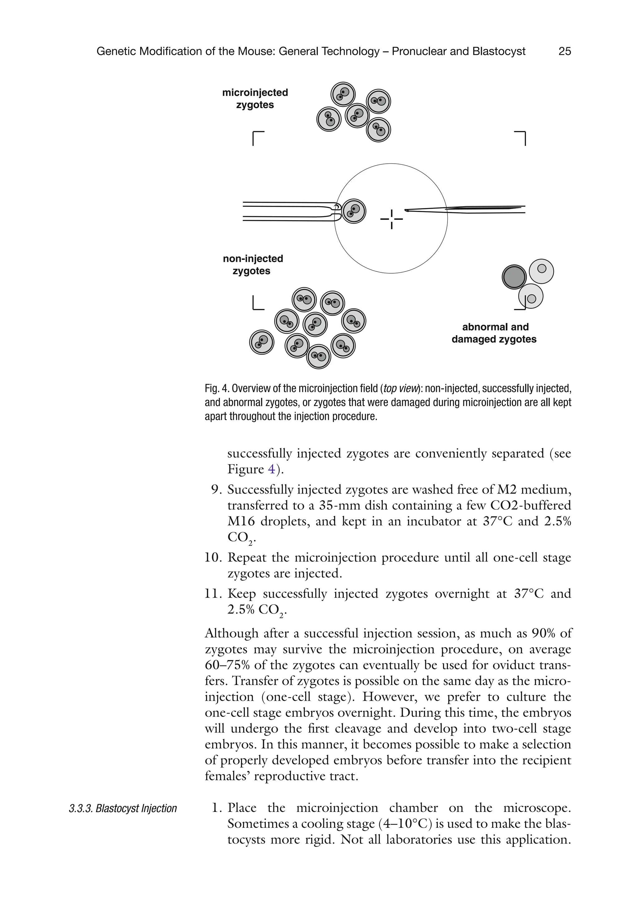
successfully injected zygotes are conveniently separated (see
Figure 4).
9. Successfully injected zygotes are washed free of M2 medium,
transferred to a 35-mm dish containing a few CO2-buffered
M16 droplets, and kept in an incubator at 37°C and 2.5%
CO2
.
10. Repeat the microinjection procedure until all one-cell stage
zygotes are injected.
11. Keep successfully injected zygotes overnight at 37°C and
2.5% CO2
.
Although after a successful injection session, as much as 90% of
zygotes may survive the microinjection procedure, on average
60–75% of the zygotes can eventually be used for oviduct trans-
fers. Transfer of zygotes is possible on the same day as the micro-
injection (one-cell stage). However, we prefer to culture the
one-cell stage embryos overnight. During this time, the embryos
will undergo the first cleavage and develop into two-cell stage
embryos. In this manner, it becomes possible to make a selection
of properly developed embryos before transfer into the recipient
females’ reproductive tract.
1. Place the microinjection chamber on the microscope.
Sometimes a cooling stage (4–10°C) is used to make the blas-
tocysts more rigid. Not all laboratories use this application.
3.3.3. Blastocyst Injection
microinjected
zygotes
non-injected
zygotes
abnormal and
damaged zygotes
Fig. 4. Overview of the microinjection field (top view): non-injected, successfully injected,
and abnormal zygotes, or zygotes that were damaged during microinjection are all kept
apart throughout the injection procedure.
43.
26 Voncken
Positioning theholding pipet and injection needle is carried
out at a magnification of 100×. Optimal needles are bevelled
to an opening of 12–14 mm at 30–35°C. Injection needles
may be siliconized and re-used.
2. Just before injection, a number of blastocysts (20–25) are
transferred into the injection droplet. Injection is carried out
in isolation medium or HEPES-buffered ES cell culturing
medium (see Chapter 8).
3. ES cell suspensions are made by trypsinizing and preplating the
suspension to get rid of feeder cells, which support ES cell
growth in culture (see Chapter 8). We typically keep a few small
ES cell cultures at hand to be able to repeat this procedure
throughout the injection day. Wash ES culture twice with
Ca2+/Mg2+m-free PBS and trypsinize the feeders plus ES
cells. Sediment cells (5 min, at 1,000–1,200 rmp at ambient
temperature) and suspend in ES cell medium; preplate on gela-
tinized culturing surface (see Chapter 8) and harvest after
15–20 min. Most feeder cells will have attached, whereas ES
cells will not. If so desired, the preplating procedure may be
repeated. Collect the ES cells, sediment, suspend well in a small
volume of Ca2+/Mg2+-free PBS, and transfer to a petri dish:
make several drop cultures in either PBS or isolation medium.
4. Transfer single ES cells into the injection chamber and load
the injection needle with a fair number of ES cells (e.g.,
75–120 ES cells). Typically, 12–15 ES cells are injected into
one blastocyst.
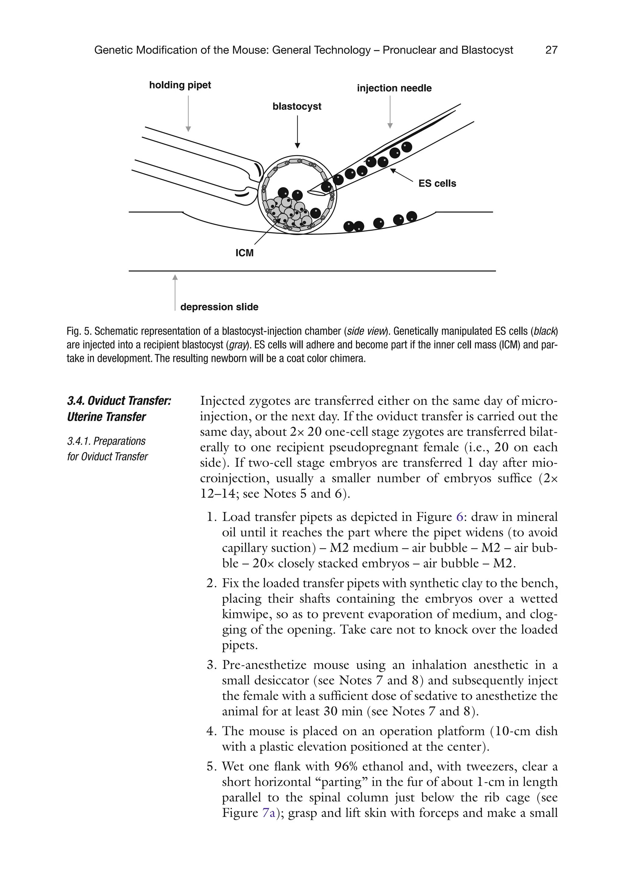
5. Fix a blastocyst in position with the holding pipet, with the
inner cell mass (ICM) located away from the injection site
(see Figure 5). Find a junction (window) between two tro-
phoblast cells and insert the needle through the zona pellu-
cida through the junction into the blastocoel; gently expel
the ES cells.
6. The injection needle is gently removed from the blastocoel,
the blastocyst will visibly collapse shortly afterwards; move
the injected blastocyst aside (as in Figure 4) and inject the
next one.
7. The successfully injected blastocysts are transferred to a
35-mm dish containing a few CO2-buffered culture medium
and kept in an incubator at 37°C and 5% CO2.
8. Repeat the injection procedure until all blastocysts are
injected. ES cell suspension will tend to aggregate; repeat the
preplating procedure with fresh suspension.
The successfully injected blastocysts are either transferred to
pseudopregnant females the same day or cultured overnight at
37°C and 5% CO2
.
44.
27
Genetic Modification ofthe Mouse: General Technology – Pronuclear and Blastocyst
Injected zygotes are transferred either on the same day of micro-
injection, or the next day. If the oviduct transfer is carried out the
same day, about 2× 20 one-cell stage zygotes are transferred bilat-
erally to one recipient pseudopregnant female (i.e., 20 on each
side). If two-cell stage embryos are transferred 1 day after mio-
croinjection, usually a smaller number of embryos suffice (2×
12–14; see Notes 5 and 6).
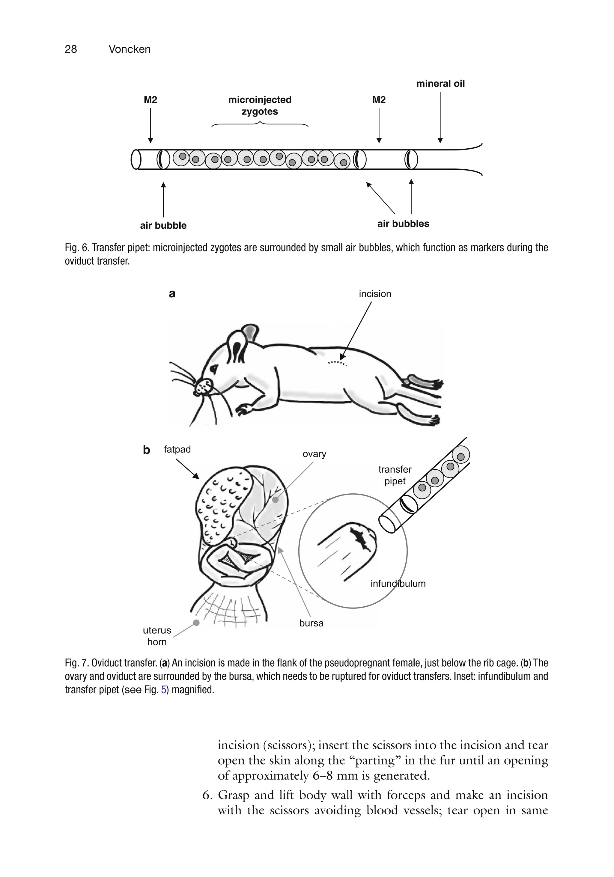
1. Load transfer pipets as depicted in Figure 6: draw in mineral
oil until it reaches the part where the pipet widens (to avoid
capillary suction) – M2 medium – air bubble – M2 – air bub-
ble – 20× closely stacked embryos – air bubble – M2.
2. Fix the loaded transfer pipets with synthetic clay to the bench,
placing their shafts containing the embryos over a wetted
kimwipe, so as to prevent evaporation of medium, and clog-
ging of the opening. Take care not to knock over the loaded
pipets.
3. Pre-anesthetize mouse using an inhalation anesthetic in a
small desiccator (see Notes 7 and 8) and subsequently inject
the female with a sufficient dose of sedative to anesthetize the
animal for at least 30 min (see Notes 7 and 8).
4. The mouse is placed on an operation platform (10-cm dish
with a plastic elevation positioned at the center).
5. Wet one flank with 96% ethanol and, with tweezers, clear a
short horizontal “parting” in the fur of about 1-cm in length
parallel to the spinal column just below the rib cage (see
Figure 7a); grasp and lift skin with forceps and make a small
3.4. Oviduct Transfer:
Uterine Transfer
3.4.1. Preparations
for Oviduct Transfer
holding pipet injection needle
depression slide
ES cells
blastocyst
ICM
Fig. 5. Schematic representation of a blastocyst-injection chamber (side view). Genetically manipulated ES cells (black)
are injected into a recipient blastocyst (gray). ES cells will adhere and become part if the inner cell mass (ICM) and par-
take in development. The resulting newborn will be a coat color chimera.
45.
28 Voncken
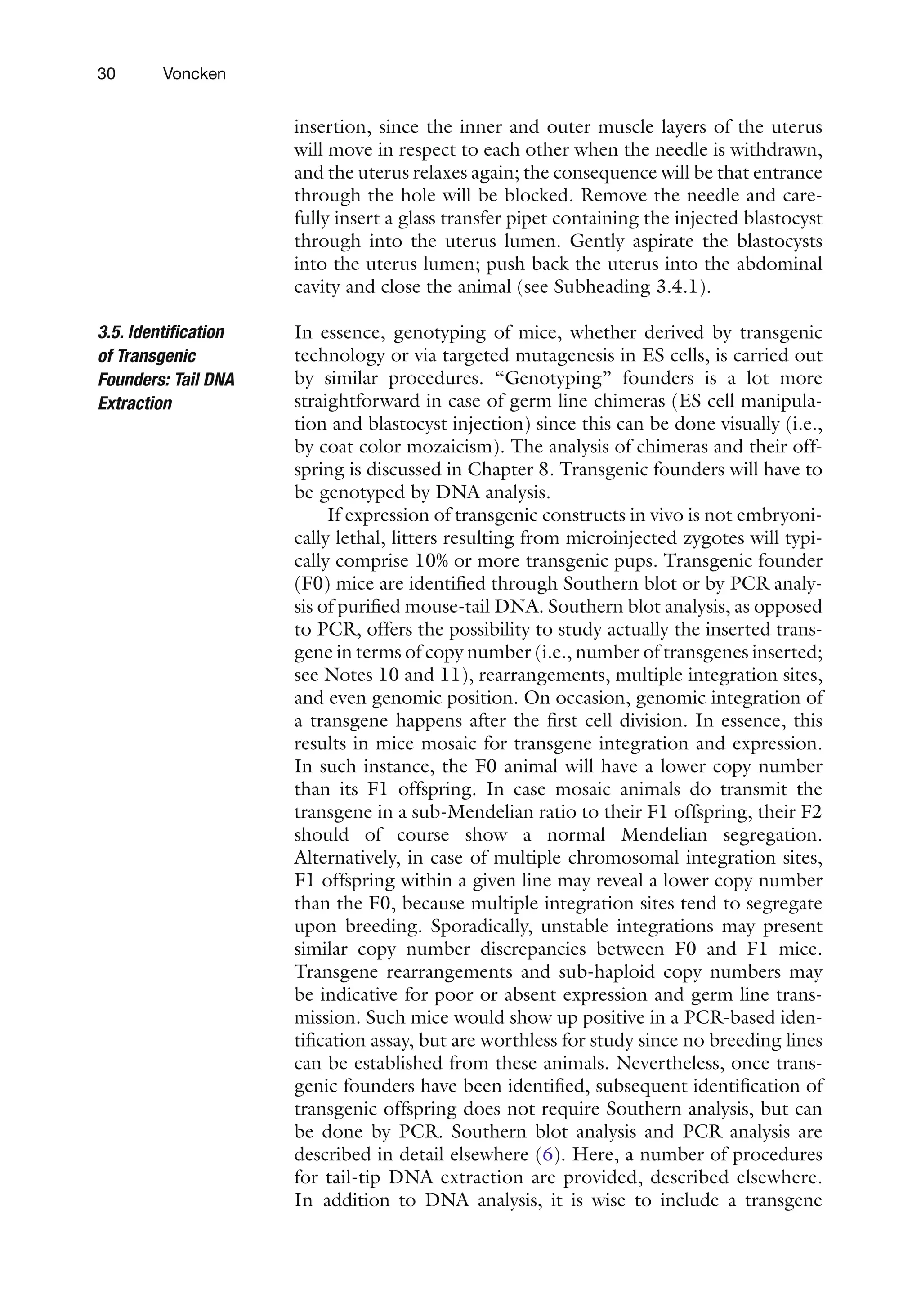
incision (scissors);insert the scissors into the incision and tear
open the skin along the “parting” in the fur until an opening
of approximately 6–8 mm is generated.
6. Grasp and lift body wall with forceps and make an incision
with the scissors avoiding blood vessels; tear open in same
M2
M2
mineral oil
air bubble air bubbles
microinjected
zygotes
Fig. 6. Transfer pipet: microinjected zygotes are surrounded by small air bubbles, which function as markers during the
oviduct transfer.
fatpad
uterus
horn
ovary
transfer
pipet
b
infundibulum
bursa
a incision
Fig. 7. Oviduct transfer. (a) An incision is made in the flank of the pseudopregnant female, just below the rib cage. (b) The
ovary and oviduct are surrounded by the bursa, which needs to be ruptured for oviduct transfers. Inset: infundibulum and
transfer pipet (see Fig. 5) magnified.
46.
29
Genetic Modification ofthe Mouse: General Technology – Pronuclear and Blastocyst
direction as the previous tear and attach suture to the upper
“lip” of the wound (body wall).
7. Pull out ovary and oviduct gently by the fat pad attached to
the ovary; attach a serafine clamp to the fat pad, pull out the
uterus a bit more, and position the clamp over the flank of the
mouse in such a manner that the infundibulum (i.e., the
entrance to the fallopian tube), still covered by bursa, is visi-
ble (see Figure 7b).
8. Transfer the mouse under a dissecting microscope; magnifica-
tion 15–20×. Use fiberoptic illuminators for lighting during
surgery.
9. At this stage, take the mouth-piece of the transfer pipet in
your mouth, while the other end remains in synthetic clay on
the microscope or table surface.
10. Gently tear an opening into the bursa covering the ovary and
infundibulum with 2× 5-tweezers, avoid tearing blood ves-
sels; if bleeding is caused inadvertently, use kimwipe tips to
soak up excessive fluids.
11. Locate the infundibulum, pick up transfer pipet, and bring
into position (i.e., line-up with the position of the infundibu-
lum and fallopian tube). Probe the infundibulum carefully
with the transfer pipet and when certain of deep enough
insertion, gently expel the injected embryos by blowing.
12. While expelling, the air bubbles surrounding the embryos
becomevisibleinthefallopiantube:iftwoormoreareobserved,
the embryo transfer was successful. Pull out the pipet com-
pletely and transfer the mouse back under the fiberoptic
illuminator.
13. Gently maneuver uterus horn back into body cavity using the
blunt tip of the serafine clamp, close the body wall with one
stitch and a tight knot, and use suture or a wound clip to
close the wound in the skin; proceed with the other side (see
Notes 5 and 6).
14. When completed, ear-mark the female, if so desired (see
Note 9), and return the animal to a cage.
15. Clean all equipment used with water and soap, and 70%
ethanol, and sterilize.
A uterine transfer is very similar to an oviduct transfer, with the
exception that injected blastocysts are transferred directly into the
uterus lumen. To this end, gently pull out the uterus horn a little
further compared to that in an oviduct transfer (see Figure 7b);
carefully insert a sterile 27-G needle into the uterus lumen
through the uterus wall. The insertion direction should be almost
parallel to the uterus horn, to avoid puncturing the uterus. It is
imperative not to stretch the uterus walls too much during needle
3.4.2. Uterine Transfer
47.
30 Voncken
insertion, sincethe inner and outer muscle layers of the uterus
will move in respect to each other when the needle is withdrawn,
and the uterus relaxes again; the consequence will be that entrance
through the hole will be blocked. Remove the needle and care-
fully insert a glass transfer pipet containing the injected blastocyst
through into the uterus lumen. Gently aspirate the blastocysts
into the uterus lumen; push back the uterus into the abdominal
cavity and close the animal (see Subheading 3.4.1).
In essence, genotyping of mice, whether derived by transgenic
technology or via targeted mutagenesis in ES cells, is carried out
by similar procedures. “Genotyping” founders is a lot more
straightforward in case of germ line chimeras (ES cell manipula-
tion and blastocyst injection) since this can be done visually (i.e.,
by coat color mozaicism). The analysis of chimeras and their off-
spring is discussed in Chapter 8. Transgenic founders will have to
be genotyped by DNA analysis.
If expression of transgenic constructs in vivo is not embryoni-
cally lethal, litters resulting from microinjected zygotes will typi-
cally comprise 10% or more transgenic pups. Transgenic founder
(F0) mice are identified through Southern blot or by PCR analy-
sis of purified mouse-tail DNA. Southern blot analysis, as opposed
to PCR, offers the possibility to study actually the inserted trans-
gene in terms of copy number (i.e., number of transgenes inserted;
see Notes 10 and 11), rearrangements, multiple integration sites,
and even genomic position. On occasion, genomic integration of
a transgene happens after the first cell division. In essence, this
results in mice mosaic for transgene integration and expression.
In such instance, the F0 animal will have a lower copy number
than its F1 offspring. In case mosaic animals do transmit the
transgene in a sub-Mendelian ratio to their F1 offspring, their F2
should of course show a normal Mendelian segregation.
Alternatively, in case of multiple chromosomal integration sites,
F1 offspring within a given line may reveal a lower copy number
than the F0, because multiple integration sites tend to segregate
upon breeding. Sporadically, unstable integrations may present
similar copy number discrepancies between F0 and F1 mice.
Transgene rearrangements and sub-haploid copy numbers may
be indicative for poor or absent expression and germ line trans-
mission. Such mice would show up positive in a PCR-based iden-
tification assay, but are worthless for study since no breeding lines
can be established from these animals. Nevertheless, once trans-
genic founders have been identified, subsequent identification of
transgenic offspring does not require Southern analysis, but can
be done by PCR. Southern blot analysis and PCR analysis are
described in detail elsewhere (6). Here, a number of procedures
for tail-tip DNA extraction are provided, described elsewhere.
In addition to DNA analysis, it is wise to include a transgene
3.5. Identification
of Transgenic
Founders: Tail DNA
Extraction
48.
31
Genetic Modification ofthe Mouse: General Technology – Pronuclear and Blastocyst
expression analysis on several tissues of F1 animals from different
founder lines. This may include both RNA and protein analyses,
siuch as for instance the detection of apolipoproteins in circula-
tion, provided that immunological detection reagents and/or
biochemical assays are available.
This method will allow for a rapid DNA extraction from mouse
tails, which can be used for PCR-based screens without prob-
lems and was even reported to yield DNA of sufficient quality
for Southern analysis (8). However, to get rid of contaminating
proteins and RNAs for an exact quantitative Southern analysis,
a more thorough DNA purification might be required (see
Notes 10 and 11). A relatively quick method for DNA isolation
is described below; in the next section (see Subheading 3.5.2),
a tail DNA purification method is outlined employing phenol
extraction. Mice are genotyped by tail DNA analysis (see Notes
10 and 11). Genotyping can be done from 10 days postpartum
(pp) onwards. To identify the individual mice following geno-
typing, animals need to be marked at the time of the tail cut.
Relatively young animals (i.e., 10–12 days pp) are toe-marked,
on older animals (3 weeks of age) ear-marking is an option.
Consult the animal facility to adhere to the marking system used
on location.
1. Collect mouse tail-tips, about 0.5 cm (10 days pp) to 1.0 cm
(3 weeks pp), in 1.5-ml reaction tubes on ice and either store
dry at –80°C or in a tail mix at –20°C if not processed imme-
diately. It is convenient to sedate mice lightly using an inhala-
tion sedative, especially when older mice are marked. It is
possible at this time to take a small blood sample for bio-
chemical analysis.
2. Add 660-ml of tail mix and 10-ml of proteinase K (20 mg/ml).
Incubate overnight at 55°C, preferably while rotating. This
will greatly help dissolving and subsequently isolating the tail
DNA.
3. Pellet the remaining debris (hairs and bone) in a microfuge at
14,000 rpm for 10–15 min at ambient temperature. Transfer
the supernatant to a fresh microfuge tube.
4. Add 0.6× volume (i.e., ±400 ml) of isopropanol and mix by
inverting the tubes gently (25×), allowing the DNA to pre-
cipitate in visible threads (vigorous shaking will hamper visi-
ble precipitation).
5. Either pellet the DNA briefly (15–20 s, 14,000 rpm) or fish the
DNA clump out using a flame-polished glass Pasteur pipet.
6. Rinse the DNA sticking to the glass tip in 70% ethanol, let the
DNA briefly air dry, and transfer to 500 ml of TE.
3.5.1. Rapid Procedure for
Tail-Tip DNA Extraction
49.
32 Voncken
7. Dissolvethe DNA thoroughly by incubating it for 10–15 min
at 55–65°C, followed by firm vortexing or shaking.
Alternatively, we use the rotator at an ambient temperature to
dissolve the DNA overnight.
8. Store the DNA samples at 4°C until needed.
9. A volume of 1.5 ml of DNA suffices for a PCR. If a Southern
analysis is attempted at this stage, 30–50 ml can be used.
1. Collect mouse tails and add 700 ml of tail mix, or store (see
Subheading 3.5.1); add 25-ml Proteinase K to each tube and
incubate at 55°C overnight (in a rotator).
2. Add 10 ml of RNase and incubate for 1–2 h at 37°C.
3. Transfer the content of the tubes to new tubes1
containing
400 ml of phenol–chloroform–isoamylalcohol using a cut blue
tip and shake firmly for 15 min (bench or wrist shaker) at an
ambient temperature.
4. Centrifuge at 14,000 rpm for 30 min, at an ambient
temperature.
5. Transfer the supernatant to tubes containing 400 ml of phe-
nol–chloroform–isoamylalcohol and shake (bench or wrist
shaker) for 15 min at an ambient temperature.
6. Centrifuge at 14,000 rpm for 30 min at an ambient
temperature.
7. Transfer the supernatant to tubes containing 400 ml of chlo-
roform to remove phenol traces and shake for 5 min at an
ambient temperature.
8. Centrifuge at 14,000 rpm for 5–10 min at an ambient
temperature.
9. Transfer the supernatant to tubes containing 0.6× vol
(±440 ml) of isopropanol.
10. Mix by inverting the tubes gently (25×), allowing the DNA to
visibly precipitate (see Subheading 3.5.1).
11. Fish out DNA with (flame-polished, slightly bent at the tip)
Pasteur pipet and wash in 1 ml of 70% ethanol. If the expected
amount of DNA is too low, to fish out, pellet at 14,000 rpm
for 10 min at an ambient temperature and wash the pellet
with 70% ethanol (2×).
12. Let the DNA air dry for a while, then place the pipet in tube
(50–100 ml of TE) until DNA comes off; DNA is dissolved by
incubating for 10–15 min at 55–65°C, and subsequent incu-
bation at 37°C, mixing firmly and repeatedly by hand until
DNA is fully dissolved.
3.5.2. Standard Procedure
for Tail-Tip DNA Extraction
1
Tubes that have been incubated o/n at 55°C with SDS inside tend to leak
when phenol–chloroform–isoamylalcohol is added.
50.
33
Genetic Modification ofthe Mouse: General Technology – Pronuclear and Blastocyst
13. Determine the DNA concentration: dilute 5 ml of DNA sam-
ple in 600 ml of water. Concentration (in mg/ml) is: OD260nm
(OD260
DNA=50 mg/ml)×6; typical yield: 50–200 mg of
DNA
14. Perform a quantitative Southern analysis (see Notes 10 and
11).
15. DNA samples may be stored at 4°C or –20°C.
Following identification of valuable transgenic founders and F1
mice, transgenic mouse lines will have to be established. It may be
useful to genotype the animals relatively early (10 days pp; see
Subheading 3.5.1), for instance when rapid onset of disease in a
given mouse necessitates early diagnosis or compromises life span.
PCR-based genotyping on 1 day pp mice and even on mouse
blood, hair, or toes has recently been described (8–12). In addi-
tion, early genotyping will allow the researcher to sort out valuable
transgenic mice from litters before weaning and cull the mouse
colony accordingly, which reduces housing expenses significantly.
If the mouse model needs to be studied in a particular genetic
background, the transgenic lines need to be backcrossed to the
strain of choice. Eight to ten backcrosses of a transgenic line to an
existing inbred strain, although not inbred by definition (see
Subheading 1.1 (1), produce a genetic background sufficiently
homogeneous for experimental purposes. A mouse line may be
bred to “homozygosity” to increase transgene expression (i.e.
double copy number); such double transgenic mice are produced
by transgenic male-to-transgenic female matings. If this is done
within one mouse line, disease or altered behavior, and even
(embryonic) lethality, all unrelated to the expression of the trans-
gene, may be the consequences: such conditions may be the result
of recessive mutations caused by transgene integration into spe-
cific loci. X chromosomal integrations can be particularly cum-
bersome in this respect, although such integrations have been
known to result in unexpected and exciting discoveries (13).
Y chromosomal transgene integrations, conversely, if not silenced,
make genotyping a fairly straightforward task. Sex-chromosome
targeting, on the contrary, can speed up the creation of null-
mutant models significantly.
Once transgenic mouse lines have been established, a typical
initial screen would include a complete analysis by a qualified
mouse pathologist, a full spectrum expressional and biochemical
analysis to evaluate the effect of transgene activity. Ideally, several
independently derived transgenic mouse lines should be studied
to validate the animal model.
The above mentioned clearly points out that establishing
mouse lines can be complex at times. It is, therefore, advisable to
keep clear records of the history of all animals used in breeding
programs (see Note 9).
3.6. Establishing
Transgenic Lines:
Breeding and Analysis
51.
34 Voncken
1. Althoughthe light period is most often 6.00–7.00 a.m. to
6.00–7.00 p.m., the day–night rhythm at which animals are
kept may be adjusted to local needs/convenience. A light
period from 2.00–3.00 a.m. to 2.00–3.00 p.m. is an option.
The authors’ experience is that fertilized oocytes, which are a
bit more advanced in development at the time of microinjec-
tion, are often more successfully microinjected: slight ruffling
of the plasma membrane makes it easier to penetrate it undam-
aged. However, since membrane ruffling signals the onset of
the first cleavage, this methodological variation may be an
option mainly for the experienced. Since mouse ovulation,
fertilization, and subsequent development of zygotes follow a
preset time course, hormone injection regimens should be
adjusted accordingly, as should the time of fertilized oocyte
isolation.
2. For matings, female mice are always placed in with a male,
not vice versa. Introducing a male into an unfamiliar environ-
ment will significantly affect their performance. Surplus of
plugged pseudopregnant females may be used again after 2
weeks. Unsuccessfully superovulated females (i.e., without a
visible copulation plug) may be kept aside for later use as fos-
ter as well.
3. Several measures are taken to reduce stickiness of the embryos
and glass injection needles. Adding BSA to incubation media
is one. Reducing the CO2
concentration in the incubator is
another factor that increases injection efficiency in our hands.
We typically use 2.5% instead of 5% CO2
.
4. In order to keep animals SPF, all equipment and working
surfaces should be sterilized before isolation of fertilized
oocytes, microinjection, and oviduct transfer. If the afore-
mentioned experimental procedures are not carried out within
the confines of the animal facility, foster mice should be trans-
ported in filter-top cages.
5. For oviduct transfers, use transfer pipets with an inner diam-
eter only slightly wider than that of zygotes. Too wide a trans-
fer pipet will be relatively hard to insert into the infundibulum.
We typically use transfer pipets with a shaft of 100–120 mm
(O.D.) maximum. Ruler slides (slides with an engraved mil-
limeter/micrometer scale) are very useful to estimate the
bore size of transfer pipets.
6. It is advisable not to pull the fallopian tube out by grabbing
the infundibulum: its fragility will cause it to be damaged very
easily. Prying and maneuvering of the infundibulum are possible
4.
Notes
52.
35
Genetic Modification ofthe Mouse: General Technology – Pronuclear and Blastocyst
if carried out extremely gently. Some laboratories prefer unilateral
oviduct transfers, other adhere to bilateral transfers, since
sometimes unilateral transfers will not result in pregnancy.
7. To reduce stress and excessive bleeding, a brief sedation with
an inhalation anesthetic may be preferred. Several inhalation
and injection sedatives are approved of by animal welfare com-
mittees and are commercially available; we use ether or meto-
fane (methoxyflurane) as inhalation anesthetic and 2.5%
avertin, described in Ref. (1), as the sedative during micro-
surgery. A volume of 20 ml of avertin/g body weight suffices,
although less is preferred and possible if proficient in fallopian
tube transfers; on the whole, 250–300 ml per average-sized
female will do. To stop bleeding, epinephrin can be used (1).
8. An indicator for full sedation is the absence of a blinking
reflex when blown (puffed) on the eyes softly. Care should be
taken to exert only minimal peri-operative stress on the ani-
mal; eyes can be covered with a paper towel wetted in 0.85%
sodium chloride, or should be closed manually at regular
intervals. Animals should be kept warm after surgery until
fully awake.
9. It is advisable to keep meticulous records of the history of all
animals (i.e., transgenic lines, individual mice within a line,
experimental and autopsy records, pedigree, etc.) at all times.
We also record success rate of superovulations, natural mat-
ings, pseudomatings and oviduct transfers, litter sizes, and %
transgenic animals obtained in a given session. A range of
software is available for this purpose.
10. To carry out a quantitative Southern analysis, 15 mg (or less)
of DNA is fragmented by restriction endonucleases (REN) in
a 50-ml reaction volume. If REN digestion of DNA is still
hampered by contaminants in solution, 1.5 ml of a 0.1-M
spermidine solution may be added (cat. # S0266; Sigma) to
the reaction mix. Alternatively, the reaction volume may be
increased to 300 ml and spermidine may be added (9 ml,
0.1 M); however, the DNA should be precipitated before
loading it onto an agarose gel. When preparing the REN
digestion in the presence of spermidine, care should be taken
to mix all components at room temperature; spermidine is
not useful in restriction buffers without salt, for it will precipi-
tate the DNA.
11. To estimate the copy number of a particular transgenic founder
or line, 1 ng of purified, linearized transgenic construct of
15 kb will roughly equal a haploid copy number of 1 in 15-mg
control tail DNA. We typically run 0.65–0.7% agarose gels.
The quality of the Southern blot after hybridization (i.e.,
straight bands and good separation) is greatly improved when
electrophoresis is carried out at low power over a few days.
53.
36 Voncken
12. Severalcompanies provide a wealth of mouse-related infor-
mation via the Internet: (http:/
/www.criver.com, http:/
/
www.harlan.com, http:/
/www.jax.org, http:/
/www.taconic.
com). A source providing a particularly extensive database on
knock-out and transgenic mice and related topics is
“TheTransgenic/Targeted Mutation DatabaseTBASE” at
http:/
/tbase.jax.org. In addition, useful web sites and Internet
links can be found at http:/
/www.bioresearchonline.com and
at Websites of transgenic facilities at a number of Universities
in Europe and USA. Important information regarding the
status of the mouse genome map, including available YAC
clones, is provided by the Whitehead Institute at http:/
/www-
genome.wi.mit.edu/cgi-bin/mouse/index; the National
Center for Biotechnology Information at http:/
/www.ncbi.
nlm.nih.gov/;andbyTheEuropeanCollaborativeInterspecific
Mouse Backcross (EUCIB) consortium at http:/
/www.hgmp.
mrc.ac.uk/MBx/MBxHomepage.html.
13. Methods and procedures described are specifically used for
blastocyst and ES cell manipulation, not for pronuclear injec-
tion and oviduct transfer.
References
1. Hogan, B., Beddington, R., Constantini, F.,
and Lacey, E. (1994) Manipulating the mouse
embryo. A laboratory manual. 2nd ed. Cold
Spring Harbor Laboratory, Cold Spring
Harbor, New York.
2. Pedersen, R. A., and Rossant, J. (1989)
Transgenic techniques in mice: a video guide.
Cold Spring Harbor Laboratory, Cold Spring
Harbor, New York.
3. Pieper, F. R., et al. (1992) Efficient generation
of functional transgenes by homologous
recombination in murine zygotes. Nucleic
Acids Res 20, 1259–1264.
4. Sigmund, C. D. (1993) Major approaches for
generating and analyzing transgenic mice. An
overview. Hypertension 22, 599–607.
5. Bronson, S. K., et al. (1996) Single-copy
transgenic mice with chosen-site integration.
Proc Natl Acad Sci USA 93, 9067–9072.
6. Sambrook, J., Fritsch E. F., and Maniatis, T.
(1989) Molecular cloning. A laboratory man-
ual. 2nd ed. (Ford, N., ed.), Cold Spring
Harbor Laboratory, Cold Spring Harbor, New
York.
7. Taketo, M., et al. (1991) FVB/N: an inbred
mouse strain preferable for transgenic analy-
ses. Proc Natl Acad Sci USA 88, 2065–2069.
8. Laird, P. W., et al. (1991) Simplified mamma-
lian DNA isolation procedure. Nucleic Acids
Res 19, 4293.
9. Chen, S. Z., and Evans, G. A. (1990) A simple
screening method for transgenic mice using the
polymerase chain reaction. Biotechniques 8, 32–33.
10. Ohhara, M., Kurosu, Y., and Esumi, M.
(1994) Direct PCR of whole blood and hair
shafts by microwave treatment. Biotechniques
17, 726–728.
11. Malumbres, M., et al. (1997) Isolation of high
molecular weight DNA for reliable genotyp-
ing of transgenic mice. Biotechniques 22,
1114–1119.
12. Henneberger, C., Grantyn, R., and Rothe, T.
(2000) Rapid genotyping of newborn gene
mutant mice. J Neurosci Meth 100, 123–126.
13. Cases, O., et al. (1995) Aggressive behavior
and altered amounts of brain serotonin and
norepinephrine in mice lacking MAOA.
Science 268, 1763–1766.
(2) Longitudinal andTransverse Striæ Nearly Equal
GYROSIGMA BALTICUM (EHR.) CL.
Valve with margins parallel nearly to the extremities, which are
suddenly unilaterally sub-conical and obtuse; raphe sigmoid;
transverse and longitudinal striæ nearly equally distant, 15 in 10 µ
(Per.). L. 200-360 µ.
Navicula baltica Ehr.
Pleurosigma balticum (Ehr.) Wm. Sm.
Common along the coast.
Pl. 23, Fig. 2.
GYROSIGMA PARKERI VAR. STAURONEIOIDES GRUN.
Valve lanceolate, slightly sigmoid, ends produced into beaks with
sub-acute apices; raphe straight in the middle part; central nodule
elliptical; transverse striæ, 21, and longitudinal, 24 in 10 µ (Per.).
An apparent stauros, variable in width, extends to the margin and, in
consequence, the median transverse striæ are more evident. L. 75
µ.
Schuylkill River. Rather rare.
Pl. 23, Fig. 7.
57.
GYROSIGMA SIMILE (GRUN.)
Valveslightly sigmoid, broad, with obtuse ends; raphe sigmoid,
nearly central; transverse striæ, 15, longitudinal, 16-17 in 10 µ
(Per.).
Pleurosigma simile Grun.
Gyrosigma balticum var. similis (Grun.) Cl.
Shark River, N. J.
Pl. 23, Fig. 4.
(3) Transverse Striæ More Distant
GYROSIGMA ACUMINATUM (KUETZ.) CL.
Valve sigmoid, tapering to the sub-acute ends; raphe central;
transverse and longitudinal striæ nearly equally distant, 17 or 18 in
10 µ (Per.).
Frustulia acuminata Kuetz.
Port Penn, Delaware River.
Pl. 23, Fig. 5.
GYROSIGMA STRIGILIS (WM. SM.) CL.
58.
Valve sigmoid, withobtuse ends; raphe doubly sigmoid; axial area
rather wide; transverse striæ, 13, and longitudinal, about 16 in 10 µ.
Long Island Sound. Not common.
Pl. 23, Fig. 1.
GYROSIGMA KUETZINGII (GRUN.) CL.
Valve sigmoid, lanceolate, with sub-acute ends; raphe central, the
central nodule elliptical; transverse striæ, 21-23, and longitudinal,
25-26 in 10 µ.
Pleurosigma spencerii var. acutiuscula Grun.
Pleurosigma spencerii var. kuetzingii Grun.
Common in fresh water.
Pl. 38, Fig. 12.
GYROSIGMA SCALPROIDES (RAB.) CL.
Valve slightly sigmoid, with obtuse ends; raphe nearly straight;
central nodule elliptical; transverse striæ, 22, slightly radiate and
more distant in the middle; longitudinal striæ, 29 in 10 µ. L. 60 µ.
Common in streams.
Pl. 38, Fig. 9.
59.
In Pl. 23,Fig. 6 represents a form more sigmoid.
GYROSIGMA SPENCERII VAR. NODIFERA GRUN.
Valve sigmoid, with obtuse ends; raphe central; central nodule
obliquely elongated; transverse striæ, 17-18 in 10 µ, curved in the
middle of the valve, longitudinal striæ, 22 in 10 µ. L. 150 µ.
Blue clay.
Pl. 23, Fig. 8.
GYROSIGMA PROLONGATUM (WM. SM.) CL.
Valve narrow, lanceolate, produced into beaks, curved in a contrary
direction; raphe central; transverse striæ, 20-21 in 10 µ, longitudinal
closer. L. 140 µ.
Along the coast, northward.
Pl. 38, Fig. 13.
I have not seen any specimens south of New England, but they will
probably occur.
(4) Striæ Alike, Extremities Produced
GYROSIGMA FASCIOLA (EHR.) CL.
Valve lanceolate, attenuated into curved beaks turned in opposite
directions; raphe central, straight, except at the beaks; transverse
60.
striæ, 22, longitudinal,24 in 10 µ (Per.).
New York Bay.
Pl. 23, Fig. 9.
Frustulia AG. (1824); em. GRUN. (1865)
(frustulum, a small piece)
Valves naviculoid, similar, usually free but sometimes enclosed in
gelatinous tubes or embedded in mucus. Median line between two
thickened ribs. Central and terminal nodules frequently elongated.
Surface of valve with fine puncta in longitudinal and transverse lines
appearing hyaline under medium powers.
Chromatophores, two, extending along the girdle. They differ from
those of Navicula in being separated from the wall in the middle by a
hemispherical mass of protoplasm. According to Pfitzer, each
chromatophore is divided in the middle, allowing a connection
between the hemispherical mass and the central plasma mass.
Schmitz states that the chromatophore is thickened in the middle
and contains a pyrenoid.
In conjugation, two frustules form two cylindrical bodies which later
become conical and from which are formed the sporangial valves
twice the usual size.
FRUSTULIA LEWISIANA (GREV.) DE TONI
Valve elliptical or linear, with rounded ends; terminal nodules
elongated, at a distance from the ends; striæ, 24 in 10 µ.
61.
Port Penn, DelawareRiver. Along the coast.
Pl. 17, Fig. 1.
FRUSTULIA RHOMBOIDES (EHR.) DE TONI
Valve lanceolate or rhombic-lanceolate, rounded at the ends; central
and terminal nodules short; striæ, 20 in 10 µ, sometimes coarser.
Common in fresh water.
Pl. 17, Fig. 2.
FRUSTULIA RHOMBOIDES VAR. AMPHIPLEUROIDES GRUN.
Valve rhombic-lanceolate; central and terminal nodules elongated;
median line somewhat excentric.
Blue clay.
Pl. 17, Fig. 3.
FRUSTULIA RHOMBOIDES VAR. SAXONICA (RAB.) DE TONI
Valve smaller than in rhomboides, with somewhat produced ends,
closer median ribs and rounded central nodule.
Fresh water.
Pl. 17, Fig. 6.
62.
FRUSTULIA VULGARIS (THWAITES)DE TONI
Valve elliptical-lanceolate, with rounded or sometimes sub-rostrate
ends; central and terminal nodules slightly elongated; striæ delicate,
closer at the ends. Frustules at first in gelatinous tubes.
Colletonema vulgaris Thwaites.
Fresh water.
Pl. 17, Fig. 4.
FRUSTULIA INTERPOSITA (LEWIS) DE TONI
Valve linear-elliptical, rounded at the ends; terminal nodules short.
Navicula interposita Lewis.
Along the coast. Port Penn, Delaware River.
Pl. 17, Fig. 5.
Amphipleura KUETZ. (1844)
(amphi, on both sides, pleura, a side)
Frustules free, in gelatinous masses or in tubes. Valve linear-
lanceolate; central nodule narrow, extending half the length of the
valve or more, then forking toward the ends. Terminal nodules
prolonged, as in Frustulia, into a porte-crayon-shaped figure.
Chromatophores two, very short.
63.
AMPHIPLEURA PELLUCIDA KUETZ.
Frustulesfree or in mucous masses. Valve fusiform; forks about one-
fourth the length of the valve; striæ transverse, punctate, 36-40 in
10 µ (J. J. Woodward).
Occasional in the Delaware River.
Pl. 17, Fig. 9.
AMPHIPLEURA RUTILANS (TRENTEPOHL) CL.
Frustules enclosed in gelatinous tubes. Valve linear-lanceolate,
obtuse at the ends; forks about one-third the length of the valve;
striæ, 28 in 10 µ.
Conferva rutilans Trentepohl.
Schizonema dillwynii Wm. Sm.
Abundant at Belmar, N. J.
Pl. 17, Fig. 10.
Fig. 11 represents a portion of the gelatinous tube containing
frustules.
Dictyoneis CLEVE (1890)
(dictyon, a net)
64.
Frustules oblong. Valvelanceolate, constricted in the middle (in our
species); an outer layer finely punctate and an inner layer of
reticulations; the margin of the valve divided into large, quadrate
cells.
The genus Dictyoneis includes species at one time ascribed to
Mastogloia and Navicula. The structure, however, is not like that of
either, as the loculi are attached to the valve and are not separable
as in Mastogloia, and the cell-wall is not like that of any Navicula.
Cleve remarks that Dictyoneis is found in warm waters. Lewis found
one specimen at Black Rock Harbor, L. I., and one in the Delaware
River blue clay. The specimens here described I found living on the
New Jersey coast.
DICTYONEIS MARGINATA VAR. TYPICA CLEVE
Valve panduriform, with cuneate lobes; axial area narrow, linear,
scarcely, or not at all, widened in the middle; terminal fissures in
contrary directions; outer stratum finely punctate, about 25 in 10 µ,
in parallel striæ; inner stratum coarsely reticulated. Four and one-
fourth times longer than broad; marginal cells, 5 in 10 µ, smaller or
obsolescent in the middle of the valve; cells of the valve in irregular
transverse rows, 10-12 in 10 µ. L. 93 µ.
Navicula marginata Lewis.
Absecon, N. J.
Pl. 20, Fig. 3.
DICTYONEIS MARGINATA VAR. COMMUTATA CLEVE
65.
Valve four andone-half times longer than broad; cells of the valve in
irregular, transverse rows about 11 in 10 µ; marginal cells nearly
equal, 6 in 10 µ. L. 125 µ.
Absecon, N. J.
Pl. 20, Fig. 2.
DICTYONEIS MARGINATA VAR. MAXIMA N. VAR.
Valve with cuneate segments; marginal cells, 4 in 10 µ; cells of the
valve, 5 in 10 µ, obsolescent in the middle and smaller; transverse
striæ, 25 in 10 µ.
Atlantic Coast. Rare.
Pl. 20, Fig. 1 (from a specimen found at Colon).
Trachyneis CLEVE (1894)
(trachys, rough, and neis (naus), named from the chief species)
Valve more or less linear or linear-lanceolate. It appears to be
composed of three strata, one an interior, coarsely dotted, an
exterior of fine puncta in longitudinal striæ, scarcely visible, and a
median of transverse anastomosing costæ forming irregular alveoli.
Chromatophores, two or four bands on the zone (Mereschkowsky).
TRACHYNEIS ASPERA VAR. INTERMEDIA GRUN.
66.
Valve linear-elliptic; axialarea a stauros widened outward and
unilateral. Striæ of the median layer of radiating rows of oblong
alveoli.
Along the coast. Not common.
Pl. 17, Fig. 15.
The type form and its numerous varieties are quite ubiquitous. Very
large specimens occur in the Antarctic regions, especially in material
from Ross Island, S. Victoria Land (Shackleton Ant. Exp.).
Brébissonia GRUN. (1860)
(named after Alphonse de Brébisson, the distinguished French
naturalist)
Frustules stipitate; valve lanceolate; striæ transverse in the middle,
radiate at the ends. Median area narrow, central nodule elongated,
terminal fissures at a distance from the ends. Valve with an outer
finely punctate stratum.
At one end of one valve in each frustule is found a conspicuous
punctum, the plasma pore of Otto Mueller, through which the
frustule is connected with the gelatinous stipe, analogous to the
pore in Diatoma connecting the zig-zag frustules.
Chromatophore single, lying on one girdle and passing over to each
valve.
BRÉBISSONIA BŒCKII (KUETZ.) GRUN.
67.
Valve lanceolate, withsub-acute apices; striæ, 3-4 in 10 µ, not
reaching the median line.
Blue clay. Very rare. Common in brackish water at Chestertown, Md.
(T. C. Palmer)
Pl. 17, Fig. 7.
BRÉBISSONIA PALMERII, N. SP.
Valve rhombic-lanceolate, with cuneate ends and produced apices.
Central nodule more elongate and terminal fissures further from the
ends than in B. bœckii.
Pavonia, N. J. (artesian well, depth of 40 ft.). Rare.
Pl. 17, Fig. 8.
I take pleasure in naming this species after Mr. T. Chalkley Palmer, of
Media, Pa., the author of numerous papers on the Diatomaceæ.
Lewis partly describes a similar form, which he does not name, as a
species of Navicula found in the blue clay at Kaighn's Point, N. J.
(Lewis, New and Intermediate Forms, etc., p. 15, Pl. 1, Fig. 8.)
Anomœoneis PFITZER (1871)
(anomoios, unlike, and neis (naus), a boat)
Valve lanceolate, axial area narrow, central area widened; transverse
striæ punctate, the puncta in longitudinal rows or interrupted by
blank lines.
68.
A single chromatophorelies along one of the girdle sides and
extends over the valves, each of the two parts being deeply notched
or slit at the ends. According to Schmitz there are two pyrenoids, but
Heinzerling thinks there is but one.
Cleve considers this genus not well founded, as it is based upon the
cell contents of but one species, the structure of the other species
not being known. As the forms here described are easily recognized
by the interrupted puncta, the genus is, at least, convenient.
ANOMŒONEIS SPHÆROPHORA (KUETZ.) CL.
Valve elliptic-lanceolate, ends rostrate-capitate. Axial area narrow,
central area rounded, larger on one side of the median line than the
other. Striæ very slightly radiate, 16 in 10 µ, punctate, the puncta
interrupted by longitudinal blank lines.
Pfitzer states that the central plasma mass is unequal on the two
sides.
Navicula sphærophora Kuetz.
Fresh and brackish water. Not common.
Pl. 40, Fig. 2.
ANOMŒONEIS SERIANS (BRÉB.) CL.
Valve lanceolate, acute; axial area lanceolate; striæ, 24 in 10 µ;
puncta elongate.
69.
Not common inthis locality, but abundant northwards; fossil in the
peat deposits of New England.
May's Landing, N. J.
Pl. 17, Fig. 12.
Forma minor—Valve rhombic-lanceolate, smaller than the type.
May's Landing, N. J.
Pl. 17, Fig. 13.
ANOMŒONEIS FOLLIS (EHR.) CL.
Valve rhomboid, tumid in the middle and obtuse at the produced
ends. Central area lanceolate; striæ radiate in the middle, transverse
at the ends.
Navicula follis Ehr.
Navicula trochus Kuetz.
Reported by Lewis as very rare in the blue clay of the Delaware
River. I have not seen it in this locality. The figure is drawn from a
specimen in the W. Bridgewater, Mass., deposit.
Pl. 17, Fig. 14.
Caloneis CLEVE (1894)
70.
(calos, beautiful)
Valve convex,linear or lanceolate in general outline, with transverse,
smooth or finely punctate striæ crossed by one or more longitudinal
lines.
Endochrome of two chromatophores lying one on each valve, entire
in some species and deeply cleft in others.
CALONEIS LIBER (WM. SM.) CL.
Valve linear, with parallel margins and rounded ends; axial area
narrow, central area orbicular; striæ transverse in the middle,
slightly divergent at the ends, 16 in 10 µ; terminal fissures slightly
curved in the same direction; longitudinal line median. L. 82 µ.
Atlantic coast, chiefly southward.
Pl. 40, Fig. 1.
CALONEIS SILICULA (EHR.) CL.
Valve linear, gibbous in the middle, with broad sub-cuneate ends;
axial area narrow, central area rounded; longitudinal line marginal;
striæ parallel or nearly so, 16 to 18 in 10 µ.
Navicula silicula Ehr.
Navicula limosa Donk.
Blue clay.
71.
Pl. 21, Fig.3 (var. genuina Cl.).
CALONEIS SILICULA VAR. INFLATA (GRUN.) CL.
Valve gibbous in the middle, with rounded ends; central area
elliptical.
Schuylkill River.
Pl. 21, Fig. 4.
C. silicula may be recognized by its yellow color when dry. Its
varieties are extremely numerous.
CALONEIS TRINODIS (LEWIS)
Valve divided into three segments of equal width; ends cuneate and
usually produced; axial area elliptical with a lunate marking on each
side; striæ radiate in the middle, elsewhere parallel, about 20 in 10
µ, finely punctate; longitudinal line marginal, scarcely visible; the
striæ become fainter toward the axial area.
Occasional in streams and in the blue clay. Abundant in a water-
trough at Ashbourne, Pa.
Pl. 21, Fig. 8.
I have retained Lewis' name as specific. Lewis, wrongly, I think,
ascribes his species to Navicula trinodis Wm. Sm., which is not
figured by Smith, but is illustrated by Van Heurck (Syn. Pl. 14, Fig.
31a), and is named by Cleve Navicula contenta var. biceps Arnott. De
Toni includes Lewis' name under Rhoiconeis trinodis (Wm. Sm.)
72.
Grun. Rhoiconeis isachnanthiform, with frustules arcuate, and the
species is named by Cleve Achnanthes trinodis (Arnott). Caloneis
schumanniana (Grun.) Cl., to which as a variety Cleve unites Lewis'
form, appears to resemble it only in the lunate marks.
Fig. 9 represents a single specimen found in the Pavonia deposit and
which I believe to be an abnormal form of C. trinodis, differing only
in the degree of inflation and in the larger central area.
Navicula trinodis var. inflata Schultze, from Staten Island, is the
same form figured by Lewis, who states that certain specimens have
produced apices.
CALONEIS PERMAGNA (BAIL.) CL.
Valve lanceolate, with produced apices; median line nearly straight;
axial area lanceolate, irregular or slightly unilateral, about half the
width of the valve; striæ, 9 in 10 µ, radiate and indistinctly punctate;
longitudinal lines double. L. 100-200 µ.
Pinnularia permagna Bail.
Common in brackish water.
Pl. 21, Fig. 1.
CALONEIS PERMAGNA VAR. LEWISIANA N. VAR.
Valve lanceolate, with undulating sides and sub-cuneate apices; axial
area less than one-third the width of the valve; striæ radiate, 12 in
10 µ, indistinctly punctate; longitudinal lines double, closer together
than in the type. L. 140 µ.
73.
Lewis illustrates thisvariety in New and Rare Species, Pl. 2, Fig.
11, and states that it is probably Navicula esox Kuetzing. This is an
error, as Kuetzing's species is Pinnularia esox Ehr., a form near P.
major.
Rather common in the Delaware River.
Pl. 21, Fig. 2.
CALONEIS FORMOSA (GREG.) CL.
Valve lanceolate, with sub-cuneate apices; axial area one-fourth to
one-fifth the width of the valve, somewhat unilateral, dilated in the
middle; striæ, 12-14 in 10 µ radiate, punctate; longitudinal lines
double, distinct. Variable in size and outline.
Abundant along the shores of the Delaware River.
Pl. 21, Fig. 18.
CALONEIS BREVIS VAR. VEXANS (GRUN.) CL.
Valve elliptical-lanceolate; apices obtuse; median fissures distant;
axial area narrow; central area large, orbicular; longitudinal lines
close together, median.
Shark River, N. J.
Pl. 21, Fig. 5.
CALONEIS WARDII CL.
74.
Valve linear, endscuneate; axial area linear; central area dilated to a
stauros reaching the margin; striæ parallel, radiate at the ends, 18 in
10 µ; longitudinal lines marginal.
Not uncommon in the Delaware River.
Pl. 21, Figs. 6 and 7.
CALONEIS POWELLII (LEWIS) CL.
Valve linear, with cuneate ends; axial area linear; central area large,
quadrate, united to the wide longitudinal lines; striæ parallel,
smooth, 8 in 10 µ.
Long Island (Lewis); Smith's Island, Delaware River.
Pl. 21, Fig. 10.
Neidium PFITZER (1871)
(neidion, dim. of naus, a boat)
Valve linear or lanceolate; median fissures turned in opposite
directions, terminal fissures appearing bifurcate (?); striæ
transverse, usually oblique, finely punctate, crossed by one or
several longitudinal blank lines.
Chromatophores, two, lying on the girdle side, in cell division each
forming a partially divided pair. A large pyrenoid is said to be found
in the middle of each chromatophore, but Mereschkowsky states
that the pyrenoids are absent, but that in N. affine four elæoplasts
are always seen in the centre of the frustule.
75.
A genus easilyrecognized by the peculiar terminal and median
fissures and by the yellowish or brownish color of the valves when
dry, darker than in Caloneis.
NEIDIUM AFFINE (EHR.) PFITZER
Valve linear, with protracted, sub-rostrate or capitate ends.
Navicula affinis Ehr.
NEIDIUM AFFINE VAR. GENUINA FORMA MAXIMA CL.
Striæ, 14 in 10 µ, punctate, oblique in the middle, convergent at the
ends; puncta, 15 in 10 µ. L. 238 µ.
Pensauken, N. J. (artesian well).
Pl. 21, Fig. 11.
Var. genuina forma minor Cl.—L. 26 µ; striæ, 24 in 10 µ.
Brandywine Creek.
Pl. 21, Fig. 12.
NEIDIUM AFFINE VAR. AMPHIRHYNCUS (EHR.) CL.
Valve linear, with protracted capitate ends; striæ transverse,
interrupted by several longitudinal lines.
Willistown, Pa.
76.
Pl. 21, Fig.13.
NEIDIUM AMPHIGOMPHUS (EHR.) PFITZER
Valve with parallel margins and cuneate ends; striæ transverse,
interrupted by several longitudinal lines; central area widened
transversely.
Navicula amphigomphus Ehr.
Wissahickon.
Pl. 21, Fig. 14.
NEIDIUM PRODUCTUM (WM. SM.) CL.
Valve linear, elongate, with capitate apices; striæ slightly oblique;
longitudinal lines marginal; axial area very narrow, central area
small.
Navicula producta Wm. Sm.
Newtown Square.
Pl. 21, Fig. 16.
NEIDIUM IRIDIS (EHR.) CL.
Valve linear or lanceolate-elliptical, with sub-cuneate or rounded
ends; striæ oblique, about 18 in 10 µ; central area orbicular.
77.
Navicula iridis Ehr.
Naviculafirma Kuetz.
Willistown, Pa.; Middletown, Delaware Co., Pa. (Palmer).
Pl. 21, Fig. 17.
The form here figured is probably the variety ampliata (Ehr.) Cl. with
less acute apices and more elliptical outline. The species occurs in
many variations, the larger being found northward, especially in the
peat deposits of New England.
NEIDIUM HITCHCOCKII (EHR.) CL.
Valve linear, with triundulate margin and cuneate ends; striæ
transverse, oblique.
Navicula hitchcockii Ehr.
Pavonia, N. J. (artesian well); Kirkwood Pond, N. J.
Pl. 21, Fig. 15.
Diploneis EHR. (1840)
(diplos, double)
Valve elliptical or panduriform; median line enclosed in strongly
siliceous horns corresponding to the lyre-shaped areas of Navicula
lyra but never punctate; central nodule, quadrate; valve costate, or
78.
striate, or both;between the horns and the outer part are thinner
spaces or sulci, and, in some species, outside of the sulci are narrow
spaces known as lunulæ.
Chromatophores, two, upon the girdle or the valves. Pyrenoids have
been found in one species only, D. interrupta.
DIPLONEIS ELLIPTICA (KUETZ.) CL.
Valve elliptical; central nodule large; sulci narrow, curved, close to
the horns; striæ punctate, in rows radiating more and more toward
the ends. Variable in size and in the coarseness of puncta which are
from 10 to 13 in 10 µ (Cleve).
Cleve describes D. ovalis Hilse as having the central nodule rounded,
but otherwise about the same as D. elliptica, and as equivalent to
Navicula ovalis A. Schmidt (Atlas, Pl. 7, Figs. 33 to 36).
Very common in fresh water and occasional in brackish.
Pl. 20, Fig. 14.
DIPLONEIS SMITHII (BRÉB.) CL.
Valve elliptical; central nodule not broad; furrows evenly curved on
the outer edge, crossed by costæ and double oblique rows of alveoli.
Variable in size and in the curvature of the furrows.
Cleve forms a new species, D. major, of the large form figured by
Schmidt (Atlas, Pl. 7, Figs. 18, 19, 21 and 22), stating that the
structure is much coarser and the form is larger with broad furrows.
79.
In the specimenhere figured the size is median and the furrows are
as in D. major.
Marine and brackish. Common.
Pl. 20, Fig. 17.
DIPLONEIS CRABRO VAR. PANDURA (BRÉB.) CL.
Valve constricted, segments tongue-shaped; central nodule small;
horns narrow, nearly parallel, with a row of large puncta; costæ, 4 in
10 µ, convergent in the middle, radiating at the ends, alternating
with a double row of puncta, 11 in 10 µ.
Pavonia, N. J. (artesian well).
Pl. 20, Fig. 4.
DIPLONEIS CRABRO VAR. EXPLETA (A. S.) CL.
Valve slightly constricted, segments tongue-shaped; costæ robust, 5
or 6 in 10 µ, alternating with double rows of rather coarse puncta. L.
56 µ.
Port Penn, Delaware River.
Pl. 20, Fig. 15.
DIPLONEIS CRABRO VAR. PANDURELLA CL.?
80.
Valve constricted, thelobes elliptical; central nodule large, with
horns parallel in the middle, convergent at the ends; furrows wide,
with faint costæ; no lunula; costæ parallel in the middle, radiate at
the ends, 9 in 10 µ, alternating with very fine double rows of puncta
(not shown in the figure). L. 65 µ.
Blue clay.
Pl. 20, Fig. 13.
DIPLONEIS CRABRO VAR.?
Valve constricted, segments elliptical; costæ, 8 in 10 µ, converging
in the middle, radiating at the ends; horns narrow; furrows wide,
costate; lunulæ indistinct. L. 75 µ.
Resembles var. pandurella except in the convergence of the costæ
and in the lunula.
Squan River. Marine.
Pl. 20, Fig. 9.
DIPLONEIS FUSCA VAR. DELICATA (A. S.) CL.
Valve elliptical; furrows broad, crossed with rows of faint costæ and
alveoli; costæ, 6 or 7 in 10 µ; alveoli, 10 in 10 µ, in short, irregular,
longitudinal rows. L. 84 µ.
Port Penn, Delaware River.
81.
Pl. 20, Fig.11.
DIPLONEIS GRUENDLERI (A. S.) CL.
Valve constricted, segments tongue-shaped, often unequal; horns
broad, divergent in the middle; furrows narrow; costæ transverse,
crossed by from 3 to 7 longitudinal costæ, interrupted in the middle
at the border.
Blue clay.
Pl. 20, Figs. 7 and 8.
DIPLONEIS PUELLA (SCHUM.) CL.
Valve elliptical, sometimes orbicular; furrows very narrow; striæ, 20
in 10 µ, indistinct. L. 15 µ.
Diploneis elliptica var. minutissima Grun.
Shark River, N. J. Brackish.
Pl. 20, Fig. 12.
DIPLONEIS EXCENTRICA, N. SP.
Valve elliptical; central nodule quadrate; furrows of the same width
throughout, nearly parallel; costæ radiating toward the ends, 10 in
10 µ, indistinct on the furrows, alternating with alveoli, 7 in 10 µ, in
irregular, longitudinal lines. One side of the valve is one and a half
times the width of the other. L. 49 µ.
82.
I can findneither description nor figure of any species to which I can
ascribe this form. It approaches D. elliptica. The alveoli are quite
distinct and distant from each other.
Brackish water. Very abundant in a gathering from Squan River, N. J.
Pl. 20, Fig. 10.
DIPLONEIS OCULATA (BRÉB.) CL.
Valve elliptical; striæ radiate at the ends, about 20 in 10 µ, coarsely
punctate. L. 23 µ.
Fresh water.
Pl. 26, Fig. 7.
The figure is drawn from Brébisson's original material in H. L. Smith's
Type Slide No. 299.
Navicula oculata Bréb.
Reported from New Jersey. I have not seen this species in this
locality. Navicula oculata, referred to by Kain as occurring in Shark
River, is not this form.
DIPLONEIS GEMMATA (GREV.) CL.
Valve oblong-linear, with cuneate ends and parallel or slightly
concave sides; central nodule large; horns parallel; furrows about
one-third the width of the valve. Costæ about 5 in 10 µ, alternating
83.
with double rowsof fine puncta; short costæ occur along the
borders of the horns.
Port Penn, Delaware River.
Pl. 20, Fig. 16.
DIPLONEIS CAMPYLODISCUS (GRUN.) CL.
Valve suborbicular; central nodule quadrate; horns divergent; costæ,
6 in 10 µ, alternating with double rows of alveoli; furrows broad,
costate near the horns.
Differs from Cleve's description in having 6, instead of 4, costæ in 10
µ.
Pensauken, N. J. (artesian well). Rare.
Pl. 20, Fig. 6.
Mastogloia THWAITES (1856)
(mastos, a breast, and gloios, gelatinous, referring to the mamillate
cushion in which the frustules are often immersed)
Frustule rectangular. Valves similar, naviculoid. Central and axial
areas usually narrow or indistinct; striæ punctate, parallel in the
middle. On each side, between the valve and the zone, is a septate
plate.
ANALYSIS OF SPECIES
84.
Striæ interrupted
by ahyaline
furrow on each
side of the
median line kinsmanii
Striæ not interrupted:
Loculi, five, or
less exigua
more
than five, equal,
ending at
distance from
the ends smithii
ending
near the ends,
distinct lanceolata
indistinct elegans
very
numerous apiculata
unequal angulata
Karsten states that there are two chromatophores, each of which
extends from the middle of one valve to the end and down the
middle of the other valve. Mereschkowsky says, however, that there
are four plates or chromatophores, sometimes on the valve,
sometimes on the zone, according to the species, and that two long
pyrenoids unite the two opposite chromatophores.
MASTOGLOIA KINSMANII LEWIS
85.
Valve lanceolate-elliptical, withsub-rostrate ends; loculi more
numerous than in M. angulata but less than in M. apiculata, the
middle ones larger. Median line with a sulcus on each side; central
area quadrate.
Mastogloia braunii Grun. (According to Cleve).
Atlantic City.
Pl. 17, Fig. 16.
MASTOGLOIA EXIGUA LEWIS
Valve elliptical- or linear-lanceolate; loculi, 2-5, usually 3, larger in
the middle and rounded; central space small; striæ, 20-24 in 10 µ.
Along the coast.
Pl. 17, Fig. 24.
MASTOGLOIA SMITHII THWAITES
Valve lanceolate, sub-rostrate; loculi forming a wide band ending at
a distance from the ends; striæ transverse, with puncta forming
longitudinal rows; central area rounded or transversely elliptical.
Along the coast.
Pl. 17, Fig. 19.
86.
MASTOGLOIA LANCEOLATA THWAITES
Valvelanceolate, with sub-rostrate apices; loculi very numerous;
median and central areas indistinct; striæ, 19 in 10 µ, punctate,
convergent at the ends.
Along the coast.
Pl. 17, Fig. 18.
MASTOGLOIA ELEGANS LEWIS
Valve lanceolate, acute; loculi indistinct or rudimentary, extending to
the ends; central area apparently quadrate, sometimes indistinct;
puncta distinct, 15 in 10 µ, in transverse and longitudinal rows.
Along the coast. Common.
Pl. 17, Fig. 20.
MASTOGLOIA APICULATA WM. SM.
Valve elliptical-lanceolate, sometimes with slightly produced apices;
median line between two ribs; central space very small; loculi
numerous; puncta in slightly radiating rows and in longitudinal lines.
Along the coast.
Pl. 17, Figs. 21, 22, 23.
MASTOGLOIA ANGULATA LEWIS
87.
Valve elliptical, withproduced apices; loculi usually less than 12,
unequal, the larger in the middle; striæ, 12 in 10 µ, puncta in
decussating rows. Differs from apiculata in its more broadly elliptical
shape, the smaller number of its loculi and the angular character of
its striation (Lewis).
Considered by Cleve as synonymous with M. apiculata Grun., not
Wm. Smith, and by De Toni as synonymous with M. apiculata Wm.
Sm. In any case, M. angulata Lewis is not the same as M. apiculata
Wm. Sm., the loculi of which are equal.
Atlantic City. H. L. Smith T. S. No. 211.
Pl. 17, Fig. 17.
Stauroneis EHR. (1843)
(stauros, a cross, and neis (naus), a boat)
Frustules free, sometimes geminate; valve as in Navicula but with a
stauros. Cell contents as in Navicula. Mereschkowsky, however, says
that the chromatophores always contain more pyrenoids than are
found in Navicula. Heinzerling gives the number as two to four in
each chromatophore.
Cleve includes under Naviculæ Microstigmaticæ all species of
Stauroneis, Pleurostauron, Schizostauron, certain Schizonemæ and
Naviculæ. As a matter of convenience, and because I have already
included certain Schizonemæ and Scoliopleura under Navicula, and
because of the small number of species in our locality, I have
arranged them under the three divisions of Cleve as follows:
Stauroneis.—Forms having a true stauros, without diaphragms.
88.
Pleurostauron.—Forms like Stauroneisbut with diaphragms at the
ends.
Schizostauron.—Forms having a bifid stauros.
STAURONEIS PHŒNICENTERON EHR.
Valve lanceolate, obtuse; striæ radiate, 18 in 10 µ, distinctly
punctate. L. usually 125 µ but sometimes 200 µ.
Common in fresh water.
Pl. 27, Fig. 1.
STAURONEIS ANCEPS EHR.
Valve lanceolate, with rostrate or capitate ends; stauros in some
cases does not reach the margin. The varieties are very numerous.
Var. gracilis (Ehr.) Cl.—Valve lanceolate, striæ very fine; margin of
stauros striated. L. 100 µ. Cape May, N. J. Pl. 27, Fig. 5.
Var. amphicephala (Kuetz.) Cl.—Valve capitate at the ends; striæ, 24
in 10 µ. L. 47 µ. Fresh water. Pl. 27, Fig. 7.
Var. ?—Valve with produced ends; striæ, 30 or more in 10 µ. L. 104
µ. Willistown, Pa. Pl. 27, Fig. 4.
Var. ?—Valve with produced ends; striæ, about 28 in 10 µ, punctate.
L. 47 µ. Newtown Square. Pl. 27, Fig. 8.
89.
Var. ?—Valve withproduced ends; striæ, 22 in 10 µ, showing a
tendency to form longitudinal rows of puncta as in Stauroneis
stodderi Greenleaf, but the rows are not so evident. L. 60 µ. Pavonia,
N. J., artesian well. Pl. 27, Fig. 9.
STAURONEIS FRICKEI VAR. ANGUSTA N. VAR.
Valve lanceolate, gradually tapering to the obtuse ends; terminal
fissures prominent, forking at a distance of 7 µ from the ends.
Frustules frequently geminate. L. 173 µ.
Newtown Square. Rare.
Pl. 26, Fig. 18.
Near Stauroneis frickei A. S. (Atlas, Pl. 242, Fig. 16), except that the
stauros is narrow at the margin.
STAURONEIS SALINA WM. SM.
Valve lanceolate, obtuse; stauros narrow, with short, scattered striæ
at the margin, 18 in 10 µ, punctate. L. 65 µ.
Along the coast. Common.
Pl. 27, Fig. 6.
STAURONEIS LEGUMEN EHR.
Valve elliptical-lanceolate, inflated in the middle, with produced sub-
capitate or rostrate ends separated by diaphragms. Stauros wide,
90.
Welcome to ourwebsite – the perfect destination for book lovers and
knowledge seekers. We believe that every book holds a new world,
offering opportunities for learning, discovery, and personal growth.
That’s why we are dedicated to bringing you a diverse collection of
books, ranging from classic literature and specialized publications to
self-development guides and children's books.
More than just a book-buying platform, we strive to be a bridge
connecting you with timeless cultural and intellectual values. With an
elegant, user-friendly interface and a smart search system, you can
quickly find the books that best suit your interests. Additionally,
our special promotions and home delivery services help you save time
and fully enjoy the joy of reading.
Join us on a journey of knowledge exploration, passion nurturing, and
personal growth every day!
ebookbell.com