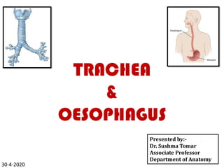
2. TRACHEA
• Also called wind pipe.
• A non-collapsible,
membrano-cartilaginous
mobile tube.
• Forms the beginning of
lower respiratory passage.
3. TRACHEA contd…
• Begins in the neck as a continuation of larynx at the lower border of
the cricoid cartilage at the level of lower border of sixth cervical
vertebra.
4. TRACHEA contd…
COURSE-
• Passes downwards through the neck and the superior mediastinum of
thorax.
• Ends by dividing into right and left principal bronchi, opposite the sternal
angle at the level of lower border of T4 vertebra.
5.
6. TRACHEA CONTD…
• Trachea lies in median plane
except at its termination.
• Near the bifurcation, trachea
deviates slightly to the right due
to the pressure of arch of aorta.
• Bifurcation of trachea
corresponds with lower border
of T4 vertebra in cadaver and in
supine position.
• Bifurcation extends to T6
vertebra in the living and and in
standing position.
• In the newborn, trachea
bifurcates at the level of T3
vertebra.
7. TRACHEA CONTD…
During expiration the bifurcation rises by
about one vertebral level.
During deep inspiration may be lowered as far
as the sixth thoracic vertebra.
8. DIMENSIONS
LENGTH: 10-11cm (4-6 inch)
BREADTH: Outer Diameter: ~2cm (in adult males)
~1.5 cm (in adult females)
Lumen is ~3mm in newborns and remain so up to the 3rd year of life.
Lumen increases by 1mm each year up to 12 years.
Lumen is smaller in living human being than in cadavers.
9. DIFFERENCES IN TRACHEA BETWEEN CHILDREN
&ADULTS
S.NO. FEATURES IN CHILDREN IN ADULTS
1 INTERNAL
DIAMETER
≥ 3mm. 12 mm.
2 PLACEMENT Deeply placed. Superficially
placed.
3 MOBILITY More movable. Less
movable.
4 LOW
TRACHEOSTOMY
Risky, because above
the supra-sternal
notch, crossed
sometimes by left
brachio-cephalic vein
and summit of arch of
aorta.
Less risky,
because
trachea is
not usually
crossed by
these
structures.
11. CARTILAGINOUS RINGS
C-shaped hyaline
cartilaginous rings.
~16-20 in number.
Deficient posteriorly in
order to allow expansion
of oesophagus during
deglutition.
The posterior free ends of
the cartilage are
connected by smooth
muscle trachealis.
First ring is the broadest.
12. CARTILAGINOUS RINGS contd…
Last ring presents a
triangular process
known as the carina.
Carina hooks
upwards from the
lower margin and
surrounds the
commencement of
two bronchi.
13. CLINICAL IMPORTANCE of CARINA
Carina presents a ridge
in the interior of
tracheal bifurcation..
Acts as a guide for the
surgeon during
bronchoscopic or other
examinations.
Mucus membrane at the
carina is one of the most
sensitive areas and is
associated with cough
reflex.
14. BI, All Rights Reserved, 2005 14
The carina
Posterior
From the head
From the front
Posterior
Note vertical RMB
15. MUCUS MEMBRANE
• Lined by ciliated
pseudo-stratified
columnar epithelium.
• Provided with
numerous goblet cells.
• Taller cells have cilia.
16. Cervical part of Trachea
• ~ 7 cm in length.
EXTENT-
• From lower border of
Cricoid cartilage to the
upper border of
manubrium sterni.
17. Relations
ANTERIOR-
• Skin.
• Superficial fascia containing anterior
jugular veins and jugular venous arch.
• Investing layer of deep cervical fascia.
• Sternothyroid and Sternohyoid muscles.
• Isthmus of Thyroid gland.
• Inferior Thyroid veins and arteria
thyroidea ima.
• Left brachiocephalic vein (in children)
• Thymus gland (in children).
• Brachiocephalic artery (in children).
20. RELATIONS (of thoracic part)
ANTERIORLY:
Sternum.
Thymus.
Left brachiocephalic vein.
Origins of the
brachiocephalic and left
common carotid arteries,
and the arch of the aorta.
21. RELATIONS OF TRACHEA CONTD…
POSTERIORLY:
Esophagus.
Left recurrent laryngeal nerve.
22. RELATIONS OF TRACHEA CONTD...
RIGHT SIDE:
Azygos vein.
Right vagus nerve.
Pleura.
LEFT SIDE:
Arch of the aorta.
Left common carotid and left
subclavian arteries.
Left vagus.
Left phrenic nerves, and the
pleura
25. NERVE SUPPLY
PARASYMPATHETIC- Recurrent laryngeal nerves.
Motor to trachealis muscles.
Secretomotor to glands.
Sensory to mucus membrane.
SYMPATHETIC-
Derived from upper 4 or 5 thoracic segments of spinal cord.
Post-ganglionic neurons are located in middle cervical sympathetic ganglion.
Vasomotor.
26. APPLIED ANATOMY
TRACHEOSTOMY-
• Life saving surgical procedure.
• Done in cases of laryngeal
obstruction.
• Comonly done in retrothyroid region.
• Trachea is opened by a vertical
incision.
27. OESOPHAGUS
• Muscular tube of 25cm
length.
• Connects the pharynx to
the stomach.
• Flattened anteroposterioly.
28. OESOPHAGUS contd…
• Begins in the neck at lower
border of cricoid cartilage
(at the lower border ofC6
vertebra).
• Pierces the diaphragm at
T10.
• Opens into the stomach at
T11.
30. OESOPHAGUS - DIVISIONS
CERVICAL PART –
• Ends at the lower border of
T1.
THORACIC PART –
• Ends at T10 where is pierces
the diaphragm.
ABDOMINAL PART –
• Ends at the cardiac end of the
stomach.
31. Cervical Part of Oesophagus
EXTENT-
• From lower border of cricoid cartilage to the superior border of
manubrium sterni.
• Begins in the midline but inclines slightly to the left as it descends.
32. Relations
Anterior-
• Trachea.
• Recurrent laryngeal
nerves.
Posterior-
• Vertebral column.
• Prevertebral fascia.
• Longus colli muscles.
On each side-
• Lobe of thyroid gland.
• Common carotid artery.
• Thoracic duct on left
side.