

Recommended
More Related Content
What's hot
What's hot (20)
Similar to Tissue and Its Types
Similar to Tissue and Its Types (20)
Recently uploaded
Recently uploaded (20)
Tissue and Its Types
- 1. Tissues Prof. Nilachal Human Anatomy & Physiology SBU, Ranchi 1 Tissue Key points • Humans—and other complex multicellular organisms—have systems of organs that work together, carrying out processes that keep us alive. • The body has levels of organization that build on each other. Cells make up tissues, tissues make up organs, and organs make up organ systems. • The function of an organ system depends on the integrated activity of its organs. For instance, digestive system organs cooperate to process food. • The survival of the organism depends on the integrated activity of all the organ systems, often coordinated by the endocrine and nervous systems. Introduction If you were a single-celled organism and you lived in a nutrient-rich place, staying alive would be pretty straightforward. For instance, if you were an amoeba living in a pond, you could absorb nutrients straight from your environment. The oxygen you would need for metabolism could diffuse in across your cell membrane, and carbon dioxide and other wastes could diffuse out. When the time came to reproduce, you could just divide yourself in two! However, odds are you are not an amoeba—given that you're using Khan Academy right now—and things aren’t quite so simple for big, many-celled organisms like human beings. Your complex body has
- 2. Tissues Prof. Nilachal Human Anatomy & Physiology SBU, Ranchi 2 over 30 trillion cells, and most of those cells aren’t in direct contact with the external environment.^11start superscript, 1, end superscript A cell deep inside your body—in one of your bones, say, or in your liver—can’t get the nutrients or oxygen it needs directly from the environment. How, then, does the body nourish its cells and keep itself running? Let's take a closer look at how the organization of your amazing body makes this possible. Multicellular organisms need specialized systems Most cells in large multicellular organisms don't directly exchange substances like nutrients and wastes with the external environment, instead, they are surrounded by an internal environment of extracellular fluid—literally, fluid outside of cells. The cells get oxygen and nutrients from this extracellular fluid and release waste products into it. Humans and other complex organisms have specialized systems that maintain the internal environment, keeping it steady and able to provide for the needs of the cells. Different systems of the body carry out different functions. For example, your digestive system is responsible for taking in and processing food, while your respiratory system—working with your circulatory system—is responsible for taking up oxygen and getting rid of carbon dioxide. The muscular and skeletal systems are crucial for movement; the reproductive system handles reproduction; and the excretory system gets rid of metabolic waste. Because of their specialization, these different systems are dependent on each other. The cells that make up the digestive, muscular, skeletal, reproductive, and excretory systems all need oxygen from the
- 3. Tissues Prof. Nilachal Human Anatomy & Physiology SBU, Ranchi 3 respiratory system to function, and the cells of the respiratory system—as well as all the other systems—need nutrients and must get rid of metabolic wastes. All the systems of the body work together to keep an organism up and running. Overview of body organization All living organisms are made up of one or more cells. Unicellular organisms, like amoebas, consist of only a single cell. Multicellular organisms, like people, are made up of many cells. Cells are considered the fundamental units of life. The cells in complex multicellular organisms like people are organized into tissues, groups of similar cells that work together on a specific task. Organs are structures made up of two or more tissues organized to carry out a particular function, and groups of organs with related functions make up the different organ systems. A tissue is a group of cells, in close proximity, organized to perform one or more specific functions. There are four basic tissue types defined by their morphology and function: epithelial tissue, connective tissue, muscle tissue, and nervous tissue. • Epithelial tissue creates protective boundaries and is involved in the diffusion of ions and molecules. • Connective tissue underlies and supports other tissue types. • Muscle tissue contracts to initiate movement in the body.
- 4. Tissues Prof. Nilachal Human Anatomy & Physiology SBU, Ranchi 4 • Nervous tissue transmits and integrates information through the central and peripheral nervous systems. Types of tissues Every organ is made up of two or more tissues, groups of similar cells that work together to perform a specific task. Humans—and other large multicellular animals—are made up of four basic tissue types: epithelial tissue, connective tissue, muscle tissue, and nervous tissue.
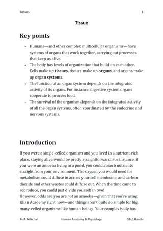
- 5. Tissues Prof. Nilachal Human Anatomy & Physiology SBU, Ranchi 5 Image credit: modified from Types of tissues: Figure 1 by OpenStax College, Anatomy & Physiology, CC BY 3.0 Epithelial tissue Epithelial tissue consists of tightly packed sheets of cells that cover surfaces—including the outside of the body—and line body cavities. For instance, the outer layer of your skin is an epithelial tissue, and so is the lining of your small intestine.
- 6. Tissues Prof. Nilachal Human Anatomy & Physiology SBU, Ranchi 6 Epithelial cells are polarized, meaning that they have a top and a bottom side. The apical, top, side of an epithelial cell faces the inside of a cavity or the outside of a structure and is usually exposed to fluid or air. The basal, bottom, side faces the underlying cells. For instance, the apical sides of intestinal cells have finger-like structures that increase surface area for absorbing nutrients. Image showing three cells lining the small intestine. Each cell contains a nucleus and is surrounded by a plasma membrane. The tops of the cells have microvilli that face the cavity from which substances will be absorbed. Image credit: Eukaryotic cells: Figure 3 by OpenStax College, Biology, CC BY 3.0 Epithelial cells are tightly packed, and this lets them act as barriers to the movement of fluids and potentially harmful microbes. Often, the cells are joined by specialized junctions that hold them tightly together to reduce leaks.
- 7. Tissues Prof. Nilachal Human Anatomy & Physiology SBU, Ranchi 7 Connective tissue Connective tissue consists of cells suspended in an extracellular matrix. In most cases, the matrix is made up of protein fibers like collagen and fibrin in a solid, liquid, or jellylike ground substance. Connective tissue supports and, as the name suggests, connects other tissues. Loose connective tissue, show below, is the most common type of connective tissue. It's found throughout your body, and it supports organs and blood vessels and links epithelial tissues to the muscles underneath. Dense, or fibrous, connective tissue is found in tendons and ligaments, which connect muscles to bones and bones to each other, respectively.
- 8. Tissues Prof. Nilachal Human Anatomy & Physiology SBU, Ranchi 8 Loose connective tissue is composed of loosely woven collagen and elastic fibers. The fibers and other components of the connective tissue matrix are secreted by fibroblasts. Image credit: Animal primary tissues: Figure 6 by OpenStax College, Biology, CC BY 4.0 Specialized forms of connective tissue include adipose tissue—body fat—bone, cartilage, and blood, in which the extracellular matrix is a liquid called plasma. Muscle tissue Muscle tissue is essential for keeping the body upright, allowing it to move, and even pumping blood and pushing food through the digestive tract. Muscle cells, often called muscle fibers, contain the proteins actin and myosin, which allow them to contract. There are three main types of muscle: skeletal muscle, cardiac muscle, and smooth muscle. From left to right. Smooth muscle cells, skeletal muscle cells, and cardiac muscle cells. Smooth muscle cells do not have striations, while skeletal muscle cells do. Cardiac muscle cells have striations, but, unlike the multinucleate skeletal cells, they have only one nucleus. Cardiac muscle tissue also has intercalated discs, specialized regions running along the plasma membrane that join adjacent cardiac muscle cells and assist in passing an electrical impulse from cell to cell. Image credit: Animal primary tissues: Figure 12 by OpenStax College, Biology, CC BY 4.0 Skeletal muscle, which is also called striated—striped—muscle, is what we refer to as muscle in everyday life. Skeletal muscle is attached to bones by tendons, and it allows you to consciously control your
- 9. Tissues Prof. Nilachal Human Anatomy & Physiology SBU, Ranchi 9 movements. For instance, the quads in your legs or biceps in your arms are skeletal muscle. Cardiac muscle is found only in the walls of the heart. Like skeletal muscle, cardiac muscle is striated, or striped. But it's not under voluntary control, so—thankfully!—you don’t need to think about making your heart beat. The individual fibers are connected by structures called intercalated disks, which allow them to contract in sync. Smooth muscle is found in the walls of blood vessels, as well as in the walls of the digestive tract, the uterus, the urinary bladder, and various other internal structures. Smooth muscle is not striped, striated, and it's involuntary, not under conscious control. That means you don't have to think about moving food through your digestive tract! Nervous tissue Nervous tissue is involved in sensing stimuli—external or internal cues—and processing and transmitting information. It consists of two main types of cells: neurons, or nerve cells, and glia. The neurons are the basic functional unit of the nervous system. They generate electrical signals called conducted nerve impulses or action potentials that allow the neurons to convey information very rapidly across long distances. The glia mainly act to support neuronal function.
- 10. Tissues Prof. Nilachal Human Anatomy & Physiology SBU, Ranchi 10 Picture of neuron. The neuron has projections called dendrites that receive signals and projections called axons that send signals. Also shown are two types of glial cells: astrocytes regulate the chemical environment of the nerve cell, and oligodendrocytes insulate the axon so the electrical nerve impulse is transferred more efficiently. Image credit: Animal primary tissues: Figure 13 by OpenStax College, Biology, CC BY 4.0
- 11. Tissues Prof. Nilachal Human Anatomy & Physiology SBU, Ranchi 11 Organs Organs, such as the heart, the lungs, the stomach, the kidneys, the skin, and the liver, are made up of two or more types of tissue organized to serve a particular function. For example, the heart pumps blood, the lungs bring in oxygen and eliminate carbon dioxide, and the skin provides a barrier to protect internal structures from the external environment. Most organs contain all four tissue types. The layered walls of the small intestine provide a good example of how tissues form an organ. The inside of the intestine is lined by epithelial cells, some of which secrete hormones or digestive enzymes and others of which absorb nutrients. Around the epithelial layer are layers of connective tissue and smooth muscle, interspersed with glands, blood vessels, and neurons. The smooth muscle contracts to move food through the gut, under control of its associated networks of neurons.^22squared
- 12. Tissues Prof. Nilachal Human Anatomy & Physiology SBU, Ranchi 12 Cross-section of the GI tract. From outside to inside: Blood vessels, networks of nerves in smooth muscle layers, connective tissue, more smooth muscle, another layer of connective tissue, epithelial tissue, and empty space in the middle as the path of digested food. Image credit: modified from Layers of the GI tract by Goran tek-en, [CC BY-SA 3.0](https://creativecommons.org/licenses/by-sa/3.0/deed.en; the modified image is licensed under a CC BY-SA 3.0 license